articol chitosan-glutaraldehida
19
Mar. Drugs 2013, 11, 1534-1552; doi:10.3390/md11051534 Marine Drugs ISSN 1660-3397 www.mdpi.com/journal/marinedrugs Article Synthesis, Characterization, and Antibacterial Activity of Cross-Linked Chitosan-Glutaraldehyde Bin Li 1,2 , Chang-Lin Shan 1 , Qing Zhou 1 , Yuan Fang 3 , Yang-Li Wang 2 , Fei Xu 4 , Li-Rong Han 5, *, Muhammad Ibrahim 1 , Long-Biao Guo 6, *, Guan-Lin Xie 1 and Guo-Chang Sun 2, * 1 State Key Laboratory of Rice Biology, Institute of Biotech nology, Z hejiang Unive rsity, Hangzhou 310058, China; E-Mails: [email protected] u.cn (B.L.); changlin_sh [email protected] (C.-L.S.); zhou-0725-qing@1 63.com ( Q.Z.); [email protected] du.pk (M.I.); [email protected] .cn (G.-L.X.) 2 State Key Laboratory B reeding Bas e for Zhejiang Sustainable P lant Pest an d Disease Control, Zhejiang Academy of Agricultural Sciences, Hangzhou 310021, China; E-Mail: wangylaa@y ahoo.com.cn 3 College of Chemistry and Life Sciences, Zhejiang Normal Unive rsity, Jinhua 321004, China; E-Mail: Fy0579@zjnu. cn 4 Institute of Digital Agriculture, Zhejiang Academy of Agricultural Sciences, Hangzhou 310021, China; E-Mail: [email protected] n 5 Research and De velopment Ce nter of Biorational Pesticides, Northwest A & F U niversity, Yangling, Shaanxi 712100, China 6 State Key Laboratory of Rice Biology, Ch ina National Rice Res earch In stitute, Hangzhou 310006, China * Authors to whom corresponden ce should be addressed; E-Ma ils: [email protected] m (L.-R.H.); [email protected] (L.-B.G.); [email protected] (G.-C.S.); Tel.: +86-29-87092122 (L.-R.H.); +86-571-63370537 (L.-B.G.); +86-571-86404273 (G.-C.S.). Received: 18 February 2013; in revised form: 1 7 April 2013 / Accepted: 26 April 201 3 / Published: 13 May 2 013 Abstract: This present study deals with synthesis, characterization and antibacterial activity of cross-linked chitosan-glutaraldehyde . Results f rom this study i ndicated that cross-linked chitosan-glutara ldehyde markedly inhibited the growth of antibiotic-resistant Burkholderia cepacia complex regardless of bacterial species and incubation time while bacterial growth was unaffected by solid chitosan. Furthermore, high temperature treated cross-linked chitosan-glutaraldehyde showed strong antibacterial activity against the selected strain 0901 although the inhibitory effects varied with different temperatures. In addition, OPEN ACCESS
-
Upload
mihaibianca -
Category
Documents
-
view
219 -
download
0
Transcript of articol chitosan-glutaraldehida
8/13/2019 articol chitosan-glutaraldehida
http://slidepdf.com/reader/full/articol-chitosan-glutaraldehida 1/19
8/13/2019 articol chitosan-glutaraldehida
http://slidepdf.com/reader/full/articol-chitosan-glutaraldehida 2/19
Mar. Drugs 2013, 11 1535
physical-chemical and structural characterization revealed that the cross-linking of chitosan
with glutaraldehyde resulted in a rougher surface morphology, a characteristic Fourier
transform infrared (FTIR) band at 1559 cm−1, a specific X-ray diffraction peak centered at
2θ = 15°, a lower contents of carbon, hydrogen and nitrogen, and a higher stability of
glucose units compared to chitosan based on scanning electron microscopic observation,FTIR spectra, X-ray diffraction pattern, as well as elemental and thermo gravimetric
analysis. Overall, this study indicated that cross-linked chitosan-glutaraldehyde is promising
to be developed as a new antibacterial drug.
Keywords: antibacterial activity; characterization; chitosan; cross-link; glutaraldehyde
1. Introduction
The Burkholderia cepacia complex (Bcc) is a collection of genetically distinct but phenotypically
similar bacteria that have emerged as life-threatening pulmonary pathogens in immunocompromised
patients, particularly individuals with cystic fibrosis (CF). Indeed, the Bcc currently comprises
17 different species and infection with Bcc often leads to a fast decline in lung function and a markedly
increased mortality [1 – 3]. The number of infections caused by the Bcc is increasing in China [4 – 6],
while some species such as Burkholderia cenocepacia, Burkholderia contaminans, Burkholderia
multivorans, Burkholderia seminalis, Burkholderia stabilis, and Burkholderia vietnamiensis have been
isolated from agricultural and hospital environments in our previous studies [6 – 10].Treatment of CF infections is very difficult due to the intrinsic resistance of Bcc bacteria to most
clinically useful antibiotics, while some isolates of Bcc even can utilize penicillin G as a sole carbon
source for growth [6,11]. Thus, it becomes important to identify newer and improved antibacterial
therapies for CF patients. Recently, chitosan, a natural nontoxic biopolymer derived by deacetylation of
chitin, a major component of the shells of crustacea such as crab, shrimp, and crawfish, has been
applied in the fields of medicine, food, chemical engineering, pharmaceuticals, nutrition,
environmental protection and agriculture [12,13]. In particular, chitosan not only has several
advantages over other types of bactericides [14], but also has strong antibacterial activity against a
variety of bacteria [15 – 20]. However, unfortunately, in our previous studies, chitosan solution possesseda limited antibacterial activity against Bcc bacteria [4,11].
Interestingly, previous studies have revealed that the biological activities of chitosan and its derivatives
could be affected by different environments [14,21] and improved by either combining chitosan with
different metal ions [22 – 25] or cross-linking chitosan with other organic compounds [26 – 28]. Indeed,
chitosan has been cross-linked with glutaraldehyde in several studies [27,29], while these cross-linked
complexes have been found to play a key role in the uptake of heavy metals [30,31]. However, to the best
of our knowledge, little is known about the antibacterial activity of these cross-linked complexes against
Bcc bacteria.
The aim of this study was to synthesize and characterize a cross-linked complex of chitosan andglutaraldehyde with strong anti-Bcc activity.
8/13/2019 articol chitosan-glutaraldehida
http://slidepdf.com/reader/full/articol-chitosan-glutaraldehida 3/19
Mar. Drugs 2013, 11 1536
2. Results and Discussion
Results from this study indicated that bacterial growth was unaffected by solid chitosan, but was
strongly inhibited by the solid cross-linked chitosan-glutaraldehyde (CLCG) with cross-linking degree
of 80.8% regardless of bacterial species and incubation time. In addition, high temperature treatedCLCG showed strong antibacterial activity against the selected strain 0901 of the Bcc although the
inhibitory effects varied with different temperatures. The differential antibacterial activity between
CLCG and chitosan may be mainly due to the difference in their physical-chemical properties, which
were evidenced by scanning electron microscopic (SEM) observation, Fourier transform infrared (FTIR)
spectra, X-ray diffraction (XRD) pattern, as well as elemental and thermo gravimetric analysis. To the
best of our knowledge, this study first synthesized the glutaraldehyde cross-linked chitosan with strong
anti-Bcc activity.
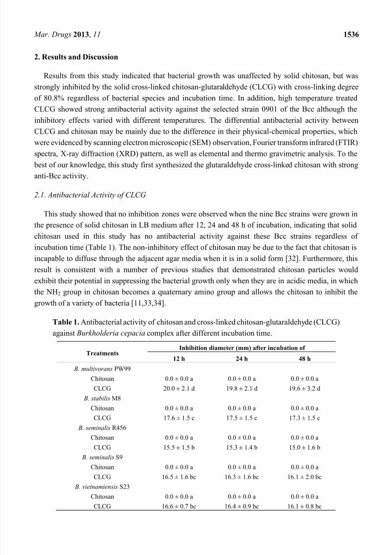
2.1. Antibacterial Activity of CLCG
This study showed that no inhibition zones were observed when the nine Bcc strains were grown in
the presence of solid chitosan in LB medium after 12, 24 and 48 h of incubation, indicating that solid
chitosan used in this study has no antibacterial activity against these Bcc strains regardless of
incubation time (Table 1). The non-inhibitory effect of chitosan may be due to the fact that chitosan is
incapable to diffuse through the adjacent agar media when it is in a solid form [32]. Furthermore, this
result is consistent with a number of previous studies that demonstrated chitosan particles would
exhibit their potential in suppressing the bacterial growth only when they are in acidic media, in which
the NH2 group in chitosan becomes a quaternary amino group and allows the chitosan to inhibit thegrowth of a variety of bacteria [11,33,34].
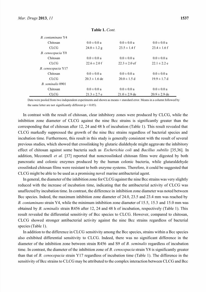
Table 1. Antibacterial activity of chitosan and cross-linked chitosan-glutaraldehyde (CLCG)
against Burkholderia cepacia complex after different incubation time.
TreatmentsInhibition diameter (mm) after incubation of
12 h 24 h 48 h
B. multivorans PW99
Chitosan 0.0 ± 0.0 a 0.0 ± 0.0 a 0.0 ± 0.0 a
CLCG 20.0 ± 2.1 d 19.8 ± 2.1 d 19.6 ± 3.2 d B. stabilis M8
Chitosan 0.0 ± 0.0 a 0.0 ± 0.0 a 0.0 ± 0.0 a
CLCG 17.6 ± 1.5 c 17.5 ± 1.5 c 17.3 ± 1.5 c
B. seminalis R456
Chitosan 0.0 ± 0.0 a 0.0 ± 0.0 a 0.0 ± 0.0 a
CLCG 15.5 ± 1.5 b 15.3 ± 1.4 b 15.0 ± 1.6 b
B. seminalis S9
Chitosan 0.0 ± 0.0 a 0.0 ± 0.0 a 0.0 ± 0.0 a
CLCG 16.5 ± 1.6 bc 16.3 ± 1.6 bc 16.1 ± 2.0 bc
B. vietnamiensis S23Chitosan 0.0 ± 0.0 a 0.0 ± 0.0 a 0.0 ± 0.0 a
CLCG 16.6 ± 0.7 bc 16.4 ± 0.9 bc 16.1 ± 0.8 bc
8/13/2019 articol chitosan-glutaraldehida
http://slidepdf.com/reader/full/articol-chitosan-glutaraldehida 4/19
Mar. Drugs 2013, 11 1537
Table 1. Cont.
B. contaminans Y4
Chitosan 0.0 ± 0.0 a 0.0 ± 0.0 a 0.0 ± 0.0 a
CLCG 24.0 ± 1.2 g 23.5 ± 1.4 f 23.4 ± 1.6 f
B. cenocepacia Y8
Chitosan 0.0 ± 0.0 a 0.0 ± 0.0 a 0.0 ± 0.0 a
CLCG 22.6 ± 2.0 f 22.3 ± 2.0 ef 22.1 ± 2.2 e
B. cenocepacia Y17
Chitosan 0.0 ± 0.0 a 0.0 ± 0.0 a 0.0 ± 0.0 a
CLCG 20.3 ± 1.6 de 20.0 ± 1.5 d 19.9 ± 1.7 d
B. seminalis 0901
Chitosan 0.0 ± 0.0 a 0.0 ± 0.0 a 0.0 ± 0.0 a
CLCG 21.3 ± 2.7 e 21.0 ± 2.9 de 20.9 ± 2.9 de
Data were pooled from two independent experiments and shown as means ± standard error. Means in a column followed by
the same letter are not significantly different ( p < 0.05).
In contrast with the result of chitosan, clear inhibitory zones were produced by CLCG, while the
inhibition zone diameter of CLCG against the nine Bcc strains is significantly greater than the
corresponding that of chitosan after 12, 24 and 48 h of incubation (Table 1). This result revealed that
CLCG markedly suppressed the growth of the nine Bcc strains regardless of bacterial species and
incubation time. Furthermore, this result in this study is generally consistent with the result of several
previous studies, which showed that crosslinking by glutaric dialdehyde might aggravate the inhibitory
effect of chitosan against some bacteria such as Escherichia coli and Bacillus subtilis [35,36]. In
addition, Mcconnell et al. [37] reported that noncrosslinked chitosan films were digested by both pancreatic and colonic enzymes produced by the human colonic bacteria, while glutaraldehyde
crosslinked chitosan films were resistant to both enzyme systems. Therefore, it could be suggested that
CLCG might be able to be used as a promising novel marine antibacterial agent.
In general, the diameter of the inhibition zone for CLCG against the nine Bcc strains was very slightly
reduced with the increase of incubation time, indicating that the antibacterial activity of CLCG was
unaffected by incubation time. In contrast, the difference in inhibition zone diameter was noted between
Bcc species. Indeed, the maximum inhibition zone diameter of 24.0, 23.5 and 23.4 mm was reached by
B. contaminans strain Y4, while the minimum inhibition zone diameter of 15.5, 15.3 and 15.0 mm was
obtained by B. seminalis strain R456 after 12, 24 and 48 h of incubation, respectively (Table 1). This
result revealed the differential sensitivity of Bcc species to CLCG. However, compared to chitosan,
CLCG showed stronger antibacterial activity against the nine Bcc strains regardless of bacterial
species (Table 1).
In addition to the difference in CLCG sensitivity among the Bcc species, strains within a Bcc species
also exhibited differential sensitivity to CLCG. Indeed, there was no significant difference in the
diameter of the inhibition zone between strain R456 and S9 of B. seminalis regardless of incubation
time. In contrast, the diameter of the inhibition zone of B. cenocepacia strain Y8 is significantly greater
than that of B. cenocepacia strain Y17 regardless of incubation time (Table 1). The difference in thesensitivity of Bcc strains to CLCG may be attributed to the complex interaction between CLCG and Bcc
8/13/2019 articol chitosan-glutaraldehida
http://slidepdf.com/reader/full/articol-chitosan-glutaraldehida 5/19
Mar. Drugs 2013, 11 1538
bacteria. However, the growth of all tested Bcc strains was significantly inhibited by CLCG compared to
chitosan (Table 1).
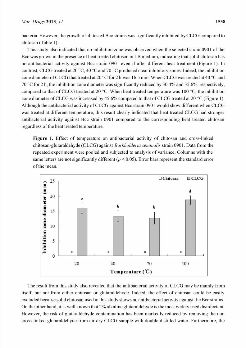
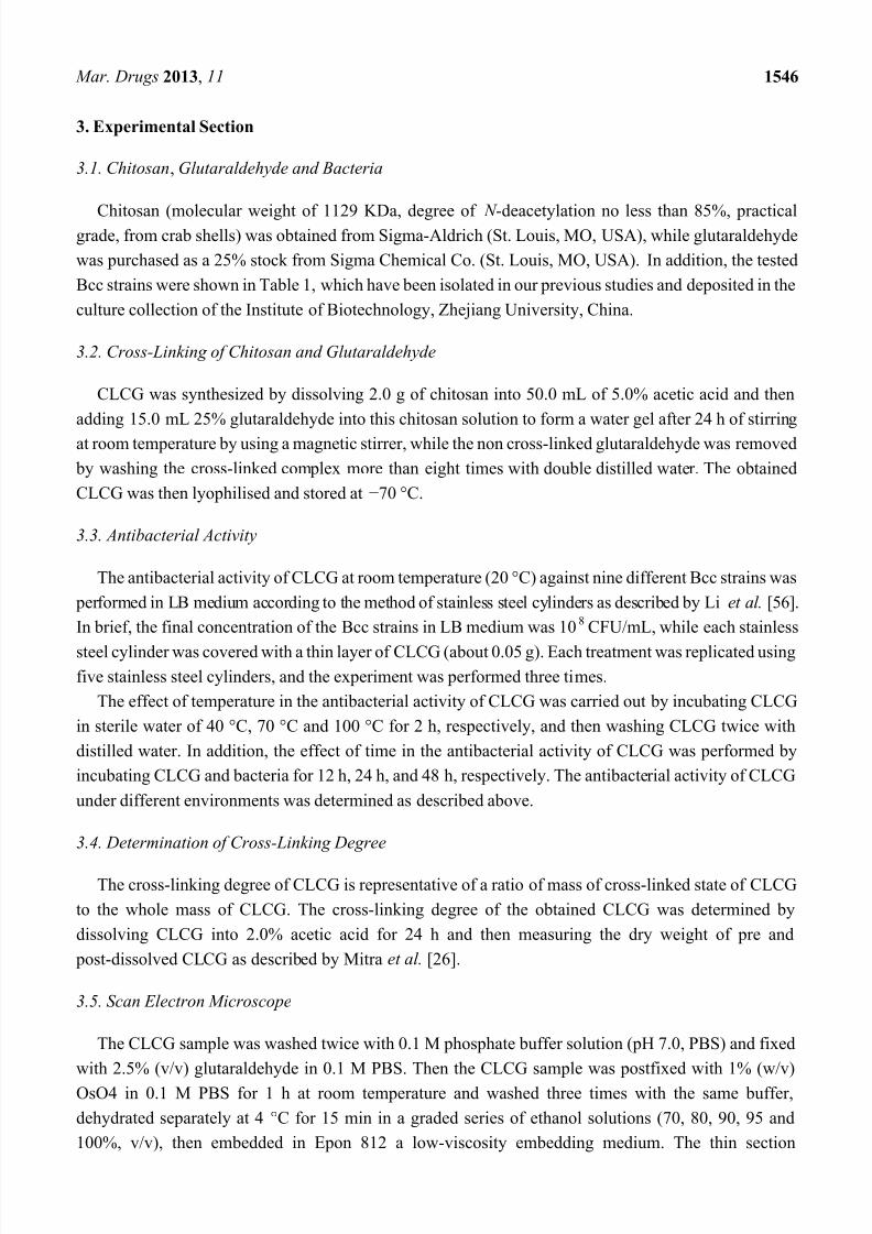
This study also indicated that no inhibition zone was observed when the selected strain 0901 of the
Bcc was grown in the presence of heat treated chitosan in LB medium, indicating that solid chitosan has
no antibacterial activity against Bcc strain 0901 even if after different heat treatment (Figure 1). Incontrast, CLCG treated at 20 °C, 40 °C and 70 °C produced clear inhibitory zones. Indeed, the inhibition
zone diameter of CLCG that treated at 20 °C for 2 h was 16.5 mm. When CLCG was treated at 40 °C and
70 °C for 2 h, the inhibition zone diameter was significantly reduced by 30.4% and 35.6%, respectively,
compared to that of CLCG treated at 20 °C. When heat treated temperature was 100 °C, the inhibition
zone diameter of CLCG was increased by 45.6% compared to that of CLCG treated at 20 °C (Figure 1).
Although the antibacterial activity of CLCG against Bcc strain 0901 would show different when CLCG
was treated at different temperature, this result clearly indicated that heat treated CLCG had stronger
antibacterial activity against Bcc strain 0901 compared to the corresponding heat treated chitosan
regardless of the heat treated temperature.
Figure 1. Effect of temperature on antibacterial activity of chitosan and cross-linked
chitosan-glutaraldehyde (CLCG) against Burkholderia seminalis strain 0901. Data from the
repeated experiment were pooled and subjected to analysis of variance. Columns with the
same letters are not significantly different ( p < 0.05). Error bars represent the standard error
of the mean.
The result from this study also revealed that the antibacterial activity of CLCG may be mainly from
itself, but not from either chitosan or glutaraldehyde. Indeed, the effect of chitosan could be easily
excluded because solid chitosan used in this study shows no antibacterial activity against the Bcc strains.
On the other hand, it is well known that 2% alkaline glutaraldehyde is the most widely used disinfectant.However, the risk of glutaraldehyde contamination has been markedly reduced by removing the non
cross-linked glutaraldehyde from air dry CLCG sample with double distilled water. Furthermore, the
8/13/2019 articol chitosan-glutaraldehida
http://slidepdf.com/reader/full/articol-chitosan-glutaraldehida 6/19
Mar. Drugs 2013, 11 1539
weak acid chitosan solution in this study may abolish the antibacterial effect of glutaraldehyde for the
glutaraldehyde activity at low concentrations is favored by an alkaline pH. In addition, the diameter of
the inhibition zone for CLCG against Bcc strain 0901 was significantly affected by heat treatment,
indicating that the antibacterial activity of CLCG mainly depends on the crosslink reaction between
chitosan and glutaraldehyde.The interaction between chitosan with glutaraldehyde have received considerable attention over the
past few decades [35,38], while the obtained data from 13C NMR, infrared and Raman spectroscopies
evidenced the formation of an ethylenic double bond in the chitosan – glutaraldehyde interaction [39]. In
addition, two main crosslinking mechanisms, involving formation of Schiff’s base structures or
Michael-type adducts, have been proposed for the reaction of chitosan and glutaraldehyde. However, the
nature of the chemical bonding between chitosan amine groups and glutaraldehyde has been subject of
controversy, which may be due to this reason that the rate of chemical gelation of chitosan chains with
glutaraldehyde depends on various parameters, namely pH, ionic strength, temperature, chitosan
concentration and degree of crosslinking [38,40].
This result indicated that the inhibition in bacterial growth should be mainly due to the direct in vitro
antibacterial activity of CLCG. However, Zhang et al. [35] found that in addition to increased
antibacterial activity, glutaric dialdehyde crosslinking also enhanced chitosan uptake on the surface of
cotton fabrics, with good durability of antibacterial properties to washing. This may be due to that
glutaric dialdehyde is a binary aldehyde compound. One aldehyde group of glutaraldehyde reacts with
amino group of chitosan, contributing to the antibacterial activity, while the other reacts with cellulose
of cotton fabrics, improving the fastness property. Therefore, it could be expected that crosslinking by
glutaraldehyde may have a direct and indirect contribution to the antibacterial activity of CLCG.
2.2. Cross-Linking Degree of CLCG
Results from this study indicated that the cross-linking degree of the synthesized CLCG with strong
anti-Bcc activity was 80.8%, while the other synthesized products with different cross-linking degrees
were discarded for limited anti-Bcc activities (data not shown). This result is in agreement with the
result of previous studies [41 – 43], which reported that in addition to degree of deacetylation in
chitosan, the degree of crosslinking also controls the properties of chitosan. In addition, a number of
studies have revealed that the aldehyde groups react immediately with the – NH2 groups along the chitosan
chains after introducing wet chitosan microspheres into glutaraldehyde solution [29,35,38,42,44,45].
Therefore, it could be suggested that CLCG might be formed by cross-linking chitosan with
glutaraldehyde at amino groups.
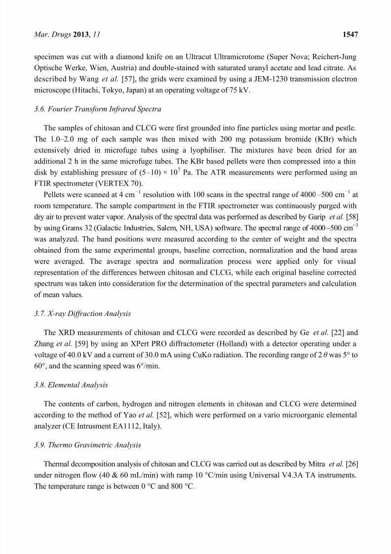
2.3. SEM of CLCG
Figure 2 presents the SEM micrographs illustrating the surface morphology of CLCG at four different
(1500×, 500×, 250× and 200×) magnifications. In general, this result clearly indicated that there is a
difference in the surface microscopic morphology between chitosan and CLCG. Indeed, compared to
the smooth, dense and flat morphology of chitosan [46], the SEM images in this study revealed thatCLCG had a rough and porous surface (Figure 2), which is consistent with the result of previous
studies [29,35,47]. In contrast, Wang et al. [48] and Kulkarni et al. [49] revealed that the microspheres of
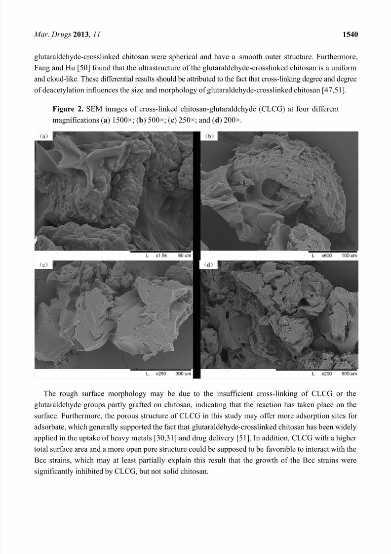
8/13/2019 articol chitosan-glutaraldehida
http://slidepdf.com/reader/full/articol-chitosan-glutaraldehida 7/19
8/13/2019 articol chitosan-glutaraldehida
http://slidepdf.com/reader/full/articol-chitosan-glutaraldehida 8/19
Mar. Drugs 2013, 11 1541
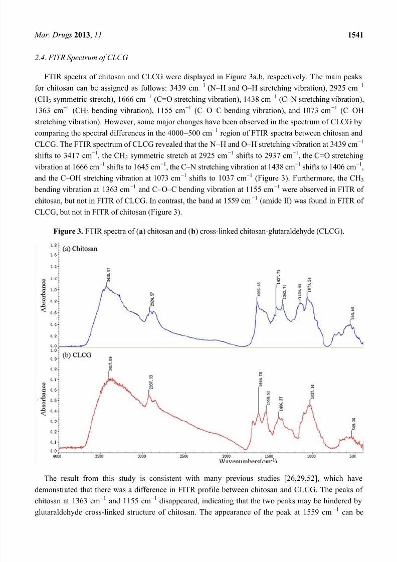
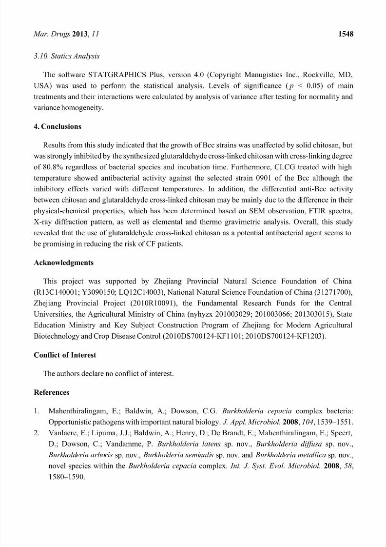
2.4. FITR Spectrum of CLCG
FTIR spectra of chitosan and CLCG were displayed in Figure 3a,b, respectively. The main peaks
for chitosan can be assigned as follows: 3439 cm−1 (N – H and O – H stretching vibration), 2925 cm−1
(CH3 symmetric stretch), 1666 cm−1 (C=O stretching vibration), 1438 cm−1 (C – N stretching vibration),1363 cm−1 (CH3 bending vibration), 1155 cm−1 (C – O – C bending vibration), and 1073 cm−1 (C – OH
stretching vibration). However, some major changes have been observed in the spectrum of CLCG by
comparing the spectral differences in the 4000 – 500 cm−1 region of FTIR spectra between chitosan and
CLCG. The FTIR spectrum of CLCG revealed that the N – H and O – H stretching vibration at 3439 cm−1
shifts to 3417 cm−1, the CH3 symmetric stretch at 2925 cm−1 shifts to 2937 cm−1, the C=O stretching
vibration at 1666 cm−1 shifts to 1645 cm−1, the C – N stretching vibration at 1438 cm−1 shifts to 1406 cm−1,
and the C – OH stretching vibration at 1073 cm−1 shifts to 1037 cm−1 (Figure 3). Furthermore, the CH3
bending vibration at 1363 cm−1 and C – O – C bending vibration at 1155 cm−1 were observed in FITR of
chitosan, but not in FITR of CLCG. In contrast, the band at 1559 cm−1 (amide II) was found in FITR ofCLCG, but not in FITR of chitosan (Figure 3).
Figure 3. FTIR spectra of (a) chitosan and (b) cross-linked chitosan-glutaraldehyde (CLCG).
The result from this study is consistent with many previous studies [26,29,52], which have
demonstrated that there was a difference in FITR profile between chitosan and CLCG. The peaks ofchitosan at 1363 cm−1 and 1155 cm−1 disappeared, indicating that the two peaks may be hindered by
glutaraldehyde cross-linked structure of chitosan. The appearance of the peak at 1559 cm−1 can be
8/13/2019 articol chitosan-glutaraldehida
http://slidepdf.com/reader/full/articol-chitosan-glutaraldehida 9/19
Mar. Drugs 2013, 11 1542
attributed to the crosslinking reaction of chitosan and glutaraldehyde. Indeed, Ramachandran et al. [53]
found that the new sharp peak at 1610 cm−1 represents stretching vibrations of C=N in Schiff’s base
formed by the reaction of glutaraldehyde and chitosan. Furthermore, Knaul et al. [45] found that the
chitosan film that reacted with glutaraldehyde exhibits a strong absorbance at 1664 cm−1, while Gupta
and Jabrail [41] reported that the IR spectra have shown a strong absorption band at 1660 cm −1 in theglutaraldehyde cross-linked chitosan microsphere. In addition, Oyrton et al. [39] revealed that the
increase of glutaraldehyde in the sequence of these modified chitosans caused a successive increase in
intensity of ethylenic bond frequency at 1562 cm−1.
The result from this study indicated that the amide II band at 1559 cm−1 that may contribute to the
amine – NH2 group was specific for CLCG. Interestingly, Tripathi et al. [32] revealed the change in the
characteristic shape of the chitosan spectrum after cross-linking while stretching vibration spectra of the
amide group of chitosan-based antimicrobial films appear at 1560 cm−1. Furthermore, Zhang et al. [35]
reported that the intensity at 1557.19 reveals the quaternary-amino band. Thus, the antibacterial activity
of CLCG may be due to the positive charge NH2+, which was expected to interact with gram-negative
bacterial surface that predominantly consisted of anionic components, such as lipopolysaccharides,
phospholipids, and lipoproteins. Indeed, previous studies have indicated that this charge interaction
could disrupt the organization of the outer membrane in bacteria and increase its permeability [11,14].
Therefore, it could be suggested that the inhibitory activity of CLCG to the Bcc strains is mainly comes
from its positive charge.
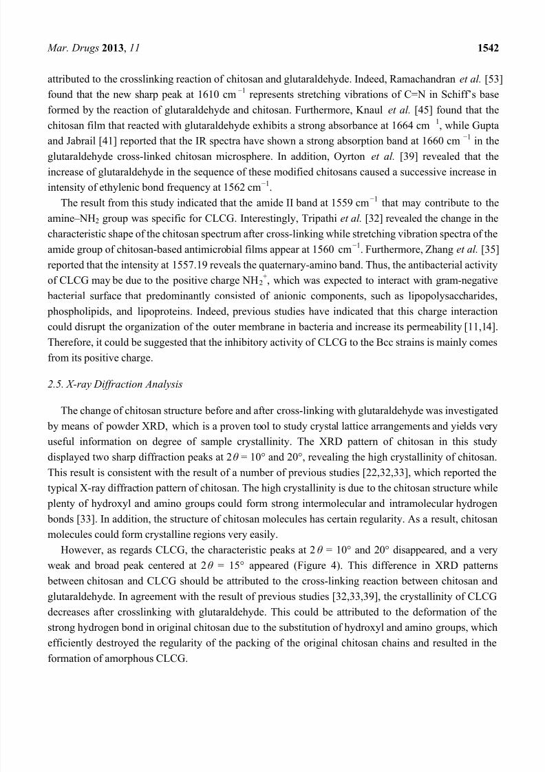
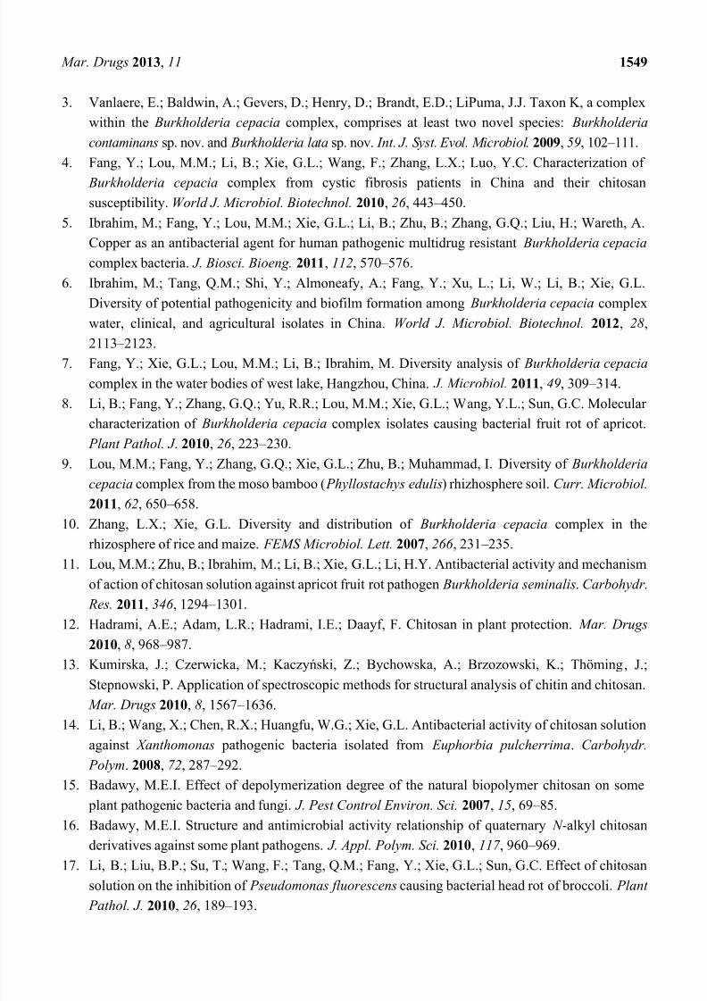
2.5. X-ray Diffraction Analysis
The change of chitosan structure before and after cross-linking with glutaraldehyde was investigated by means of powder XRD, which is a proven tool to study crystal lattice arrangements and yields very
useful information on degree of sample crystallinity. The XRD pattern of chitosan in this study
displayed two sharp diffraction peaks at 2θ = 10° and 20°, revealing the high crystallinity of chitosan.
This result is consistent with the result of a number of previous studies [22,32,33], which reported the
typical X-ray diffraction pattern of chitosan. The high crystallinity is due to the chitosan structure while
plenty of hydroxyl and amino groups could form strong intermolecular and intramolecular hydrogen
bonds [33]. In addition, the structure of chitosan molecules has certain regularity. As a result, chitosan
molecules could form crystalline regions very easily.
However, as regards CLCG, the characteristic peaks at 2θ = 10° and 20° disappeared, and a very
weak and broad peak centered at 2θ = 15° appeared (Figure 4). This difference in XRD patterns
between chitosan and CLCG should be attributed to the cross-linking reaction between chitosan and
glutaraldehyde. In agreement with the result of previous studies [32,33,39], the crystallinity of CLCG
decreases after crosslinking with glutaraldehyde. This could be attributed to the deformation of the
strong hydrogen bond in original chitosan due to the substitution of hydroxyl and amino groups, which
efficiently destroyed the regularity of the packing of the original chitosan chains and resulted in the
formation of amorphous CLCG.
8/13/2019 articol chitosan-glutaraldehida
http://slidepdf.com/reader/full/articol-chitosan-glutaraldehida 10/19
Mar. Drugs 2013, 11 1543
Figure 4. X-ray diffractograms of (a) chitosan and (b) cross-linked chitosan-glutaraldehyde (CLCG).
Recently, Mohamed and Fahmy [54] revealed that the incorporation of hydrophilic cross-linker into
chitosan allowed the synthesis of hydrogels with higher hydrophilicity, with greater positive chargedensity and with higher antimicrobial activities, which may explain the result that CLCG had a better
antibacterial activity against the Bcc strains compared to the solid chitosan. As mentioned above, the
antibacterial mechanism of CLCG may be mainly attributed to the interaction between positive
charged CLCG molecules and negatively charged bacterial cell membranes. It suggests that the greater
the number of cationized groups, the higher the antibacterial activity. Furthermore, the decrease in
intermolecular hydrogen bonds of CLCG could result in the increase of its solubility, which is able to
facilitate the penetration of CLCG into the cells of bacteria, thereby preventing the transformation of
DNA to RNA to obtain a higher antibacterial activity. In addition, the antibacterial activity of the
amorphous CLCG against the Bcc strains may partially due to its ability to chelate metal ions that bind
to nutrients essential to bacterial growth.
2.6. Elemental Analysis of CLCG
Results from this study indicated that there was a difference in the elements between chitosan and
CLCG. Indeed, the element analysis indicated that CLCG had a composition of 33.5% (w/w) carbon,
6.5% (w/w) hydrogen and 1.6% (w/w) nitrogen. In agreement with the result of Gupta et al. [41], this
study indicated that the contents of carbon, hydrogen and nitrogen in CLCG were lower than the
corresponding contents in chitosan, which reduced by 17.2%, 7.9% and 77.4%, respectively (Table 2).In contrast, Oyrton et al. [39] found that the content of carbon and hydrogen percentages is followed by
an increase of glutaraldehyde in the chitosan sequence of modified polymers, while the amount of
nitrogen decreases with the increasing degree of glutaraldehyde in this series of samples. Therefore, this
differential result may be attributed to the difference in the degree of crosslinking.
Table 2. Elementary analysis of chitosan and cross-linked chitosan-glutaraldehyde (CLCG).
SubstanceElement (%, w/w)
Carbon Hydrogen Nitrogen
ChitosanCLCG
40.4733.50
7.086.52
7.211.63
8/13/2019 articol chitosan-glutaraldehida
http://slidepdf.com/reader/full/articol-chitosan-glutaraldehida 11/19
Mar. Drugs 2013, 11 1544
From this result, it could be supposed that the cross-linking reaction happened and new functional
groups were produced, while the antibacterial activity of CLCG against the Bcc strains is associated with
the decrease in the contents of carbon, hydrogen and nitrogen. Indeed, the reduction in the elements
could be mainly attributed to the cross-linking reaction between the aldehyde groups of glutaraldehyde
and amino groups of chitosan, which resulted in the formation of amorphous CLCG with higherhydrophilicity. Furthermore, as a matter of fact, the presence of H and O from moisture is also
associated to the decrease in contents of the elements, in particular, carbon, and nitrogen. In addition,
the loosely packed structure has obviously played an important role in the antibacterial activity of CLCG
against the Bcc strains, which has been described as above.
2.7. Thermo Gravimetric Analysis of CLCG
Thermo gravimetric analysis for the experimental samples of chitosan and CLCG were illustrated in
Figure 5 and the corresponding thermal degradation values were displayed in Table 3. This resultrevealed that the thermal degradation curves of chitosan and CLCG were dependent on the temperature,
which is consistent with the above result that the antibacterial activity of CLCG was slightly changed
when it was treated under different temperature. In general, this result revealed that the thermal stability
of CLCG is lower than that of chitosan regardless of the temperature. The weight loss of CLCG is
368.1% – 384.7% higher than that of chitosan in the first stage (100 °C – 200 °C), 96.8% – 384.7% higher
than that of chitosan in the second stage (200 °C – 300 °C) and 25.7% – 96.8% higher than that of chitosan
in the third stage (300 °C – 800 °C) of thermal degradation.
Table 3. Thermal analysis of chitosan and cross-linked chitosan-glutaraldehyde (CLCG)under N2 air atmosphere.
Temperature (°C)% of weight loss (heating rate 20 °C/min)
Chitosan CLCG
100 10.13 47.42
200 11.25 54.53
300 35.06 68.99
400 59.27 76.59
500 60.08 87.80
600 68.39 89.07700 70.21 89.50
800 71.43 89.81
Previous studies have indicated that the weight loss in the first stage may be mainly attributed to
water loss, while in the second and third stage may be mainly due to the breakage of main chain and
decomposition of glucose units, respectively [32,55]. Interestingly, results from this study indicated that
the weight loss of CLCG at 800 °C is 30.2% higher than that of CLCG at 300 °C, while the weight loss of
chitosan at 800 °C is 103.7% higher than that of chitosan at 300 °C, revealing that CLCG is more stable
in the glucose units under high temperature compared to chitosan. Therefore, it could be suggested that
the weight loss of CLCG may be mainly due to the loss of water.

8/13/2019 articol chitosan-glutaraldehida
http://slidepdf.com/reader/full/articol-chitosan-glutaraldehida 12/19
Mar. Drugs 2013, 11 1545
Figure 5. Thermo gravimetric analysis of (a) chitosan and (b) cross-linked
chitosan-glutaraldehyde (CLCG).
The result from this study indicated that CLCG has a high water content, while solid chitosan used in
this study was unable to be dissolved in water. This revealed that the antibacterial activity of CLCG
may be due to its high hydrophilicity. Furthermore, Zhao et al. [55] found that the drop in the
temperature of water loss and the increase in the moisture content were related to the increase in pore
spaces resulting from crosslinking. Therefore, the increased antibacterial activity of CLCG was able to
be attributed to the difference in the thermal degradation curve between chitosan and CLCG. In addition,
this result of the thermal degradation curve also indicated that new functional groups maybe have been produced due to the cross-linking reaction, which is consistent with the results of SEM, FTIR spectra,
XRD pattern and elemental analysis.
8/13/2019 articol chitosan-glutaraldehida
http://slidepdf.com/reader/full/articol-chitosan-glutaraldehida 13/19
8/13/2019 articol chitosan-glutaraldehida
http://slidepdf.com/reader/full/articol-chitosan-glutaraldehida 14/19
Mar. Drugs 2013, 11 1547
specimen was cut with a diamond knife on an Ultracut Ultramicrotome (Super Nova; Reichert-Jung
Optische Werke, Wien, Austria) and double-stained with saturated uranyl acetate and lead citrate. As
described by Wang et al. [57], the grids were examined by using a JEM-1230 transmission electron
microscope (Hitachi, Tokyo, Japan) at an operating voltage of 75 kV.
3.6. Fourier Transform Infrared Spectra
The samples of chitosan and CLCG were first grounded into fine particles using mortar and pestle.
The 1.0 – 2.0 mg of each sample was then mixed with 200 mg potassium bromide (KBr) which
extensively dried in microfuge tubes using a lyophiliser. The mixtures have been dried for an
additional 2 h in the same microfuge tubes. The KBr based pellets were then compressed into a thin
disk by establishing pressure of (5 – 10) × 107 Pa. The ATR measurements were performed using an
FTIR spectrometer (VERTEX 70).
Pellets were scanned at 4 cm−1 resolution with 100 scans in the spectral range of 4000 – 500 cm−1 atroom temperature. The sample compartment in the FTIR spectrometer was continuously purged with
dry air to prevent water vapor. Analysis of the spectral data was performed as described by Garip et al. [58]
by using Grams 32 (Galactic Industries, Salem, NH, USA) software. The spectral range of 4000 – 500 cm−1
was analyzed. The band positions were measured according to the center of weight and the spectra
obtained from the same experimental groups, baseline correction, normalization and the band areas
were averaged. The average spectra and normalization process were applied only for visual
representation of the differences between chitosan and CLCG, while each original baseline corrected
spectrum was taken into consideration for the determination of the spectral parameters and calculation
of mean values.
3.7. X-ray Diffraction Analysis
The XRD measurements of chitosan and CLCG were recorded as described by Ge et al. [22] and
Zhang et al. [59] by using an XPert PRO diffractometer (Holland) with a detector operating under a
voltage of 40.0 kV and a current of 30.0 mA using CuKo radiation. The recording range of 2θ was 5° to
60°, and the scanning speed was 6°/min.
3.8. Elemental Analysis
The contents of carbon, hydrogen and nitrogen elements in chitosan and CLCG were determined
according to the method of Yao et al. [52], which were performed on a vario microorganic elemental
analyzer (CE Intrusment EA1112, Italy).
3.9. Thermo Gravimetric Analysis
Thermal decomposition analysis of chitosan and CLCG was carried out as described by Mitra et al. [26]
under nitrogen flow (40 & 60 mL/min) with ramp 10 °C/min using Universal V4.3A TA instruments.
The temperature range is between 0 °C and 800 °C.
8/13/2019 articol chitosan-glutaraldehida
http://slidepdf.com/reader/full/articol-chitosan-glutaraldehida 15/19
Mar. Drugs 2013, 11 1548
3.10. Statics Analysis
The software STATGRAPHICS Plus, version 4.0 (Copyright Manugistics Inc., Rockville, MD,
USA) was used to perform the statistical analysis. Levels of significance ( p < 0.05) of main
treatments and their interactions were calculated by analysis of variance after testing for normality andvariance homogeneity.
4. Conclusions
Results from this study indicated that the growth of Bcc strains was unaffected by solid chitosan, but
was strongly inhibited by the synthesized glutaraldehyde cross-linked chitosan with cross-linking degree
of 80.8% regardless of bacterial species and incubation time. Furthermore, CLCG treated with high
temperature showed antibacterial activity against the selected strain 0901 of the Bcc although the
inhibitory effects varied with different temperatures. In addition, the differential anti-Bcc activity between chitosan and glutaraldehyde cross-linked chitosan may be mainly due to the difference in their
physical-chemical properties, which has been determined based on SEM observation, FTIR spectra,
X-ray diffraction pattern, as well as elemental and thermo gravimetric analysis. Overall, this study
revealed that the use of glutaraldehyde cross-linked chitosan as a potential antibacterial agent seems to
be promising in reducing the risk of CF patients.
Acknowledgments
This project was supported by Zhejiang Provincial Natural Science Foundation of China
(R13C140001; Y3090150; LQ12C14003), National Natural Science Foundation of China (31271700),
Zhejiang Provincial Project (2010R10091), the Fundamental Research Funds for the Central
Universities, the Agricultural Ministry of China (nyhyzx 201003029; 201003066; 201303015), State
Education Ministry and Key Subject Construction Program of Zhejiang for Modern Agricultural
Biotechnology and Crop Disease Control (2010DS700124-KF1101; 2010DS700124-KF1203).
Conflict of Interest
The authors declare no conflict of interest.
References
1. Mahenthiralingam, E.; Baldwin, A.; Dowson, C.G. Burkholderia cepacia complex bacteria:
Opportunistic pathogens with important natural biology. J. Appl. Microbiol. 2008, 104, 1539 – 1551.
2. Vanlaere, E.; Lipuma, J.J.; Baldwin, A.; Henry, D.; De Brandt, E.; Mahenthiralingam, E.; Speert,
D.; Dowson, C.; Vandamme, P. Burkholderia latens sp. nov., Burkholderia diffusa sp. nov.,
Burkholderia arboris sp. nov., Burkholderia seminalis sp. nov. and Burkholderia metallica sp. nov.,
novel species within the Burkholderia cepacia complex. Int. J. Syst. Evol. Microbiol. 2008, 58,
1580 – 1590.
8/13/2019 articol chitosan-glutaraldehida
http://slidepdf.com/reader/full/articol-chitosan-glutaraldehida 16/19
Mar. Drugs 2013, 11 1549
3. Vanlaere, E.; Baldwin, A.; Gevers, D.; Henry, D.; Brandt, E.D.; LiPuma, J.J. Taxon K, a complex
within the Burkholderia cepacia complex, comprises at least two novel species: Burkholderia
contaminans sp. nov. and Burkholderia lata sp. nov. Int. J. Syst. Evol. Microbiol. 2009, 59, 102 – 111.
4. Fang, Y.; Lou, M.M.; Li, B.; Xie, G.L.; Wang, F.; Zhang, L.X.; Luo, Y.C. Characterization of
Burkholderia cepacia complex from cystic fibrosis patients in China and their chitosansusceptibility. World J. Microbiol. Biotechnol. 2010, 26 , 443 – 450.
5. Ibrahim, M.; Fang, Y.; Lou, M.M.; Xie, G.L.; Li, B.; Zhu, B.; Zhang, G.Q.; Liu, H.; Wareth, A.
Copper as an antibacterial agent for human pathogenic multidrug resistant Burkholderia cepacia
complex bacteria. J. Biosci. Bioeng. 2011, 112, 570 – 576.
6. Ibrahim, M.; Tang, Q.M.; Shi, Y.; Almoneafy, A.; Fang, Y.; Xu, L.; Li, W.; Li, B.; Xie, G.L.
Diversity of potential pathogenicity and biofilm formation among Burkholderia cepacia complex
water, clinical, and agricultural isolates in China. World J. Microbiol. Biotechnol. 2012, 28,
2113 – 2123.
7. Fang, Y.; Xie, G.L.; Lou, M.M.; Li, B.; Ibrahim, M. Diversity analysis of Burkholderia cepacia
complex in the water bodies of west lake, Hangzhou, China. J. Microbiol. 2011, 49, 309 – 314.
8. Li, B.; Fang, Y.; Zhang, G.Q.; Yu, R.R.; Lou, M.M.; Xie, G.L.; Wang, Y.L.; Sun, G.C. Molecular
characterization of Burkholderia cepacia complex isolates causing bacterial fruit rot of apricot.
Plant Pathol. J . 2010, 26 , 223 – 230.
9. Lou, M.M.; Fang, Y.; Zhang, G.Q.; Xie, G.L.; Zhu, B.; Muhammad, I. Diversity of Burkholderia
cepacia complex from the moso bamboo ( Phyllostachys edulis) rhizhosphere soil. Curr. Microbiol.
2011, 62, 650 – 658.
10. Zhang, L.X.; Xie, G.L. Diversity and distribution of Burkholderia cepacia complex in therhizosphere of rice and maize. FEMS Microbiol. Lett. 2007, 266 , 231 – 235.
11. Lou, M.M.; Zhu, B.; Ibrahim, M.; Li, B.; Xie, G.L.; Li, H.Y. Antibacterial activity and mechanism
of action of chitosan solution against apricot fruit rot pathogen Burkholderia seminalis. Carbohydr.
Res. 2011, 346 , 1294 – 1301.
12. Hadrami, A.E.; Adam, L.R.; Hadrami, I.E.; Daayf, F. Chitosan in plant protection. Mar. Drugs
2010, 8, 968 – 987.
13. Kumirska, J.; Czerwicka, M.; Kaczyński, Z.; Bychowska, A.; Brzozowski, K.; Thöming, J.;
Stepnowski, P. Application of spectroscopic methods for structural analysis of chitin and chitosan.
Mar. Drugs 2010, 8, 1567 – 1636.14. Li, B.; Wang, X.; Chen, R.X.; Huangfu, W.G.; Xie, G.L. Antibacterial activity of chitosan solution
against Xanthomonas pathogenic bacteria isolated from Euphorbia pulcherrima. Carbohydr.
Polym. 2008, 72, 287 – 292.
15. Badawy, M.E.I. Effect of depolymerization degree of the natural biopolymer chitosan on some
plant pathogenic bacteria and fungi. J. Pest Control Environ. Sci. 2007, 15, 69 – 85.
16. Badawy, M.E.I. Structure and antimicrobial activity relationship of quaternary N -alkyl chitosan
derivatives against some plant pathogens. J. Appl. Polym. Sci. 2010, 117 , 960 – 969.
17. Li, B.; Liu, B.P.; Su, T.; Wang, F.; Tang, Q.M.; Fang, Y.; Xie, G.L.; Sun, G.C. Effect of chitosan
solution on the inhibition of Pseudomonas fluorescens causing bacterial head rot of broccoli. Plant
Pathol. J. 2010, 26 , 189 – 193.
8/13/2019 articol chitosan-glutaraldehida
http://slidepdf.com/reader/full/articol-chitosan-glutaraldehida 17/19
Mar. Drugs 2013, 11 1550
18. Li, B.; Su, T.; Chen, X.L.; Liu, B.P.; Zhu, B.; Fang, Y.; Xie, G.L.; Wang, G.F.; Wang, Y.L.;
Sun, G.C. Effect of chitosan solution on the bacterial septicemia disease of Bombyx mori
(Lepidoptera: Bombycidae) caused by Serratia marcescens. Appl. Entomol. Zool. 2010, 45,
145 – 152.
19. Li, B.; Yu R.R.; Liu, B.P.; Tang, Q.M.; Zhang, G.Q.; Wang, Y.L.; Xie, G.L.; Sun, G.C.Characterization and comparison of Serratia marcescens isolated from edible cactus and from
silkworm for virulence potential and chitosan susceptibility. Braz. J. Microbiol. 2011, 42, 96 – 104.
20. Khoushab, F.; Yamabhai, M. Chitin research revisited. Mar. Drugs 2010, 8, 1988 – 2012.
21. Trimukhe, K.D.; Varma, A.J. Metal complexes of crosslinked chitosans: Correlations between
metal ion complexation values and thermal properties. Carbohydr. Polym. 2009, 75, 63 – 70.
22. Ge, H.C.; Chen, H.; Huang, S.Y. Microwave preparation and properties of O-crosslinked maleic
acyl chitosan adsorbent for Pb2+ and Cu2+. J. Appl. Polym. Sci. 2012, 125, 2716 – 2723.
23. Liu, J.H.; Wang, Q.; Wang, A.Q. Synthesis and characterization of chitosan-g-poly(acrylic
acid)/sodium humate superabsorbent. Carbohydr. Polym. 2007, 70, 166 – 173.
24. Chen, J.K.; Yeh, C.H.; Wang, L.C.; Liou, T.H.; Shen, C.R.; Liu, C.L. Chitosan, the marine
functional food, is a potent adsorbent of humic acid. Mar. Drugs 2011, 9, 2488 – 2498.
25. Guo, L.; Liu, G.; Hong, R.Y.; Li, H.Z. Preparation and characterization of chitosan poly(acrylic
acid) magnetic microspheres. Mar. Drugs 2010, 8, 2212 – 2222.
26. Mitra, T.; Sailakshmi, G.; Gnanamani, A.; Manda, A.B. Preparation and characterization of
malonic acid cross-linked chitosan and collagen 3D scaffolds: An approach on non-covalent
interactions. J. Mater. Sci. 2012, 23, 1309 – 1321.
27. Yu, Q.; Song, Y.N.; Shi, X.M.; Xu, C.Y.; Bin, Y.Z. Preparation and properties of chitosanderivative/poly(vinyl alcohol) blend film crosslinked with glutaraldehyde. Carbohydr. Polym.
2011, 84, 465 – 470.
28. Zhao, J.H.; Han, W.Q.; Chen, H.D.; Tu, M.; Zeng, R.; Shi, Y.F.; Cha, Z.G.; Zhou, C.R. Preparation,
structure and crystallinity of chitosan nano-fibers by a solid-liquid phase separation technique.
Carbohydr. Polym. 2011, 83, 1541 – 1546.
29. Jabli, M.; Baouab, M.H.V.; Sintes-Zydowicz, N.; Hassine, B.B. [Dye Molecules/Copper(II)/
Macroporous Glutaraldehyde-Chitosan] Microspheres complex: Surface characterization, kinetic,
and thermodynamic investigations. J. Appl. Polym. Sci. 2012, 123, 3412 – 3424.
30. Suguna, M.; Kumar, N.S.; Reddy, A.S.; Boddu, V.M.; Krishnaiah, A. Biosorption of lead (II) fromaqueous solution on glutaraldehyde cross-linked chitosan beads. Can. J. Chem. Eng. 2011, 89,
833 – 843.
31. Varmaa, A.J.; Deshpande, S.V.; Kennedy, J.F. Metal complexation by chitosan and its derivatives:
A review. Carbohydr. Polym. 2004, 55, 77 – 93.
32. Tripathi, S.; Mehrotra, G.K.; Dutta, P.K. Physicochemical and bioactivity of cross-linked
chitosan-PVA film for food packaging applications. Int. J. Biol. Macromol . 2009, 45, 372 – 376.
33. Saita, K.; Nagaoka, S.; Shirosaki, T.; Horikawa, M.; Matsuda, S.; Ihara, H. Preparation and
characterization of dispersible chitosan particles with borate crosslinking and their antimicrobial
and antifungal activity. Carbohydr. Res. 2012, 349, 52 – 58.
8/13/2019 articol chitosan-glutaraldehida
http://slidepdf.com/reader/full/articol-chitosan-glutaraldehida 18/19
Mar. Drugs 2013, 11 1551
34. Wiarachai, O.; Thongchul, N.; Kiatkamjornwong, S.; Hoven, V.P. Surface-quaternized chitosan
particles as an alternative and effective organic antibacterial material. Colloid Surface B 2012, 92,
121 – 129.
35. Zhang, Z.T.; Chen, L.; Ji, J.M.; Huang, Y.L.; Chen, D.H. Antibacterial properties of cotton fabrics
treated with chitosan. Text. Res. J. 2003, 73, 1103 – 1106.36. Wang, C.C.; Yang, F.L.; Zhang, H.M. Fabrication of non-woven composite membrane by chitosan
coating for resisting the adsorption of proteins and the adhesion of bacteria. Sep. Purif. Technol .
2010, 75, 358 – 365.
37. Mcconnell, E.L.; Murdan, S.; Basit, A.W. An investigation into the digestion of CHITOSAN
(noncrosslinked and crosslinked) by human colonic bacteria. J. Pharm. Sci. 2008, 97 , 3820 – 3829.
38. Argüelles-Monal, W.; Goycoolea, F.M.; Peniche, C.; Higuera-Ciapara, I. Rheological study
of the chitosan/glutaraldehyde chemical gel system. Polym. Gels Networks 1998, 6 , 429 – 440.
39. Oyrton, A.C.; Monteiro, J.; Claudio, A. Some studies of crosslinking chitosan – glutaraldehyde
interaction in a homogeneous system. Int. J. Biol. Macromol. 1999, 26 , 119 – 128.
40. Ignatova, M.; Manolova, N.; Markova, N.; Rashkov, I. Electrospun non-woven nanofibrous hybrid
mats based on chitosan and PLA for wound-dressing applications. Macromol. Biosci. 2009, 9,
102 – 111.
41. Gupta, K.C.; Jabrail, F.H. Glutaraldehyde and glyoxal cross-linked chitosan microspheres for
controlled delivery of centchroman. Carbohydr. Res. 2006, 341, 744 – 756.
42. Gonçalves, V.L.; Laranjeira, M.C.M.; Fávere, V.T. Effect of crosslinking agents on chitosan
microspheres in controlled release of diclofenac sodium. Polímeros 2005, 15, 6 – 12.
43. Dini, E.; Alexandridou, S.; Kiparissides, C. Synthesis and characterization of cross-linkedchitosan microspheres for drug delivery applications. J. Microencapsul. 2003, 20, 375 – 385.
44. Rodrigues, D.S.; Mendes, A.A.; Adriano, W.S.; Gonçalves, L.R.B.; Giordano, R.L.C. Multipoint
covalent immobilization of microbial lipase on chitosan and agarose activated by different
methods. J. Mol. Catal. B 2008, 51, 100 – 109.
45. Knaul, J.Z.; Hudson, S.M.; Creber, K.A.M. Crosslinking of chitosan fibers with dialdehydes:
Proposal of a new reaction mechanism. J. Polym. Sci. Pol. Phys. 1999, 37 , 1079 – 1094.
46. Trimukhe, K.D.; Varma, A.J. A morphological study of heavy metal complexes of chitosan and
crosslinked chitosans by SEM and WAXRD. Carbohydr. Polym. 2008, 71, 698 – 702.
47. Mirzaei, B.E.; Ramazani, S.A.A.; Shafiee, M.; Danaei, M. Studies on glutaraldehyde crosslinkedchitosan hydrogel properties for drug delivery systems. Int. J. Polym. Mater . 2013, 62, 605 – 611.
48. Wang, L.Y.; Gu, Y.H.; Zhou, Q.Z.; Ma, G.H.; Wan, Y.H.; Su, Z.G. Preparation and
characterization of uniform-sized chitosan microspheres containing insulin by membrane
emulsification and a two-step solidification process. Colloids Surfaces B 2006, 50, 126 – 135.
49. Kulkarni, V.H.; Kulkarni, P.V.; Keshavayya, J. Glutaraldehyde-crosslinked chitosan beads for
controlled release of diclofenac sodium. J. Appl. Polym. Sci. 2007, 103, 211 – 217.
50. Fang, Y.; Hu, D.D. Cross-linking of chitosan with glutaraldehyde in the presence of citric acid — A
new gelling system. Chin. J. Polym. Sci. 1999, 17 , 551 – 556.
51. Aranaz, I.; Mengíbar, M.; Harris, R.; Paños, I.; Miralles, B.; Acosta, N.; Galed, G.; Heras, Á.Functional characterization of chitin and chitosan. Curr. Chem. Biol. 2009, 3, 203 – 230.
8/13/2019 articol chitosan-glutaraldehida
http://slidepdf.com/reader/full/articol-chitosan-glutaraldehida 19/19
Mar. Drugs 2013, 11 1552
52. Yao, W.J.; Jiao, Y.; Luo, J.; Du, M.Z.; Li, Z. Practical synthesis and characterization of
mannose-modified chitosan. Int. J. Biol. Macromol. 2012, 50, 821 – 825.
53. Ramachandran, S.; Nandhakumar, S.; Dhanaraju, M.D. Formulation and characterization of
glutaraldehyde cross-linked chitosan biodegradable microspheres loaded with famotidine. Trop. J.
Pharm. Res. 2011, 10, 309 – 316.54. Mohamed, N.A.; Fahmy, M.M. Synthesis and antimicrobial activity of some novel cross-linked
chitosan hydrogels. Int. J. Mol. Sci. 2012, 13, 11194 – 11209.
55. Zhao, X.F.; Li, Z.J.; Wang, L.; Lai, X.J. Synthesis, characterization, and adsorption capacity of
crosslinked starch microspheres with N , N ′-methylene Bisacrylamide. J. Appl. Polym. Sci. 2008,
109, 2571 – 2575.
56. Li, B.; Xu, L.H.; Lou, M.M.; Li, F.; Zhang, Y.D.; Xie, G.L. Isolation and characterization of
antagonistic bacteria against bacterial leaf spot of Euphorbia pulcherrima. Lett. Appl. Microbiol.
2008, 46 , 450 – 455.
57. Wang, Y.L.; Li, L.P.; Li, B.; Wu, G.X.; Tang, Q.M.; Muhammad, I.; Liu, H.; Xie, G.L.; Sun, G.C.
Action of chitosan against Xanthomonas pathogenic bacteria isolated from Euphorbia pulcherrima.
Molecules 2012, 17 , 7028 – 7041.
58. Garip, S.; Cetin, G.A.; Severcan, F. Use of fourier transforms infrared spectroscopy for rapid
comparative analysis of Bacillus and Micrococcus isolates. Food Chem. 2009, 113, 1301 – 1307.
59. Zhang, N.Y.; Shen, Y.G.; Li, X.Q.; Cai, S.J.; Liu, M.Z. Synthesis and characterization of thermo
and pH-sensitive poly(vinyl alcohol)/poly ( N , N -diethylacrylamide-co-itaconic acid) semi-IPN
hydrogels. Biomed. Mater. 2012, 7 , 1748 – 6041.
© 2013 by the authors; licensee MDPI, Basel, Switzerland. This article is an open access article
distributed under the terms and conditions of the Creative Commons Attribution license
(http://creativecommons.org/licenses/by/3.0/).