ALEXANDRU LUCIAN BEJAN REZUMAT AL TEZEI DE DOCTORAT · Prin aceast ă tehnic ă, oochisturile apar...
32
Criptosporidioza vi eilor i iezilor: cercetări privind diagnosticul, epidemiologia i etiopatogeneza a UNIVERSITATEA DE STIINTE AGRICOLE SI MEDICINĂ VETERINARĂ CLUJ-NAPOCA SCOALA DOCTORALĂ FACULTATEA DE MEDICINĂ VETERINARĂ ALEXANDRU LUCIAN BEJAN REZUMAT AL TEZEI DE DOCTORAT CONDUCĂTOR STIINTIFIC Prof. dr. Vasile COZMA CLUJ-NAPOCA 2009
Transcript of ALEXANDRU LUCIAN BEJAN REZUMAT AL TEZEI DE DOCTORAT · Prin aceast ă tehnic ă, oochisturile apar...
Criptosporidioza vi�eilor �i iezilor: cercetări privind diagnosticul, epidemiologia �i etiopatogeneza
a
UNIVERSITATEA DE STIINTE AGRICOLE SI MEDICINĂ VETERINARĂ CLUJ-NAPOCA
SCOALA DOCTORALĂ FACULTATEA DE MEDICINĂ VETERINARĂ
ALEXANDRU LUCIAN BEJAN
REZUMAT AL TEZEI DE DOCTORAT
CONDUCĂTOR STIINTIFIC Prof. dr. Vasile COZMA
CLUJ-NAPOCA 2009
Criptosporidioza vi�eilor �i iezilor: cercetări privind diagnosticul, epidemiologia �i etiopatogeneza
b
UNIVERSITATEA DE STIINTE AGRICOLE SI MEDICINĂ VETERINARĂ CLUJ-NAPOCA
SCOALA DOCTORALĂ FACULTATEA DE MEDICINĂ VETERINARĂ
ALEXANDRU LUCIAN BEJAN
CRIPTOSPORIDIOZA VITEILOR SI IEZILOR: CERCET ĂRI PRIVIND DIAGNOSTICUL, EPIDEMIOLOGIA SI
ETIOPATOGENEZA BOLII
REZUMAT AL TEZEI DE DOCTORAT
CONDUCĂTOR �TIIN�IFIC Prof. dr. Vasile COZMA
CLUJ-NAPOCA 2009
Criptosporidioza vi�eilor �i iezilor: cercetări privind diagnosticul, epidemiologia �i etiopatogeneza
c
CRIPTOSPORIDIOZA VITEILOR SI IEZILOR: CERCT ĂRI PRIVIND DIAGNOSTICUL, EPIDEMIOLOGIA SI ETIOPATOGENEZA BOLII
REZUMAT
Criptosporidioza este o bolă întâlnită la om şi animale, cu evoluŃie favorabilă la
organismele imunocompetente şi gravă la cele imunodeficitare, caracterizată din punct de
vedere clinic, în principal, prin tulburări gastrointestinale şi mai rar prin cele respiratorii,
hepatice şi pancreatice. Boala are un caracter de zoonoză, deoarece se transmite de la
animale la om.
Pierderile economice la animale, asociate cu această boală, nu sunt reflectate doar
în mortalitate, dar şi în întârzieri în creştere, costul medicamentelor şi asistenŃa
veterinară.
Interesul faŃă de protozoarul Cryptosporidium s-a răspândit din aria sa academică
a biologilor şi parazitologilor, la medicii veterinari, medicii epidemiologi, farmacologi,
specialişti în domeniul sănătăŃii publice, a apei potabile, a fermierilor şi publicului în
general. Preocuparea pentru prevenirea şi tratamentul criptosporidiozei, se extinde de la
comunităŃile subdezvoltate la societăŃile puternic industrializate, de la persoanele
imunosupresate, la populaŃia sănătoasă.
După anul 1990, s-au efectuat numeroase cercetări şi s-a constatat că în apa
consumată de om şi animale se găsesc destul de frecvent oochisturi de Cryptosporidium.
Sunt citate 19 izbucniri de criptosporidioză cu origine acvatică, cu afectarea aproximativ
a 427000 de oameni. Cu toate tratamentele şi filtr ările apei de robinet, nu se poate garanta
absenŃa oochisturilor de Cryptosporidium parvum. Apa reprezintă vehiculul major pentru
transmiterea criptosporidiozei la animale �i oameni.
Criptosporidioza vi�eilor �i iezilor: cercetări privind diagnosticul, epidemiologia �i etiopatogeneza
d
Obiectivele lucrării
Primul capitol de cercetări proprii î�i propune să evalueze 6 tehnici diferite de
diagnostic (2 metode de colorare, o metodă de flota�ie, 2 metode imunologice �i o
metodă de biologie moleculară) din punct de vedere al eficien�ei, al sensibilită�ii �i
specificită�ii, al costului, timpului �i u�urin�ei realizării.
O dată stabilită cea mai convenabilă tehnică de diagnostic din punct de vedre
economic, al u�urin�ei realizării �i nu în ultimul rând al sensibilită�ii �i
specificită�ii, cercetările ulterioare au fost realizate doar prin această metodă.
Al doilea capitol de cercetări proprii î�i propune analiza evolu�iei
criptosporidiozei la diferite categorii de vi�ei �i iezi, urmărindu-se câteva direc�ii
principale. Astfel, s-a realizat un studiu epidemiologic descriptiv, pentru cunoa�terea
frecven�ei �i prevalen�ei bolii la vi�ei �i iezi; s-a urmărit influen�a vârstei sexului
�i a varia�iilor sezoniere asupra evolu�iei bolii; s-a identificat specia de
Cryptosporidium ce afectează vi�eii prin micromăsurători, dar �i prin tehnici de
biologie moleculară; nu în ultimul rând s-a realizat un studiu al parazitozelor ce afectează
vi�eii cu vârsta cuprinsă între o zi �i 8 săptămâni.
Al treilea capitol de cercetări proprii vizează stabilirea locului criptosporidiozei în
etiologia diareei neonatale la vi�ei. Astfel, s-a urmărit frecven�a eliminărilor de
cryptosporidium spp., E. coli factorul F5, rotavirus �i coronavirus la vi�eii din centrul
�i nord-vestul României �i s-a încercat identificarea unor corela�ii existente între
gravitatea manifestărilor clinice, agen�ii etiologici biotici �i severitatea anomaliilor
biochimice �i hematologice la vi�eii afecta�i de diaree neonatală.
Al patrulea capitol de cercetări proprii î�i propune semnalarea unui focar de
criptosporidioză caprină, de la manifestările clinice apărute până la descrierea integrală a
patogenezei criptosporidiozei intestinale la tineretul caprin.
Ultimul capitol de cercetări proprii urmăre�te evaluarea dinamicii
coproeliminărilor de oochisturi de Cryptosporidium �i evaluarea parametrilor
hematologici în vederea determinării răspunsului sistemic, la iezii infecta�i experimental
cu oochisturi de Cryptosporidium spp.
Criptosporidioza vi�eilor �i iezilor: cercetări privind diagnosticul, epidemiologia �i etiopatogeneza
e
EVALUAREA COMPARATIV Ă A METODELOR DE DIAGNOSTIC UTILIZATE PENTRU DETECTAREA OOCHISTURILOR DE
CRYPTOSPORIDIUM SPP.
Acest studiu �i-a propus să evalueze 6 tehnici de diagnostic (colora�ia
Henriksen, colora�ia Gunther, metoda flota�iei cu zaharoză, metoda ELISA, metoda
imunocromatografică �i tehnica PCR).
Au fost luate în studiu un număr total de 195 de probe de fecale, provenite de la
vi�ei cu vârsta cuprinsă între o zi �i 6 săptămâni. Probele au fost recoltate individual de
la nivel rectal �i păstrate la 4ºC.
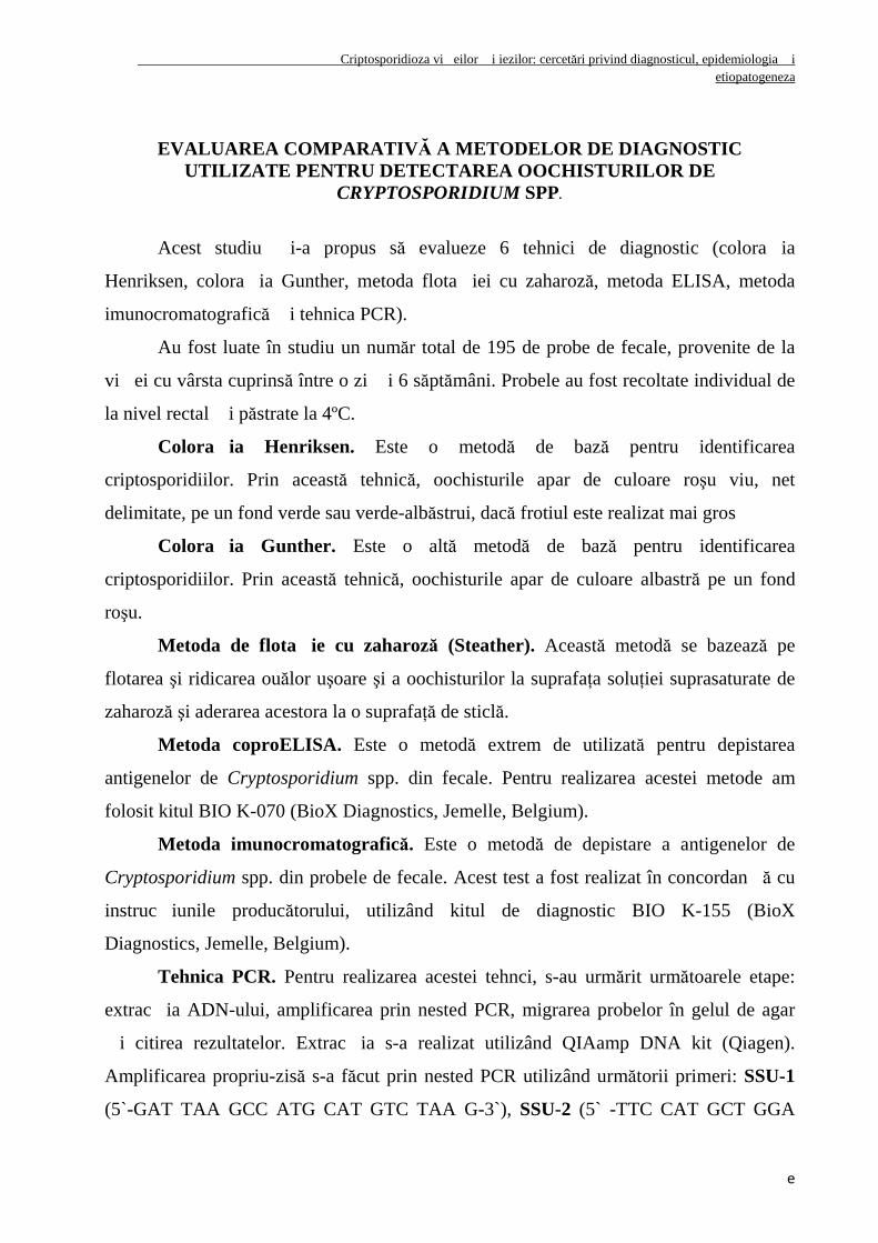
Colorația Henriksen. Este o metodă de bază pentru identificarea
criptosporidiilor. Prin această tehnică, oochisturile apar de culoare roşu viu, net
delimitate, pe un fond verde sau verde-albăstrui, dacă frotiul este realizat mai gros
Colorația Gunther. Este o altă metodă de bază pentru identificarea
criptosporidiilor. Prin această tehnică, oochisturile apar de culoare albastră pe un fond
roşu.
Metoda de flotație cu zaharoză (Steather). Această metodă se bazează pe
flotarea şi ridicarea ouălor uşoare şi a oochisturilor la suprafaŃa soluŃiei suprasaturate de
zaharoză şi aderarea acestora la o suprafaŃă de sticlă.
Metoda coproELISA. Este o metodă extrem de utilizată pentru depistarea
antigenelor de Cryptosporidium spp. din fecale. Pentru realizarea acestei metode am
folosit kitul BIO K-070 (BioX Diagnostics, Jemelle, Belgium).
Metoda imunocromatografică. Este o metodă de depistare a antigenelor de
Cryptosporidium spp. din probele de fecale. Acest test a fost realizat în concordan�ă cu
instruc�iunile producătorului, utilizând kitul de diagnostic BIO K-155 (BioX
Diagnostics, Jemelle, Belgium).
Tehnica PCR. Pentru realizarea acestei tehnci, s-au urmărit următoarele etape:
extrac�ia ADN-ului, amplificarea prin nested PCR, migrarea probelor în gelul de agar
�i citirea rezultatelor. Extrac�ia s-a realizat utilizând QIAamp DNA kit (Qiagen).
Amplificarea propriu-zisă s-a făcut prin nested PCR utilizând următorii primeri: SSU-1
(5`-GAT TAA GCC ATG CAT GTC TAA G-3`), SSU-2 (5` -TTC CAT GCT GGA
Criptosporidioza vi�eilor �i iezilor: cercetări privind diagnosticul, epidemiologia �i etiopatogeneza
f
GTA TTC AAG- 3`), SSU-3 (5`-CAG TTA TAG TTT ACT TGA TAA TC-3`) �i SSU-
4 (5`-CCT GCT TTA AGC ACT CTA ATT TTC-3`). Migrarea produ�ilor de
amplificare s-a realizat prin electroforeză orizontală în gel de agaroză 1,5%, la 80 V, timp
de 60 de minute. După migrarea probelor în gelul de agaroză, imaginea gelului cu
fragmentele de ADN după migrare a fost fotografiată, utilizând aparatul BioDoc-ITTM
Imaging System
Analiza statistică a rezultatelor a fost făcută cu ajutorul programelor Epi Info
3.5.1, Win Episcope 2.0 �i Statistica. Astfel, s-au calculat: sensibilitatea, specificitatea,
valoarea predictivă pozitivă, valoarea predictivă negativă, prevalen�a adevărată,
prevalen�a aparentă, coeficientul de concordan�ă între teste k, indicele Youden �i
indicele Spearman.
Rezultate
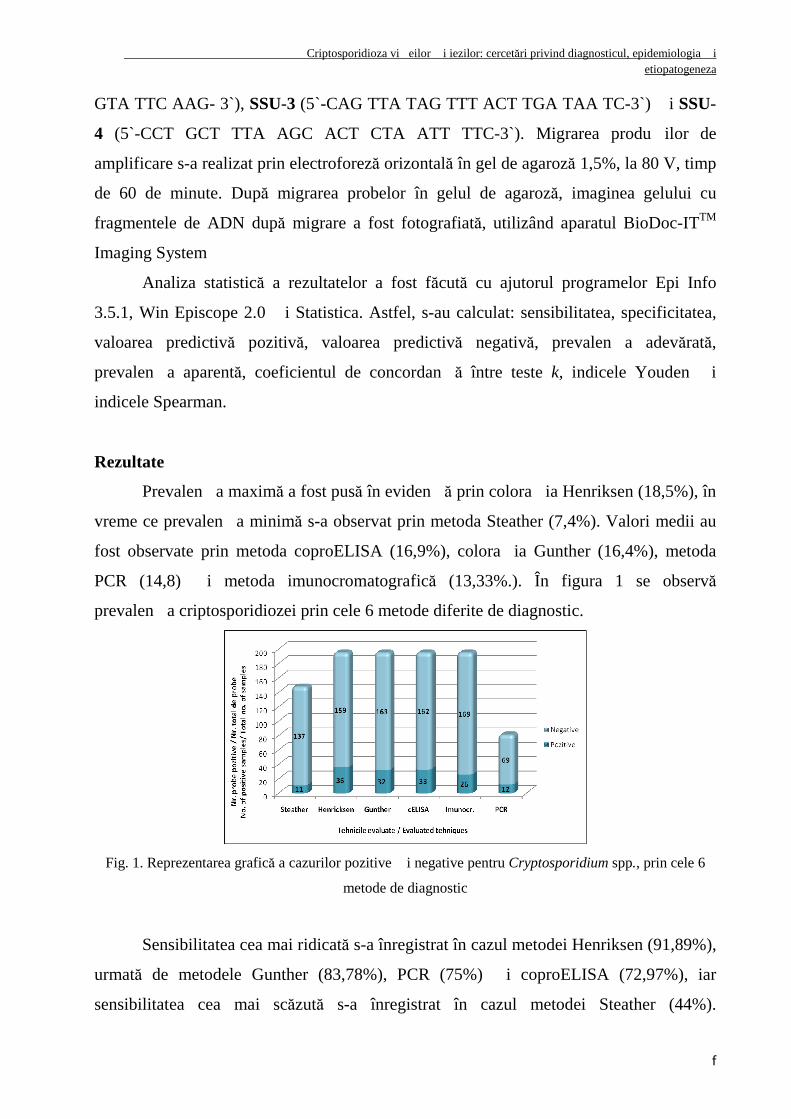
Prevalen�a maximă a fost pusă în eviden�ă prin colora�ia Henriksen (18,5%), în
vreme ce prevalen�a minimă s-a observat prin metoda Steather (7,4%). Valori medii au
fost observate prin metoda coproELISA (16,9%), colora�ia Gunther (16,4%), metoda
PCR (14,8) �i metoda imunocromatografică (13,33%.). În figura 1 se observă
prevalen�a criptosporidiozei prin cele 6 metode diferite de diagnostic.
Fig. 1. Reprezentarea grafică a cazurilor pozitive �i negative pentru Cryptosporidium spp., prin cele 6
metode de diagnostic
Sensibilitatea cea mai ridicată s-a înregistrat în cazul metodei Henriksen (91,89%),
urmată de metodele Gunther (83,78%), PCR (75%) �i coproELISA (72,97%), iar
sensibilitatea cea mai scăzută s-a înregistrat în cazul metodei Steather (44%).
Criptosporidioza vi�eilor �i iezilor: cercetări privind diagnosticul, epidemiologia �i etiopatogeneza
g
Specificitatea tehnicilor studiate a fost peste 95%, fiind chiar 100% pentru metodele
Steather �i PCR.
Metodele coproscopice de depistare a oochisturilor de Cryptosporidium spp. sunt
cele mai ieftine, în vreme ce tehnicile coproELISA �i PCR au costul consumabilelor
foarte ridicat �i necesită investi�ii mari în aparatură. Cea mai expeditivă metodă este
metoda imunocromatografică, în vreme ce metoda PCR este metoda cea mai laborioasă.
Dintre metodele cu sensibilitate ridicată, colora�ia Ziehl-Neelsen, modificată de
Henriksen, este cea mai convenabilă din punct de vedere economic, dar �i din punct de
vedere al u�urin�ei �i timpului necesar realizării.
STUDII EPIDEMIOLOGICE ÎN CRIPTOSPORIDIOZ Ă LA VI țEI țI IEZI DIN
CENTRUL țI NORD-VESTUL ROMÂNIEI
Acest studiu a avut drept scop analiza evolu�iei criptosporidiozei la diferite
categorii de vi�ei �i iezi, urmărindu-se prevalen�a, influen�a vârstei, sexului �i a
varia�iilor sezoniere asupra evolu�iei bolii �i identificarea speciilor de
Cryptosporidium ce afectează vi�eii �i iezii prin micromăsurători �i tehnici de biologie
moleculară.
Au fost lua�i în studiu un număr total de 708 vi�ei, proveni�i din 29 de ferme de
bovine �i 412 iezi proveni�i din 12 ferme de caprine din centrul �i nord-vestul
României. Pentru punerea în eviden�ă a oochisturilor de Cryptosporidium spp. s-a
utilizat colora�ia Henriksen. Au fost efectuate micromăsurători pe un număr de 20 de
oochisturi cu ajutorul programului AdobePhotoshop CS 4, pe imagini obŃinute la
microscopul Olympus BSX430 cu obiectivul cu imersie. Pentru determinarea speciilor de
Cryptosporidium prin tehnici de biologie moleculară s-a utilizat tehnica Multiplex PCR
folosind gena Hsp 70. Pentru aceasta, s-au folosit primeri specifici pentru următoarele 4
genotipuri investigate: Cryptosporidium parvum (genotipul uman), Cryptosporidium
parvum (genotipul bovin), Cryptosporidium canis �i Cryptosporidium felis.
Rezultate
Criptosporidioza vi�eilor �i iezilor: cercetări privind diagnosticul, epidemiologia �i etiopatogeneza
h
A. La vi�ei. Din cei 708 viŃei cu vârsta cuprinsă între o zi şi 8 săptămâni luaŃi în
studiu, 198 de viŃei au fost eliminatori de oochisturi de Cryptosporidium spp., adică
27,96% (95% IC =24,7%- 31,5%).
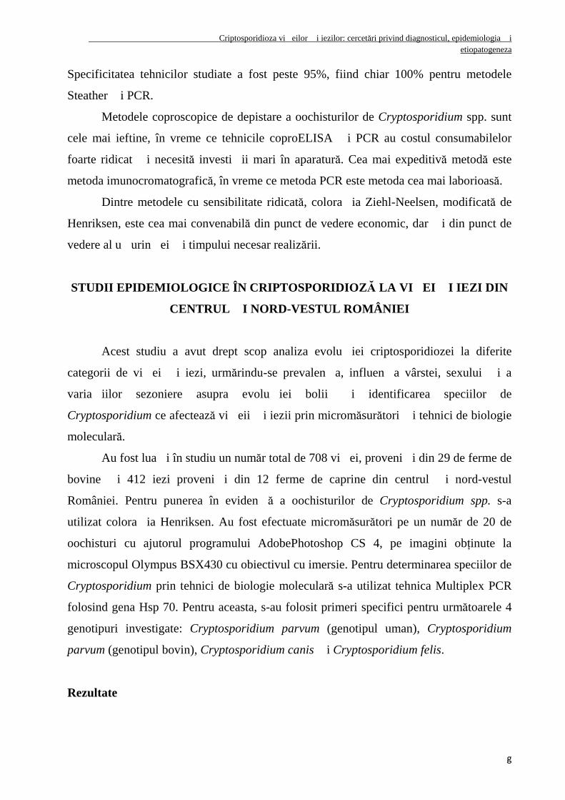
Eliminări de oochisturi de Cryptosporidium spp. s-au înregistrat încă de la vârsta
de 4 zile, prevalen�a cea mai mare constatându-se între 2 şi 3 săptămâni (fig. 2). Faptul
că infec�ia cu Cryptosporidium spp. a fost diagnosticată atât de timpuriu, sugerează o
contaminare foarte mare a acestei zone. Cea mai mare intensivitate a parazitării cu
Cryptosporidium spp. s-a observat la categoria de vârstă 1-2 săptămâni, unde 54% din
cazuri au prezentat intensivitate medie şi 38% din cazuri intensivitate crescută, în vreme
ce la categoriile de vârstă sub o săptămână şi peste 3 săptămâni, s-a înregistrat o
intensivitate scăzută (fig. 3).
Fig. 2. Prevalen�a criptosporidiozei (%) la viŃei
în funcŃie de vârstă
Fig. 3. Intensivitatea parazitării cu
Cryptosporidium spp. în funcŃie de vârstă
Cele mai mari valori ale inciden�ei infec�iei cu Cryptosporidium spp. s-au
înregistrat la sfâr�itul iernii - începutul primăverii, când au ajuns la 70,21%, în vreme ce
în restul anului, valorile au fost de 16,80 ± 5,78%.
Prin micromăsurătorile oochisturilor de Cryptosporidium spp. s-a depistat
eliminarea unui singur tip de oochist cu dimensiunile de 4,51 ± 0,41 µm lungime �i 4,07
± 0,33 µm lă�ime, având o formă u�or ovală.
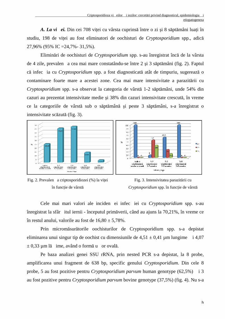
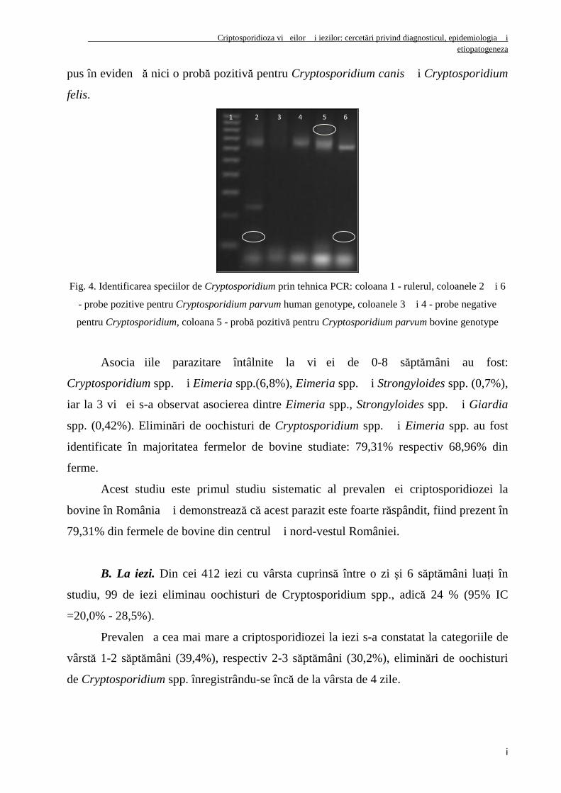
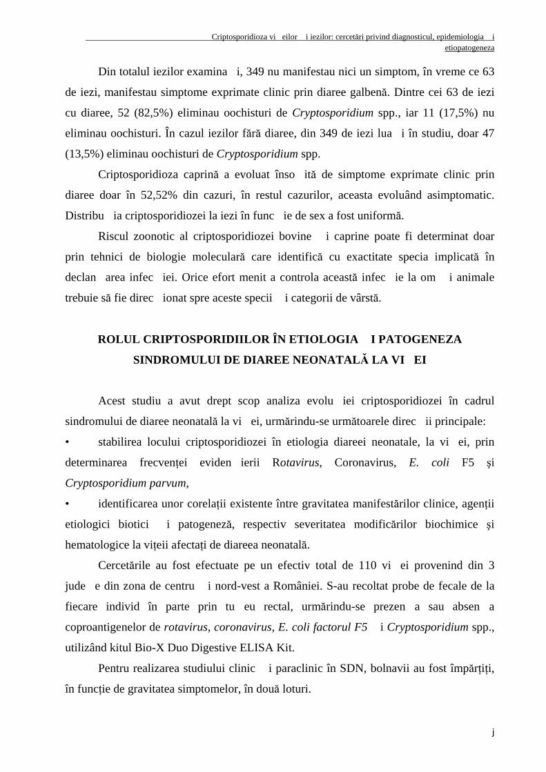
Pe baza analizei genei SSU rRNA, prin nested PCR s-a depistat, la 8 probe,
amplificarea unui fragment de 638 bp, specific genului Cryptosporidium. Din cele 8
probe, 5 au fost pozitive pentru Cryptosporidium parvum human genotype (62,5%) �i 3
au fost pozitive pentru Cryptosporidium parvum bovine genotype (37,5%) (fig. 4). Nu s-a
Criptosporidioza vi�eilor �i iezilor: cercetări privind diagnosticul, epidemiologia �i etiopatogeneza
i
pus în eviden�ă nici o probă pozitivă pentru Cryptosporidium canis �i Cryptosporidium
felis.
Fig. 4. Identificarea speciilor de Cryptosporidium prin tehnica PCR: coloana 1 - rulerul, coloanele 2 �i 6
- probe pozitive pentru Cryptosporidium parvum human genotype, coloanele 3 �i 4 - probe negative
pentru Cryptosporidium, coloana 5 - probă pozitivă pentru Cryptosporidium parvum bovine genotype
Asocia�iile parazitare întâlnite la vi�ei de 0-8 săptămâni au fost:
Cryptosporidium spp. �i Eimeria spp.(6,8%), Eimeria spp. �i Strongyloides spp. (0,7%),
iar la 3 vi�ei s-a observat asocierea dintre Eimeria spp., Strongyloides spp. �i Giardia
spp. (0,42%). Eliminări de oochisturi de Cryptosporidium spp. �i Eimeria spp. au fost
identificate în majoritatea fermelor de bovine studiate: 79,31% respectiv 68,96% din
ferme.
Acest studiu este primul studiu sistematic al prevalen�ei criptosporidiozei la
bovine în România �i demonstrează că acest parazit este foarte răspândit, fiind prezent în
79,31% din fermele de bovine din centrul �i nord-vestul României.
B. La iezi. Din cei 412 iezi cu vârsta cuprinsă între o zi şi 6 săptămâni luaŃi în
studiu, 99 de iezi eliminau oochisturi de Cryptosporidium spp., adică 24 % (95% IC
=20,0% - 28,5%).
Prevalen�a cea mai mare a criptosporidiozei la iezi s-a constatat la categoriile de
vârstă 1-2 săptămâni (39,4%), respectiv 2-3 săptămâni (30,2%), eliminări de oochisturi
de Cryptosporidium spp. înregistrându-se încă de la vârsta de 4 zile.
1 2 3 4 5 6
Criptosporidioza vi�eilor �i iezilor: cercetări privind diagnosticul, epidemiologia �i etiopatogeneza
j
Din totalul iezilor examina�i, 349 nu manifestau nici un simptom, în vreme ce 63
de iezi, manifestau simptome exprimate clinic prin diaree galbenă. Dintre cei 63 de iezi
cu diaree, 52 (82,5%) eliminau oochisturi de Cryptosporidium spp., iar 11 (17,5%) nu
eliminau oochisturi. În cazul iezilor fără diaree, din 349 de iezi lua�i în studiu, doar 47
(13,5%) eliminau oochisturi de Cryptosporidium spp.
Criptosporidioza caprină a evoluat înso�ită de simptome exprimate clinic prin
diaree doar în 52,52% din cazuri, în restul cazurilor, aceasta evoluând asimptomatic.
Distribu�ia criptosporidiozei la iezi în func�ie de sex a fost uniformă.
Riscul zoonotic al criptosporidiozei bovine �i caprine poate fi determinat doar
prin tehnici de biologie moleculară care identifică cu exactitate specia implicată în
declan�area infec�iei. Orice efort menit a controla această infec�ie la om �i animale
trebuie să fie direc�ionat spre aceste specii �i categorii de vârstă.
ROLUL CRIPTOSPORIDIILOR ÎN ETIOLOGIA țI PATOGENEZA
SINDROMULUI DE DIAREE NEONATAL Ă LA VI țEI
Acest studiu a avut drept scop analiza evolu�iei criptosporidiozei în cadrul
sindromului de diaree neonatală la vi�ei, urmărindu-se următoarele direc�ii principale:
• stabilirea locului criptosporidiozei în etiologia diareei neonatale, la vi�ei, prin
determinarea frecvenŃei eviden�ierii Rotavirus, Coronavirus, E. coli F5 şi
Cryptosporidium parvum,
• identificarea unor corelaŃii existente între gravitatea manifestărilor clinice, agenŃii
etiologici biotici �i patogeneză, respectiv severitatea modificărilor biochimice şi
hematologice la viŃeii afectaŃi de diareea neonatală.
Cercetările au fost efectuate pe un efectiv total de 110 vi�ei provenind din 3
jude�e din zona de centru �i nord-vest a României. S-au recoltat probe de fecale de la
fiecare individ în parte prin tu�eu rectal, urmărindu-se prezen�a sau absen�a
coproantigenelor de rotavirus, coronavirus, E. coli factorul F5 �i Cryptosporidium spp.,
utilizând kitul Bio-X Duo Digestive ELISA Kit.
Pentru realizarea studiului clinic �i paraclinic în SDN, bolnavii au fost împărŃiŃi,
în funcŃie de gravitatea simptomelor, în două loturi.
Criptosporidioza vi�eilor �i iezilor: cercetări privind diagnosticul, epidemiologia �i etiopatogeneza
k
Lotul nr. 1 a fost alcătuit din 10 viŃei prezentând simptome de enterită catarală.
Lotul nr. 2 a fost alcătuit din 9 viŃei prezentând simptome de enterită hemoragică.
Rezultate
Cryptosporidium parvum este cel mai frecvent agent patogen implicat în SDN cu
o prevalen�ă de 35,45%, în 16,36% din cazuri ca unic agent patogen �i, în 19,09% din
cazuri, în asocia�ie cu al�i enteropatogeni. Infec�ia cu rotavirus a fost identificată în
17,27% din cazuri, din care în 7,27% din cazuri ca unic agent patogen �i în 10% în
asocia�ie cu al�i enteropatogeni. Infec�ia cu coronavirus, a fost identificată în 17,27%
din cazuri, din care în 2,73% din probe ca unic agent patogen �i în 14,54% din probe în
asocia�ie cu Cryptosporidium parvum. Infec�ia cu E.coli factorul F5 a fost identificată
doar în 1,82% din cazuri. 52,27% din probe au fost negative pentru to�i cei 4
enteropatogeni cerceta�i.
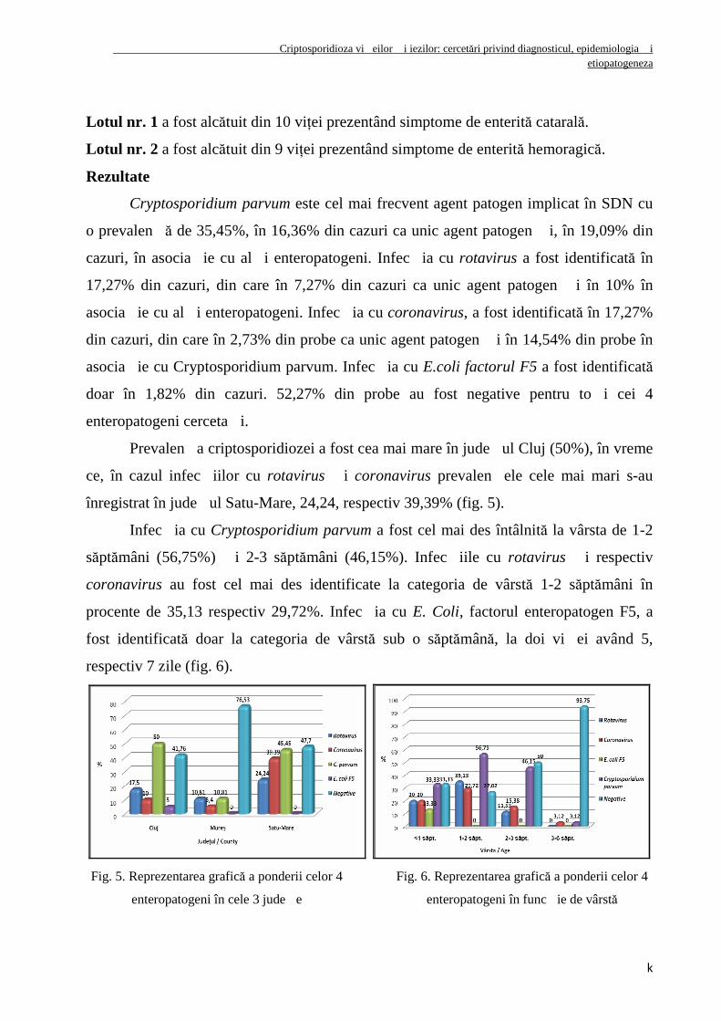
Prevalen�a criptosporidiozei a fost cea mai mare în jude�ul Cluj (50%), în vreme
ce, în cazul infec�iilor cu rotavirus �i coronavirus prevalen�ele cele mai mari s-au
înregistrat în jude�ul Satu-Mare, 24,24, respectiv 39,39% (fig. 5).
Infec�ia cu Cryptosporidium parvum a fost cel mai des întâlnită la vârsta de 1-2
săptămâni (56,75%) �i 2-3 săptămâni (46,15%). Infec�iile cu rotavirus �i respectiv
coronavirus au fost cel mai des identificate la categoria de vârstă 1-2 săptămâni în
procente de 35,13 respectiv 29,72%. Infec�ia cu E. Coli, factorul enteropatogen F5, a
fost identificată doar la categoria de vârstă sub o săptămână, la doi vi�ei având 5,
respectiv 7 zile (fig. 6).
Fig. 5. Reprezentarea grafică a ponderii celor 4
enteropatogeni în cele 3 jude�e
Fig. 6. Reprezentarea grafică a ponderii celor 4
enteropatogeni în func�ie de vârstă
Criptosporidioza vi�eilor �i iezilor: cercetări privind diagnosticul, epidemiologia �i etiopatogeneza
l
Sindromul de diaree neonatală la viŃei are o exprimare clinică de gravitate
variabilă care rezultă din interacŃiunea complexă dintre factorii de risc, factorii
declanşatori şi reactivitatea organismului nou-născut. Formele clinice cele mai grave au
fost cele consecutive enteritelor hemoragice în care, alături de deshidratare şi acidoză
metabolică, au evoluat cu efecte agravante anemia şi endotoxemia.
În etiologia SDN la viŃeii cu simptome de enterită catarală nu s-au identificat
agenŃi biotici. La cazurile cu simptome de enterită hemoragică, agenŃii patogeni implicaŃi
au fost reprezentaŃi de Cryptosporidium spp. (66,7%), Eimeria spp. (22,22%), Rotavirus
(11,1%), Coronavirus (11,1%).
Deshidratarea hipertonă şi acidoza metabolică lactică au fost dominantele
patogenetice majore care au marcat sindromul umoral, atât în formele uşoare, cât şi în
cele grave ale sindromului de diaree neonatală. Hipoglicemia evidenŃiată la ambele loturi
de viŃei constituie un element patogenetic major care rezultă în urma anorexiei,
maldigestiei şi malabsorbŃiei.
Fibrinogenul plasmatic reprezintă un indicator umoral foarte sensibil şi fiabil în
afecŃiunile inflamatorii gastro-intestinale la viŃeii nou născuŃi.
CRIPTOSPORIDIOZA NATURAL Ă LA IEZI, ÎNTR-UN FOCAR DIN JUDE țUL CLUJ – OBSERVAțII EPIDEMIOLOGICE, CLINICE, MORFOPATOLOGICE
țI ELECTRONOMICROSCOPICE
Scopul acestui studiu a fost de a descrie un focar natural de criptosporidioză a
iezilor, de la modificările clinice apărute, până la descrierea patogenezei bolii.
În februarie 2009, a fost identificat un focar de criptosporidioză cu diaree apoasă
galbenă, la iezi cu vârsta cuprinsă între o zi �i 3 săptămâni, la o fermă de caprine din
jude�ul Cluj. Fecalele a 58 de iezi mor�i �i 83 de iezi în via�ă au fost prelucrate prin
metode coproparazitologice, în vederea identificării agen�ilor patogeni implica�i. La 10
cadavre s-a făcut examenul necropsic constând în: examen macroscopic, examen
histopatologic �i examen electronomicoscopic.
Rezultate
Criptosporidioza vi�eilor �i iezilor: cercetări privind diagnosticul, epidemiologia �i etiopatogeneza
m
Prevalen�a criptosporidiozei la iezi în cadrul focarului semnalat a fost de 61,70%
(87 / 141).
Manifestările clinice observate au fost diaree la 68,68% din iezi (57 / 83), cifoză,
la 9,64% din iezi (8 / 83) �i deshidratare la 44,58% din iezi (37/83). Mortalitatea iezilor,
în focarul studiat a fost de 41,13%.
Coproeliminări de oochisturi de Cryptosporidium spp. au fost observate la 81,6%
din efectiv (115 / 141).
La examenul anatomopatologic s-au observat cadavre cahectice cu trenul posterior
murdărit cu fecale. Macroscopic, s-a observat conŃinutul intestinal apos, cu fecale
galbene �i ansele intestinale destinse de gaze.
Modificările histologice observate în ileon �i colon au inclus alteraŃii ale
vilozităŃilor caracterizate prin atrofia �i denudarea acestora.
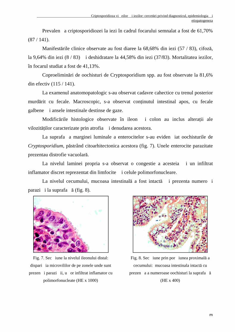
La suprafa�a marginei luminale a enterocitelor s-au eviden�iat oochisturile de
Cryptosporidium, păstrând citoarhitectonica acestora (fig. 7). Unele enterocite parazitate
prezentau distrofie vacuolară.
La nivelul laminei propria s-a observat o congestie a acesteia �i un infiltrat
inflamator discret reprezentat din limfocite �i celule polimorfonucleare.
La nivelul cecumului, mucoasa intestinală a fost intactă �i prezenta numero�i
parazi�i la suprafa�ă (fig. 8).
Fig. 7. Sec�iune la nivelul ileonului distal:
dispari�ia microvililor de pe zonele unde sunt
prezen�i parazi�ii, u�or infiltrat inflamator cu
polimorfonucleate (HE x 1000)
Fig. 8. Sec�iune prin por�iunea proximală a
cecumului: mucoasa intestinala intactă cu
prezen�a a numeroase oochisturi la suprafa�ă
(HE x 400)
Criptosporidioza vi�eilor �i iezilor: cercetări privind diagnosticul, epidemiologia �i etiopatogeneza
n
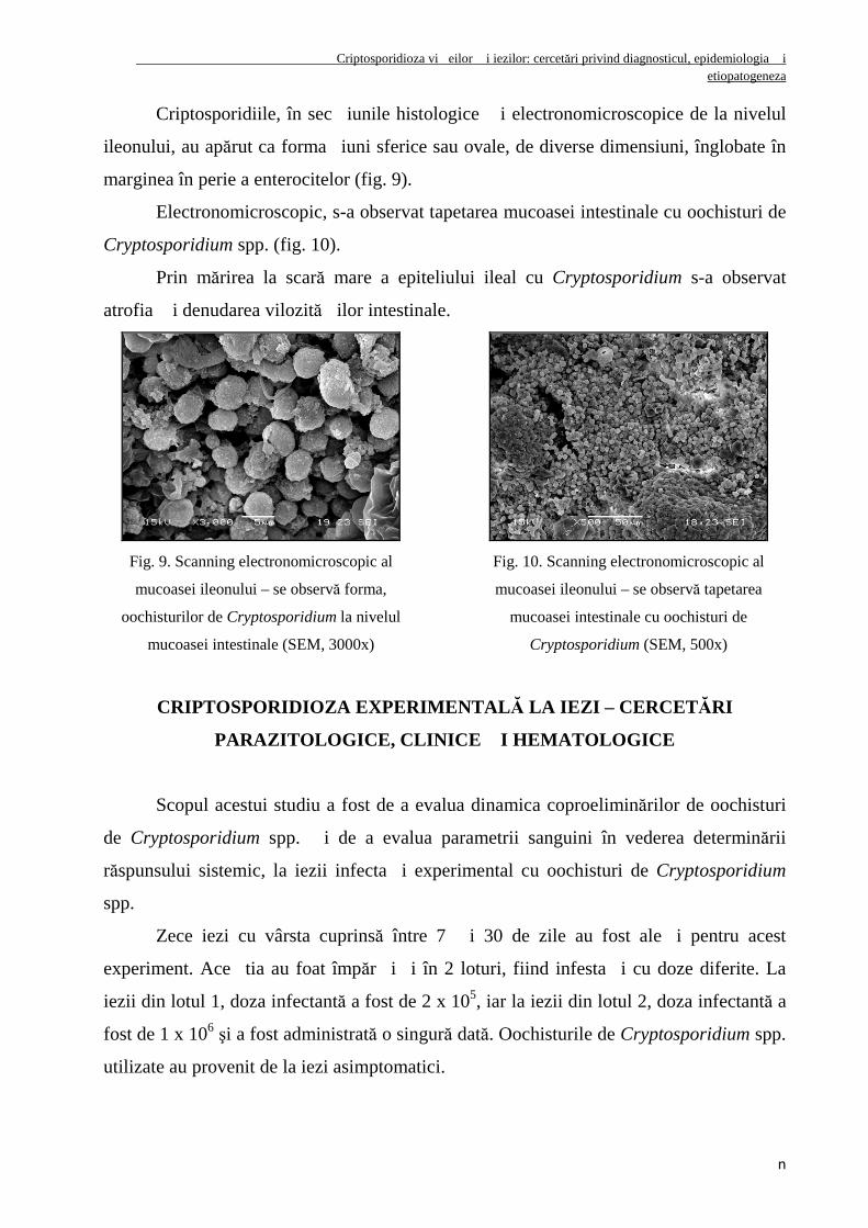
Criptosporidiile, în sec�iunile histologice �i electronomicroscopice de la nivelul
ileonului, au apărut ca forma�iuni sferice sau ovale, de diverse dimensiuni, înglobate în
marginea în perie a enterocitelor (fig. 9).
Electronomicroscopic, s-a observat tapetarea mucoasei intestinale cu oochisturi de
Cryptosporidium spp. (fig. 10).
Prin mărirea la scară mare a epiteliului ileal cu Cryptosporidium s-a observat
atrofia �i denudarea vilozită�ilor intestinale.
Fig. 9. Scanning electronomicroscopic al
mucoasei ileonului – se observă forma,
oochisturilor de Cryptosporidium la nivelul
mucoasei intestinale (SEM, 3000x)
Fig. 10. Scanning electronomicroscopic al
mucoasei ileonului – se observă tapetarea
mucoasei intestinale cu oochisturi de
Cryptosporidium (SEM, 500x)
CRIPTOSPORIDIOZA EXPERIMENTAL Ă LA IEZI – CERCET ĂRI
PARAZITOLOGICE, CLINICE țI HEMATOLOGICE
Scopul acestui studiu a fost de a evalua dinamica coproeliminărilor de oochisturi
de Cryptosporidium spp. �i de a evalua parametrii sanguini în vederea determinării
răspunsului sistemic, la iezii infecta�i experimental cu oochisturi de Cryptosporidium
spp.
Zece iezi cu vârsta cuprinsă între 7 �i 30 de zile au fost ale�i pentru acest
experiment. Ace�tia au foat împăr�i�i în 2 loturi, fiind infesta�i cu doze diferite. La
iezii din lotul 1, doza infectantă a fost de 2 x 10⁵, iar la iezii din lotul 2, doza infectantă a
fost de 1 x 10⁶ şi a fost administrată o singură dată. Oochisturile de Cryptosporidium spp.
utilizate au provenit de la iezi asimptomatici.
Criptosporidioza vi�eilor �i iezilor: cercetări privind diagnosticul, epidemiologia �i etiopatogeneza
o
Examenul coproparazitologic s-a efectuat utilizând coloraŃia Henriksen, iar
dinamica coproeliminările de oochisturi de Cryptosporidium spp. a fost evaluată folosind
o metodă proprie, şi anume numărarea oochisturilor existente în 100 de câmpuri
microscopice. Pentru determinarea parametrilor hematologici, s-a recoltat sânge de la
fiecare ied în parte, de la nivelul venei jugulare în prima zi anteinfectant �i în ziua a 7-a
postinfectant. Pentru evaluarea diferenŃelor statistice între cele două loturi s-a folosit
testul Wilcoxon Rank Sum Test (Statistix 9.0).
Rezultate
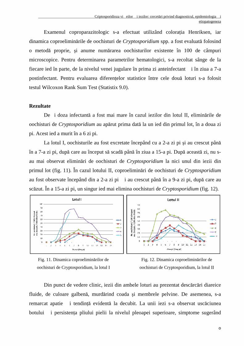
De�i doza infectantă a fost mai mare în cazul iezilor din lotul II, eliminările de
oochisturi de Cryptosporidium au apărut prima dată la un ied din primul lot, în a doua zi
pi. Acest ied a murit în a 6 zi pi.
La lotul I, oochisturile au fost excretate începând cu a 2-a zi pi şi au crescut până
în a 7-a zi pi, după care au început să scadă până în ziua a 15-a pi. După această zi, nu s-
au mai observat eliminări de oochisturi de Cryptosporidium la nici unul din iezii din
primul lot (fig. 11). În cazul lotului II, coproeliminări de oochisturi de Cryptosporidium
au fost observate începând din a 2-a zi pi �i au crescut până în a 9-a zi pi, după care au
scăzut. În a 15-a zi pi, un singur ied mai elimina oochisturi de Cryptosporidium (fig. 12).
Fig. 11. Dinamica coproeliminărilor de
oochisturi de Cryptosporidium, la lotul I
Fig. 12. Dinamica coproeliminărilor de
oochisturi de Cryptosporidium, la lotul II
Din punct de vedere clinic, iezii din ambele loturi au prezentat descărcări diareice
fluide, de culoare galbenă, murdărind coada şi membrele pelvine. De asemenea, s-a
remarcat apatie �i tendinŃă evidentă la decubit. La unii iezi s-a observat uscăciunea
botului �i persistenŃa pliului pielii la nivelul pleoapei superioare, simptome sugerând
Cryptosporidiosis in calves and goat kids: research concerning diagnosis, epidemiology, and etiopathogenesis
p
diferite grade de deshidratare. Un aspect deosebit de important este acela că nici unul din
subiecŃii luaŃi în studiu nu a prezentat modificări ale temperaturii corporale, nici chiar
aceia care prezentau diaree severă.
Tabloul umoral la iezii cu criptosporidioză, infecta�i experimental, nu aduce
elemente patognomonice de diagnostic pozitiv, dar furnizează elemente valoroase de
prognostic şi mai ales de orientare şi fundamentare a unei conduite terapeutice raŃionale.
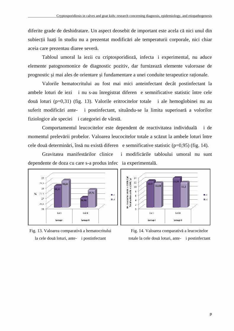
Valorile hematocritului au fost mai mici anteinfectant decât postinfectant la
ambele loturi de iezi �i nu s-au înregistrat diferen�e semnificative statistic între cele
două loturi (p=0,31) (fig. 13). Valorile eritrocitelor totale �i ale hemoglobinei nu au
suferit modificări ante- �i postinfectant, situându-se la limita superioară a volorilor
fiziologice ale speciei �i categoriei de vârstă.
Comportamentul leucocitelor este dependent de reactivitatea individuală �i de
momentul prelevării probelor. Valoarea leucocitelor totale a scăzut la ambele loturi între
cele două determinări, însă nu există diferen�e semnificative statistic (p=0,95) (fig. 14).
Gravitatea manifestărilor clinice �i modificările tabloului umoral nu sunt
dependente de doza cu care s-a produs infec�ia experimentală.
Fig. 13. Valoarea comparativă a hematocritului
la cele două loturi, ante- �i postinfectant
Fig. 14. Valoarea comparativă a leucocitelor
totale la cele două loturi, ante- �i postinfectant
Cryptosporidiosis in calves and goat kids: research concerning diagnosis, epidemiology, and etiopathogenesis
q
UNIVERSITY OF AGRICULTURAL SCIENCES AND VETERINARY MEDICINE CLUJ-NAPOCA
DOCTORAL SCHOOL COLLEGE OF VETERINARY MEDICINE
ALEXANDRU LUCIAN BEJAN
SUMMARY OF Ph.D THESIS
PROJECT LEADER Vasile COZMA, Professor Ph.D
CLUJ-NAPOCA 2009
Cryptosporidiosis in calves and goat kids: research concerning diagnosis, epidemiology, and etiopathogenesis
r
UNIVERSITY OF AGRICULTURAL SCIENCES AND VETERINARY MEDICINE CLUJ-NAPOCA
DOCTORAL SCHOOL COLLEGE OF VETERINARY MEDICINE
ALEXANDRU LUCIAN BEJAN
CRYPTOSPORIDIOSIS IN CALVES AND GOAT KIDS: RESEARCH CONCERNING DIAGNOSIS, EPIDEMIOLOGY,
AND ETIOPATHOGENESIS
SUMMARY OF Ph.D. THESIS
Scientific Coordinator Vasile COZMA, Professor Ph.D
CLUJ-NAPOCA 2009
Cryptosporidiosis in calves and goat kids: research concerning diagnosis, epidemiology, and etiopathogenesis
s
SUMMARY
Criptosporidiosis is a disease found in humans and animals, with a favorable
evolution in the organisms immunocompetents and serious in immunodeficiency
organisms characterized clinically, mainly, by gastrointestinal disorders and occasionally
by respiratory, hepatic and pancreatic disorders. The disease is a zoonosis because is
transmitted from animals to humans.
The zootehnic economical looses, associated with this disease, are not correlated
with mortality, but are related with growth retardation, cost of therapies and veterinary
services.
The attention in protozoan Cryptosporidium is expending from biological and
parasitological academic areas to veterinaries, epidemiologists, pharmacists, specialists in
public health, public water, farmers and common people. The concern in prevention and
treatment of criptosporidiosis is extended from underdeveloped community to
industrialized society, from immunodepressed people to healthy population.
After 1990, resulting from many researchers was found that in water commonly
used by humans and animals, there are frequently Cryptosporidium oocysts. 19 outbreaks
of Cryptosporidium with water origin are cited and approximatively 427000 humans
were affected. All the water treatment and filtration can`t guarantee the absence of
Cryptosporidium oocysts. Water represents the most important vehicle for the
transmission of criptosporidiosis to animals and humans.
Research goals
The first chapter of the personal research plans to evaluate six different diagnostic
techniques (two staining methods, one flotation method, two immunological methods,
and one molecular biology method) in terms of efficiency, sensitivity, and specificity of
cost, time, and ease of completion.
Cryptosporidiosis in calves and goat kids: research concerning diagnosis, epidemiology, and etiopathogenesis
t
Once established the most suitable diagnostic technique from the economic, ease
of achieving, and sensitivity and specificity point of view, further research was carried
out only through this method.
The second chapter of the personal research aims to analyze the evolution of
Cryptosporidiosis in different categories of calves and goat kids, following a few main
directions. Thus, a descriptive epidemiological study was developed, in order to
understand the frequency and prevalence of the disease in calves and goat kids; the
influence of age, sex, and seasonal variations on the disease was followed; the
Cryptosporidium species that affects calves was identified through micro-measurements,
and also through molecular biology techniques; last but not least a study concerning the
parasitosis that affect calves aged one day through eight weeks was completed.
The third chapter of the personal research aimed at determining the place of
Cryptosporidiosis in the etiology of neonatal diarrhea in calves. The research tracked the
frequency of Cryptosporidium spp., E. coli factor F5, Rotavirus and Coronavirus
discharge in calves from the center and northwest regions of Romania and tried to
identify a correlation between the seriousness of clinical manifestations, biotic and
etiologic agents, and the severity of biochemical and hematological anomalies in calves
presenting neonatal diarrhea.
The fourth chapter of the personal research aims to report an outbreak of goat
Cryptosporidiosis, from clinical manifestations to full description of the pathogenesis of
the intestinal Cryptosporidiosis in goat youth.
The last chapter of the personal research intends to assess the dynamics of the
coproelimination of oocysts and the hematological parameters, in order to determine the
systemic response in experimentally infected goat kids with Cryptosporidium spp.
oocysts.
COMPARATIVE ASSESSMENT OF DIAGNOSTIC METHODS UTILIZED IN THE DETECTION OF CRYPTOSPORIDIUM SPP. OOCYSTS
Cryptosporidiosis in calves and goat kids: research concerning diagnosis, epidemiology, and etiopathogenesis
u
This study aims to evaluate six diagnostic techniques: Henriksen stain, Guthner
stain, sucrose flotation method, ELISA method, immunochromatographic method, and
the PCR technique.
A number of 195 fecal samples were studied, coming from calves aged one day
through six weeks. Samples were collected individually from rectal level and kept at a
temperature of 4ºC.
Henriksen Stain. It is a basic method for identifying different types of
Cryptosporidiosis. Through this technique, oocysts appear a very deep red color, well
defined, on a blue-green background, if the smear is made thicker.
Gunther Stain. It is another basic method for identifying different types of
Cryptosporidiosis. Through this technique, oocysts appear blue on a red background.
Sucrose Flotation Method (Steather). This method is based on the flotation and
rising of light oocysts to the surface of the supersaturated sucrose solution, and their
adherence to a glass surface.
Copro-ELISA Method. It is a highly used method for detecting antigens of
Cryptosporidium spp. in feces. To implement this method, the BIO K-070 kit was used
(BioX Diagnostics, Jemelle, Belgium).
Immunochromatographic Method. It is a method of detecting antigens of
Cryptoporidium spp. in fecal samples. This test was performed according to the
manufacturer’s instructions, utilizing BIO K-155 diagnostic kit (BioX Diagnostics,
Jemelle, Belgium).
PCR Technique. To accomplish this technique, these steps were followed: DNA
extraction, amplification through nested PCR, migration of samples in agarose gel, and
reading of results. Extraction was performed using the QIAamp DNA kit (Qiagen). The
amplification itself was performed through nested PCR, utilizing the following primers:
SSU-1 (5`-GAT TAA GCC ATG CAT GTC TAA G-3`), SSU-2 (5` -TTC CAT GCT
GGA GTA TTC AAG- 3`), SSU-3 (5`-CAG TTA TAG TTT ACT TGA TAA TC-3`) �i
SSU-4 (5`-CCT GCT TTA AGC ACT CTA ATT TTC-3`). Movement of amplification
products was achieved through horizontal electrophoresis in 1.5% agarose gel at 80V for
60 minutes. After the movement of samples in the agarose gel, the image of the gel with
Cryptosporidiosis in calves and goat kids: research concerning diagnosis, epidemiology, and etiopathogenesis
v
the DNA fragments was photographed, utilizing the BioDoc-ITTM Imaging System
camera.
The statistical analysis of results was performed using Epi Info 3.5.1, Win
Episcope 2.0, and Statistica software. Thus were calculated: sensitivity, specificity,
positive predictive value, true prevalence, and apparent prevalence, the correlation
coefficient between k tests, Youden index, and Spearman index.
Results
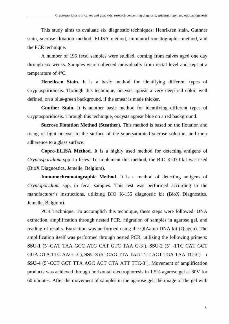
Maximum prevalence was evidenced through Henricksen stain (18.5%), while
minimum prevalence was observed through Steather method (7.4%). Median values were
observed through Copro-ELISA method (16.9%), Gunther stain (16.4%), PCR method
(14.8%), and immunochromatographic method (13.33%). Figure 1 notes the prevalence
of Cryptosporidiosis through the six different diagnostic methods.
Fig. 1. Graphic representation of positive and negative cases of Cryptosporidium spp.,
through the six different diagnostic methods
The highest sensitivity was achieved through the Henriksen stain method
(91.89%), followed by Gunther method (83.78%), PCR method (75%), and copro-ELISA
method (72.97%), and the lowest sensitivity was observed through the Steather method
(44%). The specificity of studied techniques was over 95%, with even 100% for the PCR
and Steather methods.
The coproscopic methods used in the detection of Cryposporidium spp. oocysts
are the cheapest, while the copro-ELISA technique and the PCR method involve high
cost of consumables and require large investment in equipment. The most expedient
Cryptosporidiosis in calves and goat kids: research concerning diagnosis, epidemiology, and etiopathogenesis
w
method is the immunochromatographic method, while the PCR method is the most
laborious.
Between the high sensitivity methods, the stain Ziehl-Neelsen, modified by
Henriksen, is the most convenient from the economic point of view, but also in terms of
ease and time required to achieve.
EPIDEMIOLOGICAL STUDIES CONCERNING CRYPTOSPORIDIOSI S
IN CALVES AND GOAT KIDS
IN THE CENTER AND NORTHWEST REGIONS OF ROMANIA
This study aimed to analyze the evolution of Cryptosporidiosis in calves and goat
kids, following the prevalence, the influence of age, sex and seasonal variations on the
disease, and the identification of Cryptosporidium species that affect calves and goat kids
through micro-measurements and molecular biology techniques.
A total of 708 calves originating from 29 cattle farms and 412 goat kids coming
from 12 goat farms from the center and northwest regions of Romania were studied. The
Henriksen stain was used for the highlight of Cryptosporidium spp. oocysts. Micro-
measurements were conducted on 20 oocysts, with the use of Adobe Photoshop CS 4
software, on images obtained with the Olympus BSX430 microscope with immersion
objective. The Multiplex PCR technique using the gene Hsp 70 was employed in order to
determine the Cryptosporidium species through molecular biology techniques. Specific
primers were used for the following four studied genotypes: Cryptosporidium parvum
(human genotype), Cryptosporidium parvum (bovine genotype), Cryptosporidium canis,
and Cryptosporidium felis.
Results
A. In calves. Between the 708 studies calves, aged from one day to eight
weeks, 198 calves, or about 27.96% (95% IC=24.7%-31.5%) were eliminating
Cryptosporidium spp. oocysts.
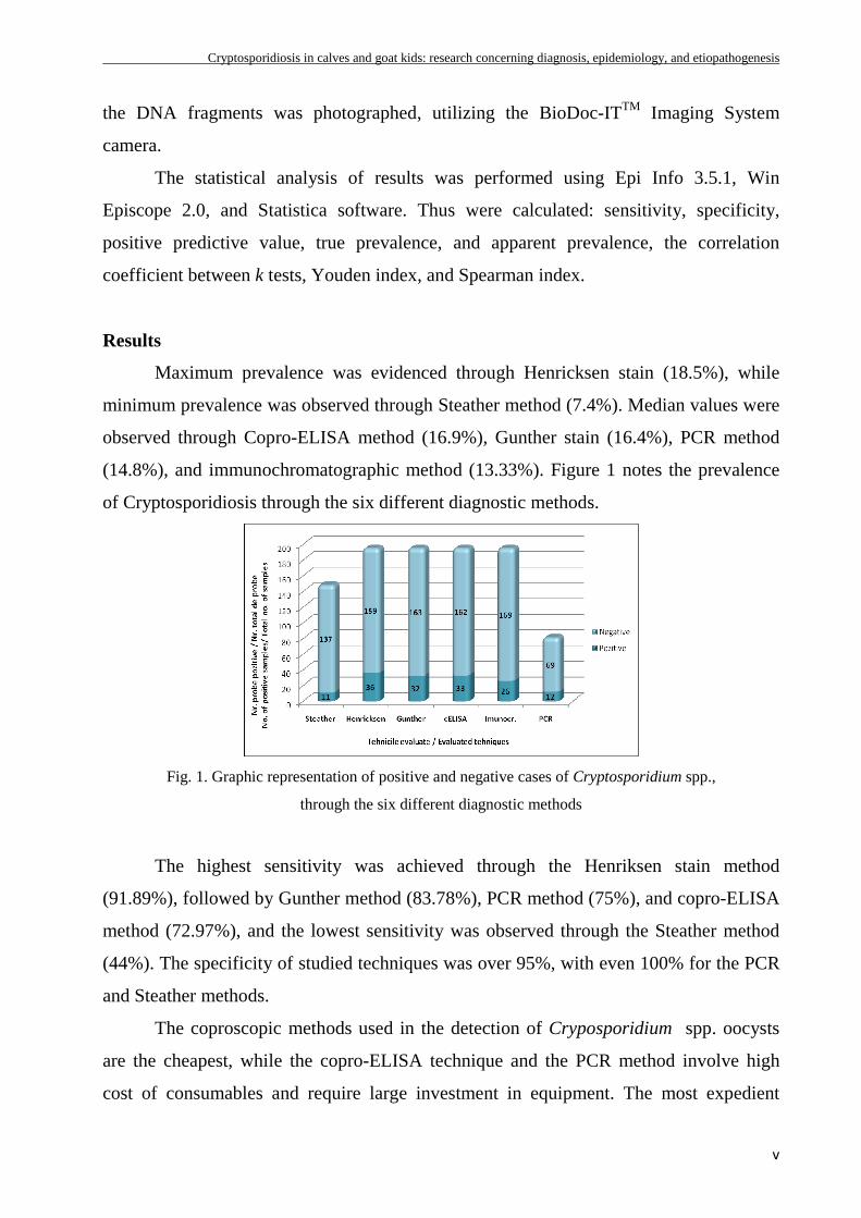
Cryptosporidium spp. oocysts were eliminated starting at 4 days old, the highest
prevalence being observed between 2-3weeks (figure 2). The fact that Cryptosporidium
Cryptosporidiosis in calves and goat kids: research concerning diagnosis, epidemiology, and etiopathogenesis
x
spp. infection was diagnosed at such an early age, suggests a high contamination of this
area. The highest intensity of the parasitization with Cryptosporidium spp. was observed
between 1-2 weeks, where 54% of the cases presented average intensity, and 38%
presented elevated intensity, while at ages under one week and over three weeks, a low
intensity was observed (figure 3).
Fig. 2. Cryptosporidiosis prevalence (%) in
calves by age
Fig 3. Intensity of the parasitization with
Cryptosporidium spp. by age
The highest values of Cryptosporidium spp. infection incidence were observed at
the end of winter – beginning of spring, when they reached 70.21%, while at other times
during the year , the incidence was 16.80% ± 5.78%.
Through Cryptosporidium spp. micro-measurements it was detected the
elimination of one single type of oocyst measuring 4.51 ± 0.41 µm in lenght and 4.07 ±
0.33 µm in width, having a slightly oval shape.
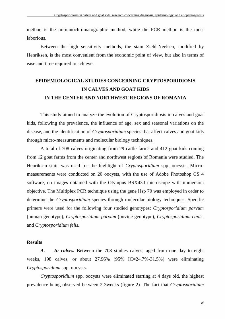
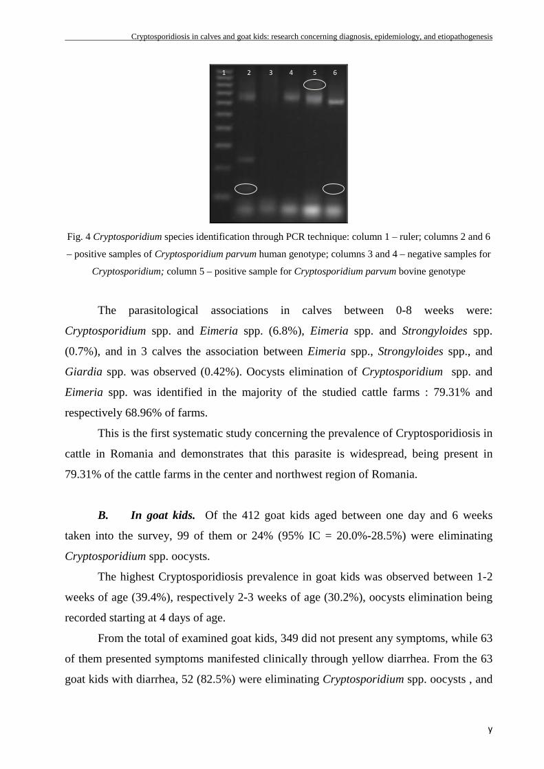
Based on the analysis of SSU rRNA gene, trough nested PCR, it was detected in 8
samples, the amplification of a fragment of 638 bp, specific to the Cryptosporidium
genus. Out of the 8 samples, 5 were positive for Cryptosporidium parvum, human
genotype (62.5%), and 3 were positive for the Cryptosporidium parvum bovine genotype
(37.5%) (figure 4). No positive sample was revealed for the Cryptosporidium canis and
Cryptosporidium felis.
Cryptosporidiosis in calves and goat kids: research concerning diagnosis, epidemiology, and etiopathogenesis
y
Fig. 4 Cryptosporidium species identification through PCR technique: column 1 – ruler; columns 2 and 6
– positive samples of Cryptosporidium parvum human genotype; columns 3 and 4 – negative samples for
Cryptosporidium; column 5 – positive sample for Cryptosporidium parvum bovine genotype
The parasitological associations in calves between 0-8 weeks were:
Cryptosporidium spp. and Eimeria spp. (6.8%), Eimeria spp. and Strongyloides spp.
(0.7%), and in 3 calves the association between Eimeria spp., Strongyloides spp., and
Giardia spp. was observed (0.42%). Oocysts elimination of Cryptosporidium spp. and
Eimeria spp. was identified in the majority of the studied cattle farms : 79.31% and
respectively 68.96% of farms.
This is the first systematic study concerning the prevalence of Cryptosporidiosis in
cattle in Romania and demonstrates that this parasite is widespread, being present in
79.31% of the cattle farms in the center and northwest region of Romania.
B. In goat kids. Of the 412 goat kids aged between one day and 6 weeks
taken into the survey, 99 of them or 24% (95% IC = 20.0%-28.5%) were eliminating
Cryptosporidium spp. oocysts.
The highest Cryptosporidiosis prevalence in goat kids was observed between 1-2
weeks of age (39.4%), respectively 2-3 weeks of age (30.2%), oocysts elimination being
recorded starting at 4 days of age.
From the total of examined goat kids, 349 did not present any symptoms, while 63
of them presented symptoms manifested clinically through yellow diarrhea. From the 63
goat kids with diarrhea, 52 (82.5%) were eliminating Cryptosporidium spp. oocysts , and
1 2 3 4 5 6
Cryptosporidiosis in calves and goat kids: research concerning diagnosis, epidemiology, and etiopathogenesis
z
11 (17.5%) were not eliminating oocysts. For the goat kids without diarrhea, from the
349 goat kids taken into the study, only 47 (13.5%) were eliminating Cryptosporidium
spp. oocysts.
Goat Cryptosporidiosis evolved with symptoms expressed clinically through
diarrhea in only 52.52% of the cases, for the rest of the cases, the disease developed
asymptomatically. Cryptosporidiosis distribution in goat kids by gender was uniform.
The zoonotic risk of Cryptosporidiosis in cattle and goats can only be determined
through molecular biology techniques that identify the exact species involved in the onset
of the infection. Any effort designed to control this disease in humans and cattle has to be
directed toward these species and age categories.
THE PLACE OF CRYPTOSPORIDIOSIS IN THE ETIOLOGY OF T HE
NEONATAL DIARRHEA IN CALVES
This study aimed to analyze the evolution of Cryptosporidiosis in the Neonatal
Diarrhea Syndrome in calves, by following these main directions:
• Determining the place of Crytposporidiosis in the etiology of the neonatal diarrhea
in calves, by determining the isolation frequency of Rotavirus, Coronavirus, F5 E.
coli, and Cryptosporidium parvum
• Identifying the correlations between the severity of the clinical manifestations,
biotic etiological agents, and the severity of the hematological and biochemical
anomalies in calves affected by neonatal diarrhea
The research was conducted on 110 calves coming from three different counties in
the center and northwest region of Romania. Fecal samples were collected from each
individual through rectal exam, trying to find the presence or absence of copro-antigenes
of Rotavirus, Coronavirus, F5 E. Coli, and Cryptosporidium spp. using the Bio-X Duo
Digestive ELISA kit.
To accomplish the clinical and paraclinical study in Neonatal Diarrhea Syndrome,
the patients were divided in two groups, based on the severity of their symptoms:
Cryptosporidiosis in calves and goat kids: research concerning diagnosis, epidemiology, and etiopathogenesis
aa
Group No. 1 was made of 10 calves presenting symptoms of catarrhal enteritis
Group No. 2 was made of 9 calves presenting symptoms of hemorrhagic enteritis
Results
Cryptosporidium parvum is the most common pathogen involved in the Neonatal
Diarrhea Syndrome, with a prevalence of 35.45% of the total cases, in 16.36% of them as
a sole pathogen, and in 19.09% of the cases in association with other enteropathogens.
The Rotavirus infection was found in 17.27% of the total cases, from which in 7.27% as a
sole pathogen, and in 10% in association with other enteropathogens. The infection with
Coronavirus was identified in 17.27% of the total cases, from which in 2.73% as a sole
pathogen, and in 14.54% in association with Cryptosporidium parvum. The Factor 5 E.
coli infection was identified in only 1.82% of the total cases. Out of the total sample
cases, 52.27% were negative for all 4 of the studied enteropathogens.
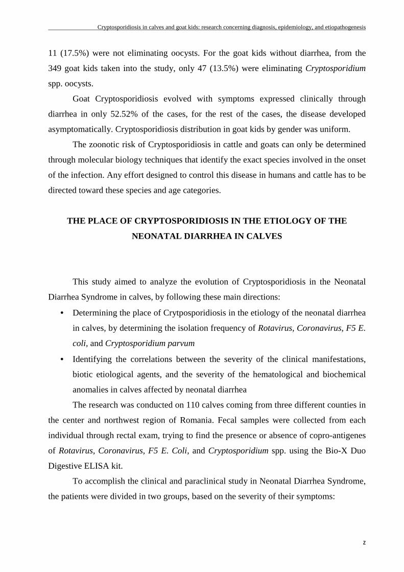
The prevalence of Cryptosporidiosis was highest in the Cluj County (50%), while
the Rotavirus and Coronavirus infections had the highest prevalence in Satu Mare
County, with 24.24% and respectively 39.39% (figure 5).
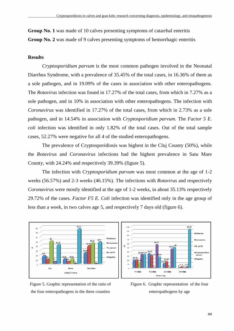
The infection with Cryptosporidium parvum was most common at the age of 1-2
weeks (56.57%) and 2-3 weeks (46.15%). The infections with Rotavirus and respectively
Coronavirus were mostly identified at the age of 1-2 weeks, in about 35.13% respectively
29.72% of the cases. Factor F5 E. Coli infection was identified only in the age group of
less than a week, in two calves age 5, and respectively 7 days old (figure 6).
Figure 5. Graphic representation of the ratio of
the four enteropathogens in the three counties
Figure 6. Graphic representation of the four
enteropathogens by age
Cryptosporidiosis in calves and goat kids: research concerning diagnosis, epidemiology, and etiopathogenesis
bb
The Neonatal Diarrhea Syndrome on calves has a variable clinical expression,
which results from the complex interaction between the risk factors, the onset factors, and
the newborn’s reactivity. The most severe clinical forms were those consecutive to the
hemorrhagic enteritis in which, besides the dehydration and metabolic acidosis, evolved
anemia and endotoxemia.
In the Neonatal Diarrhea Syndrome in calves with symptoms of catarrhal enteritis,
no biotic agents were identified. In the cases with symptoms of catarrhal enteritis, the
pathogenic agents involved were represented by Cryptosporidium spp. (66.7%), Eimeria
spp. (22.22%), Rotavirus (11.1%), and Coronavirus (11.1%).
Hypertonic dehydration and metabolic lactic acidosis were the major pathogenetic
elements that marked the humoral syndrome, both in the mild and severe forms of the
Neonatal Diarrhea Syndrome. Hypoglycemia, evidenced in both groups of calves,
constitutes a major pathogenetic element that results from the anorexia, maldigestion, and
malabsorption.
The plasmatic fibrinogen represents a very sensitive and reliable humoral indicator
in the gastrointestinal inflammatory diseases in newborn calves.
RECORDING OF AN OUTBREAK OF GOAT CRYPTOSPORIDIOSIS
IN CLUJ COUNTY
The purpose of this study was to describe an outbreak of goat Cryptosporidiosis,
from the clinical manifestations through the full description of the pathogenesis of
intestinal Cryptosporidiosis in young goats.
In February 2009, an outbreak of Cryptosporidiosis with aqueous yellow diarrhea
was identified, in goat kids aged one day through three weeks, in a goat farm in Cluj
County. The feces of 57 dead goat kids and 83 living goat kids were processed through
coproparasitological techniques, in order to identify the pathogens involved. On 10
corpses a necropsic exam was performed, consisting of macroscopic exam,
histopatological examination, and electronomicroscopic exam.
Cryptosporidiosis in calves and goat kids: research concerning diagnosis, epidemiology, and etiopathogenesis
cc
Results
The prevalence of Cryptosporidiosis in goat kids in the outbreak was 61.70% (87
out of 141).
The clinical manifestations observed were diarrhea in 68.68% of goat kids (57 out
of 83), kyphosis in 9.64% of the goat kids (8 out of 83), and dehydration in 44.58% of the
goat kids (37 out of 83). Mortality rate in the studied outbreak was 41.13%.
Coproelimination of Cryptosporidium spp. was observed in 81.6% o the total goat
kids (115 out of 141).
At the anatomical-pathological exam, cachectical corpses with hindquarters soiled
with feces were observed. Macroscopically, there was watery intestinal content, with
yellow feces and the intestines presented relaxed from gases.
Histological changes observed in the ileum and colon include alterations of the
intestinal villosities, characterized mainly through atrophy and denudation.
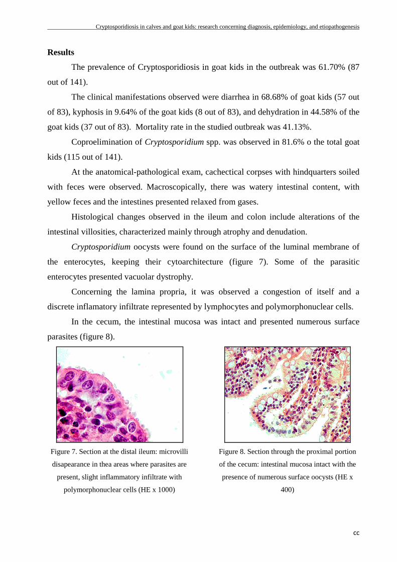
Cryptosporidium oocysts were found on the surface of the luminal membrane of
the enterocytes, keeping their cytoarchitecture (figure 7). Some of the parasitic
enterocytes presented vacuolar dystrophy.
Concerning the lamina propria, it was observed a congestion of itself and a
discrete inflamatory infiltrate represented by lymphocytes and polymorphonuclear cells.
In the cecum, the intestinal mucosa was intact and presented numerous surface
parasites (figure 8).
Figure 7. Section at the distal ileum: microvilli
disapearance in thea areas where parasites are
present, slight inflammatory infiltrate with
polymorphonuclear cells (HE x 1000)
Figure 8. Section through the proximal portion
of the cecum: intestinal mucosa intact with the
presence of numerous surface oocysts (HE x
400)
Cryptosporidiosis in calves and goat kids: research concerning diagnosis, epidemiology, and etiopathogenesis
dd
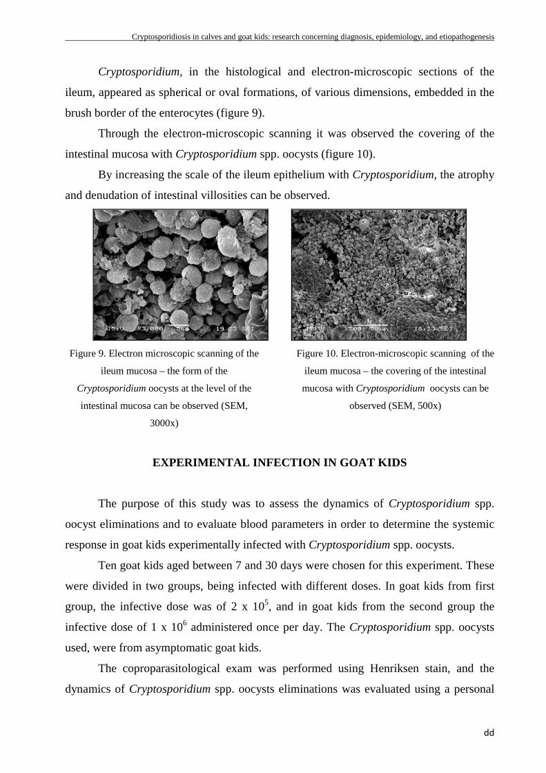
Cryptosporidium, in the histological and electron-microscopic sections of the
ileum, appeared as spherical or oval formations, of various dimensions, embedded in the
brush border of the enterocytes (figure 9).
Through the electron-microscopic scanning it was observed the covering of the
intestinal mucosa with Cryptosporidium spp. oocysts (figure 10).
By increasing the scale of the ileum epithelium with Cryptosporidium, the atrophy
and denudation of intestinal villosities can be observed.
Figure 9. Electron microscopic scanning of the
ileum mucosa – the form of the
Cryptosporidium oocysts at the level of the
intestinal mucosa can be observed (SEM,
3000x)
Figure 10. Electron-microscopic scanning of the
ileum mucosa – the covering of the intestinal
mucosa with Cryptosporidium oocysts can be
observed (SEM, 500x)
EXPERIMENTAL INFECTION IN GOAT KIDS
The purpose of this study was to assess the dynamics of Cryptosporidium spp.
oocyst eliminations and to evaluate blood parameters in order to determine the systemic
response in goat kids experimentally infected with Cryptosporidium spp. oocysts.
Ten goat kids aged between 7 and 30 days were chosen for this experiment. These
were divided in two groups, being infected with different doses. In goat kids from first
group, the infective dose was of 2 x 10⁵, and in goat kids from the second group the
infective dose of 1 x 10⁶ administered once per day. The Cryptosporidium spp. oocysts
used, were from asymptomatic goat kids.
The coproparasitological exam was performed using Henriksen stain, and the
dynamics of Cryptosporidium spp. oocysts eliminations was evaluated using a personal
Cryptosporidiosis in calves and goat kids: research concerning diagnosis, epidemiology, and etiopathogenesis
ee
method, namely counting existing oocysts in 100 microscopical fields. In order to
determine the hematological parameters, blood was drawn from each goat kid, from the
jugular vein in the first day before the infection was started and in the 7th day after the
infection began. In order to evaluate the statistical differences between the two groups,
the Wilcoxon Rank Sum Test (Statistix 9.0) was used.
Results
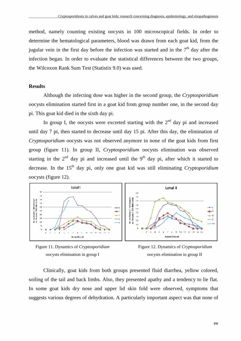
Although the infecting dose was higher in the second group, the Cryptosporidium
oocysts elimination started first in a goat kid from group number one, in the second day
pi. This goat kid died in the sixth day pi.
In group I, the oocysts were excreted starting with the 2nd day pi and increased
until day 7 pi, then started to decrease until day 15 pi. After this day, the elimination of
Cryptosporidium oocysts was not observed anymore in none of the goat kids from first
group (figure 11). In group II, Cryptosporidium oocysts elimination was observed
starting in the 2nd day pi and increased until the 9th day pi, after which it started to
decrease. In the 15th day pi, only one goat kid was still eliminating Cryptoporidium
oocysts (figure 12).
Figure 11. Dynamics of Cryptosporidium
oocysts elimination in group I
Figure 12. Dynamics of Cryptosporidium
oocysts elimination in group II
Clinically, goat kids from both groups presented fluid diarrhea, yellow colored,
soiling of the tail and back limbs. Also, they presented apathy and a tendency to lie flat.
In some goat kids dry nose and upper lid skin fold were observed, symptoms that
suggests various degrees of dehydration. A particularly important aspect was that none of
Cryptosporidiosis in calves and goat kids: research concerning diagnosis, epidemiology, and etiopathogenesis
ff
the subjects taken into study showed changes in body temperature, not even those which
presented severe diarrhea.
The humoral picture in goat kids experimentally infected with Cryptosporidiosis
does not bring any pathognomonic elements of positive diagnosis, but it does provide
valuable prognostic factors and especially the guidance and foundation of a rational
therapeutic behavior.
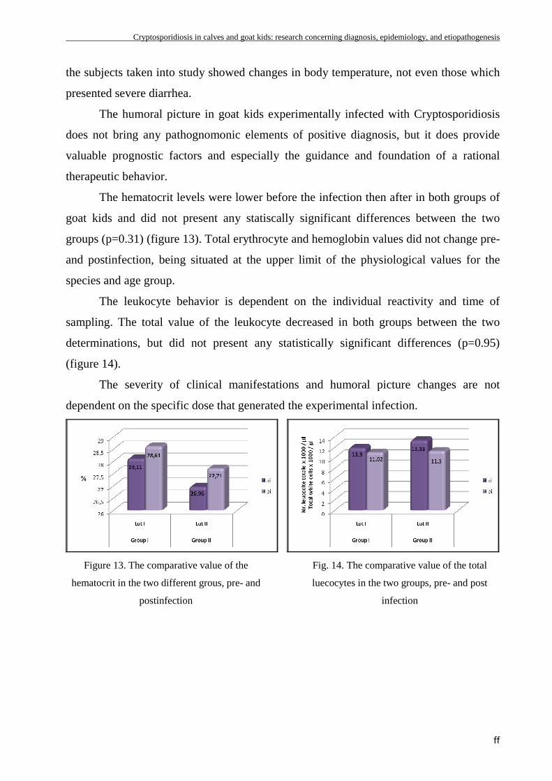
The hematocrit levels were lower before the infection then after in both groups of
goat kids and did not present any statiscally significant differences between the two
groups (p=0.31) (figure 13). Total erythrocyte and hemoglobin values did not change pre-
and postinfection, being situated at the upper limit of the physiological values for the
species and age group.
The leukocyte behavior is dependent on the individual reactivity and time of
sampling. The total value of the leukocyte decreased in both groups between the two
determinations, but did not present any statistically significant differences (p=0.95)
(figure 14).
The severity of clinical manifestations and humoral picture changes are not
dependent on the specific dose that generated the experimental infection.
Figure 13. The comparative value of the
hematocrit in the two different grous, pre- and
postinfection
Fig. 14. The comparative value of the total
luecocytes in the two groups, pre- and post
infection