Rezumatele lucrarilor trimise pentru Conferinta de...
46
FIZIOLOGIA physiology CHIEF EDITOR FRANCISC SCHNEIDER CO-CHIEF EDITORS IOANA SISKA CARMEN TATU ASSOCIATE EDITORS MIHAI NECHIFOR SORIN RIGA EXECUTIVE EDITORS FLORINA BOJIN GABRIELATANASIE DACIANA NISTOR CALIN MUNTEAN EDITORIAL BOARD ARDELEAN AUREL (Arad) BADIU GHEORGHE (Constanţa) BĂDĂRĂU ANCA (Bucureşti) BENEDEK GYORGY (Szeged) BENGA GHEORGHE (Cluj) BUNU CARMEN (Timişoara) COJOCARU MANOLE (Bucureşti) CUPARENCU BARBU (Oradea) CONSTANTIN NICOLAE (Bucureşti) HAULICĂ ION (Iaşi) IANCAU MARIA (Craiova) MIHALAŞ GEORGETA (Timişoara) MUNTEAN DANINA (Timişoara) MUREŞAN ADRIANA (Cluj) NESTIANU VALERIU (Craiova) OPREA TUDOR (New Mexico) PĂUNESCU VIRGIL (Timişoara) PETROIU ANA (Timişoara) POPESCU LAURENJIU (Bucureşti) RÂCZ OLIVER (Kosice) RIGA DAN (Bucureşti) SABĂU MARIUS (Tg. Mureş) SIMIONESCU MAIA (Bucureşti) SIMON ZENO (Timişoara) SAULEA I. AUREL (Chişinău) SWYNGHEDAUW BERNARD (Paris) TANGUAY M. ROBERT (Canada) TATU FABIAN ROMULUS (Timişoara) VLAD AURELIAN (Timişoara) VOICU VICTOR (Bucureşti) ZĂGREAN LEON (Bucureşti) ACCREDITED BY CNCSIS - B+CATEGORY ■ CODE 240 http://journals.indexcopemicus.com/karta.php?action=masterlist&id=4929 http://www.ebscohost.com/titleLists/a9h-journals.pdf Publication data: Fiziologia (Physiology) is issued quarterly Subscription rates: Subscriptions run a full calendar year. Prices are give per volume, surface postage induded. Personal subscription: Romania - 100 RON, Outside Romania - 35$ (must be in the name of, billed to, and paid by an individual. Order must be marked „personal subscription”) Instituţional subscription: 50$ (regular rate) Single issues and back volumes: Information on availability and prices can be obtained through the Publisher. Change of address: Both old and new address should be stated and send to the subscription source. Bibliographic indices: We hope this journal will be regularly listed in bibliographic services, induding „Current Contents” Book Reviews: Books are accepted for review by special agreement. Advertising: Correspondence and rate requests should be addressed to the Publisher. 1. FOR SUBSCRIPTION ADDRESS HVB Bank TIMIŞOARA RO 21 BACX 0000000218508250 TIMIŞOARA-ROMANIA PENTRU REVISTA „FIZIOLOGIA-PHYSIOLOGY” 2. CORRESPONDENCE SHOULD BE ADDRESSED TO THE CHIEF EDITOR PROF. DR.FRANCISC SCHNEIDER PO BOX 135 300024 - TIMIŞOARA - ROMANIA e-mail: [email protected] Editura EUROSTAMPA www.eurostampa.ro Bd. Revoluţiei din 1989 nr. 26, Timişoara Tel/fax: 0256-204816 ISSN 1223-2076
Transcript of Rezumatele lucrarilor trimise pentru Conferinta de...
FIZIOLOGIA
p h y s i o l o g y
CHIEF EDITOR FRANCISC SCHNEIDER
CO-CHIEF EDITORS IOANA SISKA
CARMEN TATU ASSOCIATE EDITORS MIHAI NECHIFOR
SORIN RIGA EXECUTIVE EDITORS FLORINA BOJIN
GABRIELATANASIE DACIANA NISTOR CALIN MUNTEAN
E D I T O R I A L B O A R D
ARDELEAN AUREL (Arad) BADIU GHEORGHE (Constanţa) BĂDĂRĂU ANCA (Bucureşti) BENEDEK GYORGY (Szeged) BENGA GHEORGHE (Cluj) BUNU CARMEN (Timişoara) COJOCARU MANOLE (Bucureşti) CUPARENCU BARBU (Oradea) CONSTANTIN NICOLAE (Bucureşti)
HAULICĂ ION (Iaşi)
IANCAU MARIA (Craiova) MIHALAŞ GEORGETA (Timişoara) MUNTEAN DANINA (Timişoara) MUREŞAN ADRIANA (Cluj) NESTIANU VALERIU (Craiova) OPREA TUDOR (New Mexico)
PĂUNESCU VIRGIL (Timişoara) PETROIU ANA (Timişoara) POPESCU LAURENJIU (Bucureşti) RÂCZ OLIVER (Kosice) RIGA DAN (Bucureşti) SABĂU MARIUS (Tg. Mureş) SIMIONESCU MAIA (Bucureşti) SIMON ZENO (Timişoara) SAULEA I. AUREL (Chişinău) SWYNGHEDAUW BERNARD (Paris) TANGUAY M. ROBERT (Canada) TATU FABIAN ROMULUS (Timişoara) VLAD AURELIAN (Timişoara) VOICU VICTOR (Bucureşti) ZĂGREAN LEON (Bucureşti)
ACCREDITED BY CNCSIS - B+CATEGORY ■ CODE 240
http://journals.indexcopemicus.com/karta.php?action=masterlist&id=4929 http://www.ebscohost.com/titleLists/a9h-journals.pdf
Publication data: Fiziologia (Physiology) is issued quarterly
Subscription rates: Subscriptions run a full calendar year. Prices
are give per volume, surface postage induded.
Personal subscription: Romania - 100 RON, Outside
Romania - 35$ (must be in the name of, billed to, and paid by an
individual. Order must be marked „personal subscription”)
Instituţional subscription: 50$ (regular rate)
Single issues and back volumes: Information on availability
and prices can be obtained through the Publisher.
Change of address: Both old and new address should be stated
and send to the subscription source.
Bibliographic indices: We hope this journal will be regularly listed
in bibliographic services, induding „Current Contents”
Book Reviews: Books are accepted for review by special
agreement.
Advertising: Correspondence and rate requests should be
addressed to the Publisher.
1. FOR SUBSCRIPTION ADDRESS HVB Bank TIMIŞOARA
RO 21 BACX 0000000218508250
TIMIŞOARA-ROMANIA PENTRU REVISTA
„FIZIOLOGIA-PHYSIOLOGY”
2. CORRESPONDENCE SHOULD BE ADDRESSED TO THE CHIEF EDITOR
PROF. DR.FRANCISC SCHNEIDER
PO BOX 135 300024 - TIMIŞOARA - ROMANIA
e-mail: [email protected]
Editura EUROSTAMPA www.eurostampa.ro
Bd. Revoluţiei din 1989 nr. 26, Timişoara Tel/fax: 0256-204816
ISSN 1223-2076
2 Fiziologia - Physiology • 2016.26.1(89)
Instructions to Authors
Submission: Only original papers in English are corisidered and should be sentto:
Prof. dr. Francisc Schneider Chief Editor of "Fiziologia" PO Box 135 300024, TIMIŞOARA, ROMANIA TeUFax: 40-256/490507
Manuscripts should be submitted in triplicate sets of illustrations (of which one is an original), typewritten doublespaced on one side of the paper, with a wide margin.
Conditions: AII manuscripts are subject to editorial review. Manuscripts are received with the explicit understanding that they are not under simultaneous consideration by any other publication. Submission of an artide for publication implies the transfer of the Copyright from the author to the publisher upon acceptance. Accepted papers become the permanent property of "Fiziologia" (Physiology) and may not be reproduced by any means, in-whole or in part, without the written consent of the publisher. It is the author's responsibility to obtain permission to reproduce illustrations, tables, etc. from other publications.
Arrangement: Title page: The first of each paper should indicate
the title (main title underlined), the authors' names, and the institute where the work was conducted. A short title for use as running head is also required.
Keywords: for indexing purposes, a list of 3-10 keywords in English and Romanian is essential.
Abstract: Each paper needs abstract and title in Romanian and English language, fonts size 9, Arial Narrow.
Bady text: fonts size 10, Arial Narrow. Small type: Paragraphs which can or must be set in
smaller type (case histories, test methods, etc.) should be indicated with a „p" (petit) in the margin on the left-hand side.
Footnotes: Avoid footnotes. When essential, they are numbered consecutively and typed at the foot of the appropriate page, fonts size 8, Arial Narrow.
Tables and illustrations: Tables (numbered in Roman numerals) and illustrations (numbered in Arabic numerals) should be prepared on separate sheets, fonts size 9, Arial Narrow. Tables require a heading, and ligures a legend, also prepared on a separate sheet. For the reproduction of illustrations, only good drawings and original photographs can be accepted; negatives or photocopies cannot be used. When possible, group several illustrations on one blockfor reproduction (max. size 140x188 mm) or provide crop marks. On the back of each illustration indicate its number, the author's name,
and artide title. Colour illustration are reproduced at the author's expense.
References: In the text identify references by Arabic figures, (in brackets), fonts size 9, Arial Narrow. Material submitted for publication but not yet accepted should be noted as "unpublished data" and not be induded in the reference list. The list of references should include only those publications which are cited in the text. The references should be numbered and arranged alphabetically by the authors' names. The surnames of the authors followed by initials should be given. There should be no punctuation signs other than a comma to separate the authors. When there are more than 3 authors, the names of the 3 only are used, followed by "et al" abbreviate journal names according to the Index Medicus system. (also see International Committee of Medical Journal Editors: Uniform Requirements for manuscripts submitted to biomedical journals. Ann Intern Med 1982; 96: 766-771).
Examples:
(a) Papers published in periodicals: Kauffman HF, van der Heide S, Beaumont F, et al: Class-apecific antibody determination against Aspergillus fumigatus by mean of the enzyme-linked immunosorbent assay. III. Comparative study: IgG, IgA, IgM, ELISA titers, precipitating antibodies and IGE biding after fractionation of the antigen. Int Arch Allergy Appl Immunol 1986; 80:300 - 306.
(b) Monographs; Matthews DE, Farewell VT: Using and Understanding Medical Statistics. Basel, Karger, 1985.
(c) Edited books: Hardy WD Jr, Essex M:. FeLV-inducted feline acquired immune deficiency syndrome: A model for human AIDS; in Klein E(ed): Acquired Immunodeficiency Syndrome. Prag Allergy, Busel, Karger, 1986, vol 37,353 - 376.
Full address: The exact postal address complete with postal code of the senior author must be given; if correspondence is handled by someone else, indicate this accordingly. Add the E-mail address if possible.
Page charges: There is no page charge for papers of 4 or fewer printed pages (induding tables, illustrations and references).
Galley proofs: unless indicated otherwise, galley proofs are sent to the first-named author and should be retumed with the least possible delay. Alternations
made in galley proofs, other than the corrections of printer's errors, are charged to the author. No page proofs are supplied.
Reprints: Order forms and a price list are sent with the galley proofs. Orders submitted after the issue is printed are subject to considerably higher prices. Allow five weeks from date of publication for delivery of reprints.
Fiziologia - Physiology • 2016.26.1(89) 3
FIZIOLOGIA
phys io logy
CONTENTS 1. Vitex doniana has Restorative Effect on Sperm Parameters and Hormonal Production in
Male Wistar Rats following Testicular Torsion ................................................................................................................................. 4 Adelodun ST, Adewole OS, Olatunji SY, Odukoya SA, Adekomi AD, Adalumo OA, Adeyeye OB
2. CYP2D6 Enzymatic Deficiency and Extrapyramidal Sides Effects in an Autistic Patient Treated with Risperidone .................. 9 Gradinaru R, Andreescu N, Puiu M
3. Effects of Chronic Copper Sulphate Administration on Feeding Pattern and Markers of Renal and Liver Functions of Wistar Rats ................................................................................................................................................. 13
Akomolafe RO, Olukiran OS, Imafidon CE, Ayannuga OA, Oyekunle JA, Oladele AA
4. Contribution of Self-Perceived Stress to Postsurgical Life Quality Change of Patients Undergoing Catharact Surgery ........ 20 Ciobotea D, Șerban C, Putnoky S, Fira Mladinescu C, Tuță Sas I, Băcean Miloicov C, Vlaicu B
5. Biocomposite Processing Technology and Medical Applications. A Review ................................................................................ 25 Voisan R, Gingu O
6. Assessment of Obsessive-Compulsive Behavior in Patients with Haemophilia ........................................................................... 31 Hogea L, Nussbaum L, Dumache R
7. Characterization of the Effects of SarcKATP And MitoKATP Modulators on Reperfusion-Induced Arrhythmias in Isolated Rat Hearts ................................................................................................................................................. 35
Petruș A, Sturza A, Duicu O, Jost N, Muntean D, Baczko I
8. Stem cells from amniotic fluid, an essential tool for regenerative medicine ................................................................................. 40 Honcea A
CUPRINS
1. Vitex doniana are efect restaurator asupra parametrilor spermici și producției hormonale la șobolanii Wistar după torsiunea testiculară ..................................................................................................................................... 4
Adelodun ST, Adewole OS, Olatunji SY, Odukoya SA, Adekomi AD, Adalumo OA, Adeyeye OB
2. Deficitul enzimatic de CYP2D6 şi efectele adverse extrapiramidale la un pacient cu autism tratat cu risperidonă .................... 9 Gradinaru R, Andreescu N, Puiu M
3. Efectele administrării cronice de sulfat de cupru aupra modalităților de hrănire și a markerilor funcționali renali și hepatici la șobolanii Wistar ............................................................................................................................................... 13
Akomolafe RO, Olukiran OS, Imafidon CE, Ayannuga OA, Oyekunle JA, Oladele AA
4. Contribuția stresului autoperceput la modificarea postoperatorie a calității vieții la pacienți supuși operației de cataractă ......... 20 Ciobotea D, Șerban C, Putnoky S, Fira Mladinescu C, Tuță Sas I, Băcean Miloicov C, Vlaicu B
5. Tehnologia procesării materialelor biocompozite și aplicabilitatea în domeniul medical ............................................................ 25 Voisan R, Gingu O
6. Evaluarea comportamentului obsesiv-compulsiv la pacienții cu hemofilie .................................................................................. 31 Hogea L, Nussbaum L, Dumache R
7. Caracterizarea efectelor modulatorilor sarcKATP și mitoKATP asupra aritmiilor induse de reperfuzie pe inimi izolate de șobolan ............................................................................................................................................................... 35
Petruș A, Sturza A, Duicu O, Jost N, Muntean D, Baczko I
8. Celulele stem din lichidul amniotic – un instrument esențial pentru medicina regenerativă ...................................................... 40 Honcea A
4 Fiziologia - Physiology • 2016.26.1(89)
Received January 5th, 2016. Accepted February 12th, 2016. Address for correspondence: Stephen Taiye Adelodun, Department of Anatomy and Cell Biology, Obafemi Awolowo University, Ile-Ife, Nigeria; tel: +2348069823009, e-mail [email protected].
VITEX DONIANA HAS RESTORATIVE EFFECT ON SPERM
PARAMETERS AND HORMONAL PRODUCTION IN MALE
WISTAR RATS FOLLOWING TESTICULAR TORSION
STEPHEN TAIYE ADELODUN*1
, OLARINDE STEPHEN ADEWOLE1
, SUNDAY YINKA
OLATUNJI2
, SAMSON AYODEJI ODUKOYA1
, ADEDAYO DAMILARE ADEKOMI3
,
OLUSOJI ADEOLA ADALUMO4
AND OPEYEMI BLESSING ADEYEYE4
1Department of Anatomy and Cell Biology, Obafemi Awolowo University, Ile-Ife, Nigeria. 2Department of Anatomy, Ben Carson (Snr.) School of Medicine, Babcock University, Ilisan-Remo, Nigeria. 3Department of Anatomy, Osun State University, Osogbo, Nigeria. 4Department of Physiological Sciences, Obafemi Awolowo University, Ile-Ife, Nigeria.
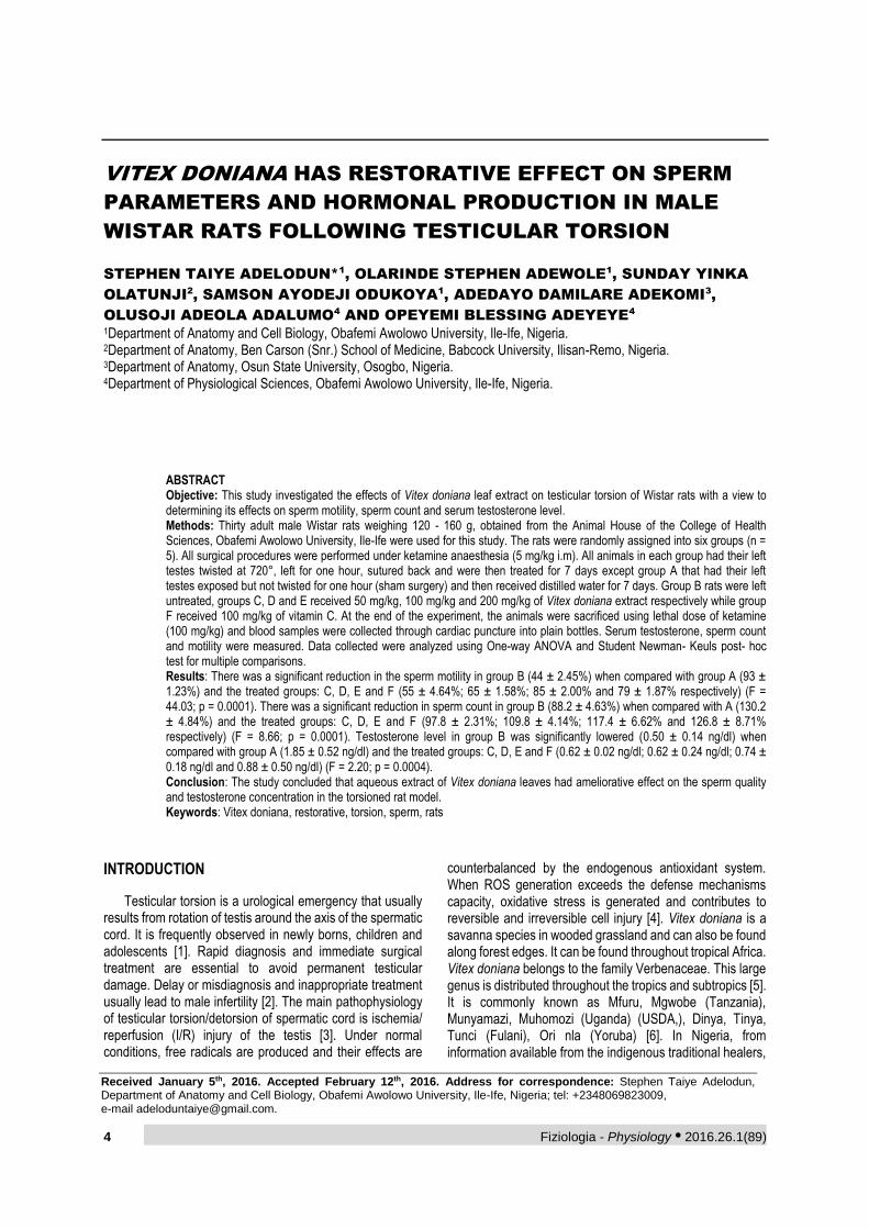
ABSTRACT Objective: This study investigated the effects of Vitex doniana leaf extract on testicular torsion of Wistar rats with a view to determining its effects on sperm motility, sperm count and serum testosterone level. Methods: Thirty adult male Wistar rats weighing 120 - 160 g, obtained from the Animal House of the College of Health Sciences, Obafemi Awolowo University, Ile-Ife were used for this study. The rats were randomly assigned into six groups (n = 5). All surgical procedures were performed under ketamine anaesthesia (5 mg/kg i.m). All animals in each group had their left testes twisted at 720°, left for one hour, sutured back and were then treated for 7 days except group A that had their left testes exposed but not twisted for one hour (sham surgery) and then received distilled water for 7 days. Group B rats were left untreated, groups C, D and E received 50 mg/kg, 100 mg/kg and 200 mg/kg of Vitex doniana extract respectively while group F received 100 mg/kg of vitamin C. At the end of the experiment, the animals were sacrificed using lethal dose of ketamine (100 mg/kg) and blood samples were collected through cardiac puncture into plain bottles. Serum testosterone, sperm count and motility were measured. Data collected were analyzed using One-way ANOVA and Student Newman- Keuls post- hoc test for multiple comparisons. Results: There was a significant reduction in the sperm motility in group B (44 ± 2.45%) when compared with group A (93 ± 1.23%) and the treated groups: C, D, E and F (55 ± 4.64%; 65 ± 1.58%; 85 ± 2.00% and 79 ± 1.87% respectively) (F = 44.03; p = 0.0001). There was a significant reduction in sperm count in group B (88.2 ± 4.63%) when compared with A (130.2 ± 4.84%) and the treated groups: C, D, E and F (97.8 ± 2.31%; 109.8 ± 4.14%; 117.4 ± 6.62% and 126.8 ± 8.71% respectively) (F = 8.66; p = 0.0001). Testosterone level in group B was significantly lowered (0.50 ± 0.14 ng/dl) when compared with group A (1.85 ± 0.52 ng/dl) and the treated groups: C, D, E and F (0.62 ± 0.02 ng/dl; 0.62 ± 0.24 ng/dl; 0.74 ± 0.18 ng/dl and 0.88 ± 0.50 ng/dl) (F = 2.20; p = 0.0004). Conclusion: The study concluded that aqueous extract of Vitex doniana leaves had ameliorative effect on the sperm quality and testosterone concentration in the torsioned rat model. Keywords: Vitex doniana, restorative, torsion, sperm, rats
INTRODUCTION
Testicular torsion is a urological emergency that usually results from rotation of testis around the axis of the spermatic cord. It is frequently observed in newly borns, children and adolescents [1]. Rapid diagnosis and immediate surgical treatment are essential to avoid permanent testicular damage. Delay or misdiagnosis and inappropriate treatment usually lead to male infertility [2]. The main pathophysiology of testicular torsion/detorsion of spermatic cord is ischemia/ reperfusion (I/R) injury of the testis [3]. Under normal conditions, free radicals are produced and their effects are
counterbalanced by the endogenous antioxidant system. When ROS generation exceeds the defense mechanisms capacity, oxidative stress is generated and contributes to reversible and irreversible cell injury [4]. Vitex doniana is a savanna species in wooded grassland and can also be found along forest edges. It can be found throughout tropical Africa. Vitex doniana belongs to the family Verbenaceae. This large genus is distributed throughout the tropics and subtropics [5]. It is commonly known as Mfuru, Mgwobe (Tanzania), Munyamazi, Muhomozi (Uganda) (USDA,), Dinya, Tinya, Tunci (Fulani), Ori nla (Yoruba) [6]. In Nigeria, from information available from the indigenous traditional healers,
Fiziologia - Physiology • 2016.26.1(89) 5
a decoction of the chopped stem bark part of V. doniana is prepared and taken orally for treatment of gastroenteritis. It is administered for ailments including diarrhea and dysentery. It is also taken to improve fertility and the juice may be squeezed into the eyes to treat eye troubles. It is also used in the treatment of liver disease.
MATERIALS AND METHODS
Extraction of Vitex doniana Leaves
Vitex doniana leaves were collected from the lawn behind Moremi Hall, Obafemi Awolowo University, Ile-Ife and were taken to a taxonomist at the Department of Botany, Obafemi Awolowo University, Ile-Ife for authentication after which a voucher specimen was deposited at Ife Herbarium for reference with a reference number IFE-17377433. The leaves were cleaned and air-dried at room temperature. The dried leaves were pulverized using an electric blender. The extraction was done using percolation method which involved continuous washing of the pulverized leaves with water at 80 °C and 30 extraction cycles for 3 hours using Soxhlet extractor. The extract was concentrated in vacuo at 65 °C using a vacuum rotary evaporator and freeze-dried in a lyophilizer. The extract was stored in a desiccator until used.
Animal Care and Management
Thirty young male Wistar rats weighing between 120 - 160 g were obtained from the Animal House of the College of Health Sciences Obafemi Awolowo University Ile-Ife, and used for this research. The rats were randomly assigned into six groups of five rats per group (Groups A, B, C, D, E and F). Animals were housed in clean plastic cages under natural light and dark cycles and at room temperature. Animals in all groups were fed on normal laboratory chow, and had access to water ad libitum.
Surgical Procedure and Experimental Protocol
Animals in group A had their testes exposed for one hour without twisting the spermatic cords, sutured back (sham surgery) and then were given distilled water for 7 days. Group B had their spermatic cords twisted at 720°, left for one hour, sutured back and then received distilled water for 7 days. Group C, D and E also had their spermatic cords twisted at 720°, left for one hour, sutured back and then were orally administered with V. doniana extract (50 mg/kg, 100 mg/kg and 200 mg/kg per body weight) respectively for 7 days. Group F had their spermatic cords twisted at 720°, left for one hour, sutured back and then were orally administered with vitamin C (100 mg/kg per day) for 7 days. All surgical procedures were performed under ketamine anaesthesia (5 mg/kg i.m). V. doniana extract and vitamin C dissolved in distilled water were administered orally on a daily basis for
the period of administration using suitable oral cannula. Vitamin C was obtained from KUNIMED PHARMACHEM LTD Nigeria.
Sperm Counts and Motility Analysis
The sperm concentration was determined using the haematocytometer method. 1:20 dilution from each well mixed sample was prepared by diluting 50 µl of liquefied semen with 950 µl diluents. The diluents were prepared by adding 50 g of sodium hydrogen trioxocarbonate (NaHCO3), 10 ml of 35% (v/v) formalin and 0.25 g of trypan blue to distilled water and making up the solution to final volume of 1000 ml. A fixed volume of the sample was withdrawn with micro-pipette and delivered onto the edges of Neubauer chamber of haematocytometer and covered with 22x22 mm cover slip. The weight of the cover slip spread the sample which made the semen to move to center of Neubauer by capillary action and standardized so that the analyses were carried out in a preparation with fixed depth. Both chambers of haematocytometer were scored and the average count was calculated.
A fixed volume of semen was collected from harvested epididymis and put in normal saline. Not more than 10 µl of the semen was withdrawn with micro-pipette and delivered onto clean glass slide covered by 22 x 22 mm cover slip and standardized so that the analyses were carried out in a preparation with depth (i.e., 20 µl). This depth allowed full expression of the rotating movement of normal spermatozoa. The weight of the cover slip spread the sample for optimal viewing. The freshly made, wet preparation was left to stabilize for approximately one minute and the procedure was carried out at a room temperature between 18 and 24oC in the laboratory. The microscopic field was scanned systematically and the motility of each spermatozoon was graded as being motile or non- motile. Spermatozoa graded motile displayed rapid progressive motility along a linear track, covering a distance of at least half the length of the spermatozoon per second.
Testosterone Assay
Blood obtained from the left ventricular through cardiac puncture was assayed for serum testosterone. The level of testosterone was estimated using Bio-Inteco KIT by Inteco Diagnostics UK Ltd.
Desired numbers of coated wells were secured in the holder after which 10 µL of standards, specimens and controls were dispensed into appropriate wells. Then 50 µL of rabbit anti-Testosterone reagent was dispensed into each well. The mixtures were thoroughly mixed for 30 minutes. 100 µL of Testosterone-HRP Conjugate Reagent was dispensed into each well and it was incubated at 37oC for 90 minutes. The microwells were rinsed and flicked 5 times with washing Buffer (1x). 100 µL of TMB Substrate was dispensed into each well and gently mixed for 10 seconds, incubated at room temperature (18–22 oC) for 20 minutes.
6 Fiziologia - Physiology • 2016.26.1(89)
The reaction was stopped by adding 100µL of Stop Solution to each well. It was gently mixed for 30 seconds and it was made sure that all the blue color changed to yellow color completely. Absorbance was read at 450nm with a microliter well reader within 15 minutes.
Statistical Analysis
Results were expressed as mean ± standard deviation. One-way ANOVA was used to analyze data, followed by Student Newman-Keuls (SNK) test for multiple comparisons. GraphPad Prism5 (Version 5.03, GraphPad Inc USA.) was the statistical package used for data analysis. The results were considered significant when p < 0.05.
RESULTS
Testicular torsion was accompanied by weight loss even with the administration of Vitex doniana and vitamin C for the first 2-3 days in the experimental animals. Weight loss continued in the control (sham surgery) and torted untreated group (Group A and B) respectively. There was a slight weight gain in the treated groups C, D, E and F towards the end of the experimental period (Table I).
Table I. Absolute Organ Weight and Difference in Body Weight of Male
Wistar Rats Treated with Vitex donina following Testicular Torsion
Treatment Groups
Initial Body
Weight (g)
Final Body
Weight (g)
Absolute Testicular Weight (g)
Relative Testicular
Weight (%)
A 176.00 ±
7.97 161.60 ±
7.97 1.25 ± 0.07 0.78 ± 0.04
B 225.00 ±
5.00* 175.30 ±
21.59 0.79 ± 0.14*
0.44 ± 0.05*
C 149.00 ±
8.86 148.20 ±
9.90* 0.97 ± 0.04*
0.67 ± 0.04
D 156.30 ±
8.75 152.60 ±
8.41 0.90 ± 0.05*
0.59 ± 0.05
E 173.80 ±
11.43 159.60 ±
9.46 1.41 ± 0.10 0.89 ± 0.08
F 153.00 ±
9.03 143.90 ±
9.59* 1.08 ± 0.07 0.78 ± 0.09
Results are presented as Mean ± SEM (n =5). * Significantly different from Control at p < 0.05.
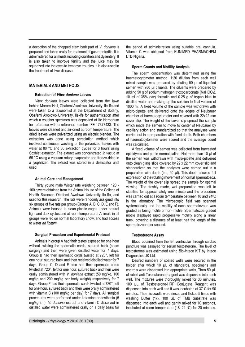
The mean sperm motility in the torted untreated group B
is (44.0 ± 4.47%) which is significantly (p < 0.05) lower when compared with the control (93.0 ± 1.23 %) and the torted and treated groups C, D, E and F (58.0 ± 4.64, 59.0 ± 2.45, 49.0 ± 5.57 and 56.0 ± 6.00 %) (Figure 1).
Fig. 1. Effects of Vitex doniana on Sperm Motility of Male Wistar Rats following Testicular Torsion
* significantly different from control at p < 0.05.
# significantly different from B at p < 0.05.
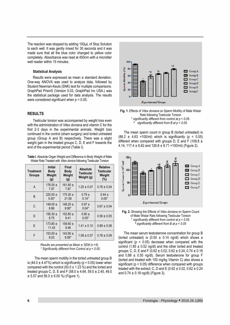
The mean sperm count in group B (torted untreated) is
(88.2 ± 4.63 ×100/ml) which is significantly (p < 0.05) different when compared with groups D, E and F (109.8 ± 4.14, 117.4 ± 6.62 and 126.8 ± 8.71 ×100/ml) (Figure 2).
Fig. 2. Showing the Effects of Vitex doniana on Sperm Count of Male Wistar Rats following Testicular Torsion * significantly different from control at p < 0.05.
# significantly different from B at p < 0.05
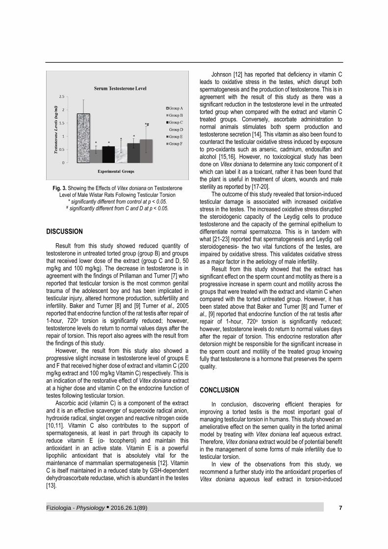
The mean serum testosterone concentration for group B
(torted untreated) is (0.50 ± 0.14 ng/dl) which shows a significant (p < 0.05) decrease when compared with the control (1.85 ± 0.52 ng/dl) and the other torted and treated groups; C, D, E and F (0.62 ± 0.02, 0.62 ± 0.24, 0.74 ± 0.18 and 0.88 ± 0.50 ng/dl). Serum testosterone for group F (torted and treated with 100 mg/kg Vitamin C) also shows a significant (p < 0.05) difference when compared with groups treated with the extract; C, D and E (0.62 ± 0.02, 0.62 ± 0.24 and 0.74 ± 0.18 ng/dl) (Figure 3).
Fiziologia - Physiology • 2016.26.1(89) 7
Fig. 3. Showing the Effects of Vitex doniana on Testosterone Level of Male Wistar Rats Following Testicular Torsion
* significantly different from control at p < 0.05. # significantly different from C and D at p < 0.05.
DISCUSSION
Result from this study showed reduced quantity of testosterone in untreated torted group (group B) and groups that received lower dose of the extract (group C and D, 50 mg/kg and 100 mg/kg). The decrease in testosterone is in agreement with the findings of Prillaman and Turner [7] who reported that testicular torsion is the most common genital trauma of the adolescent boy and has been implicated in testicular injury, altered hormone production, subfertility and infertility. Baker and Turner [8] and [9] Turner et al., 2005 reported that endocrine function of the rat testis after repair of 1-hour, 720o torsion is significantly reduced; however, testosterone levels do return to normal values days after the repair of torsion. This report also agrees with the result from the findings of this study.
However, the result from this study also showed a progressive slight increase in testosterone level of groups E and F that received higher dose of extract and vitamin C (200 mg/kg extract and 100 mg/kg Vitamin C) respectively. This is an indication of the restorative effect of Vitex doniana extract at a higher dose and vitamin C on the endocrine function of testes following testicular torsion.
Ascorbic acid (vitamin C) is a component of the extract and it is an effective scavenger of superoxide radical anion, hydroxide radical, singlet oxygen and reactive nitrogen oxide [10,11]. Vitamin C also contributes to the support of spermatogenesis, at least in part through its capacity to reduce vitamin E (α- tocopherol) and maintain this antioxidant in an active state. Vitamin E is a powerful lipophilic antioxidant that is absolutely vital for the maintenance of mammalian spermatogenesis [12]. Vitamin C is itself maintained in a reduced state by GSH-dependent dehydroascorbate reductase, which is abundant in the testes [13].
Johnson [12] has reported that deficiency in vitamin C leads to oxidative stress in the testes, which disrupt both spermatogenesis and the production of testosterone. This is in agreement with the result of this study as there was a significant reduction in the testosterone level in the untreated torted group when compared with the extract and vitamin C treated groups. Conversely, ascorbate administration to normal animals stimulates both sperm production and testosterone secretion [14]. This vitamin as also been found to counteract the testicular oxidative stress induced by exposure to pro-oxidants such as arsenic, cadmium, endosulfan and alcohol [15,16]. However, no toxicological study has been done on Vitex doniana to determine any toxic component of it which can label it as a toxicant, rather it has been found that the plant is useful in treatment of ulcers, wounds and male sterility as reported by [17-20].
The outcome of this study revealed that torsion-induced testicular damage is associated with increased oxidative stress in the testes. The increased oxidative stress disrupted the steroidogenic capacity of the Leydig cells to produce testosterone and the capacity of the germinal epithelium to differentiate normal spermatozoa. This is in tandem with what [21-23] reported that spermatogenesis and Leydig cell steroidogenesis- the two vital functions of the testes, are impaired by oxidative stress. This validates oxidative stress as a major factor in the aetiology of male infertility.
Result from this study showed that the extract has significant effect on the sperm count and motility as there is a progressive increase in sperm count and motility across the groups that were treated with the extract and vitamin C when compared with the torted untreated group. However, it has been stated above that Baker and Turner [8] and Turner et al., [9] reported that endocrine function of the rat testis after repair of 1-hour, 720o torsion is significantly reduced; however, testosterone levels do return to normal values days after the repair of torsion. This endocrine restoration after detorsion might be responsible for the significant increase in the sperm count and motility of the treated group knowing fully that testosterone is a hormone that preserves the sperm quality.
CONCLUSION
In conclusion, discovering efficient therapies for improving a torted testis is the most important goal of managing testicular torsion in humans. This study showed an ameliorative effect on the semen quality in the torted animal model by treating with Vitex doniana leaf aqueous extract. Therefore, Vitex doniana extract would be of potential benefit in the management of some forms of male infertility due to testicular torsion.
In view of the observations from this study, we recommend a further study into the antioxidant properties of Vitex doniana aqueous leaf extract in torsion-induced
8 Fiziologia - Physiology • 2016.26.1(89)
testicular damage as induction of oxidative stress is the main mechanism of damage in this urological challenge. Conflicts of Interest The authors declare that they have no conflict of interest.
REFERENCES
1. Arbonnier M. Trees shrubs and lianas dry areas of West Africa, CIRAD- MNHN, Montpellier, France 2002.
2. Atawodi SE, Bulus T, Ibrahim S, Ameh DA, Nok AJ, Mamman M and Galadima M. In vitro trypanocidal effect of methanolic extract of some Nigerian savannah plants. Afr. J. Biotech 2003; 2(9): 317-321.
3. Baker LA and Turner TT. Leydig cell function after experimental testicular torsion despite loss of spermatogenesis. J Androl 1995; 16:12-7.
4. Bulger EM and Maier RV. Antioxidants in critical illness. Arch Surg 2001; 136: 1201-1207.
5. Chen H, Liu J and Luo L. Vitamin E, aging and Leydig cell steroidogenesis. Exp Gerontol 2005; 40:728-736.
6. Ergur BU, Kiray M, Pekcetin C, Bagriyanik HA and Erbil G. Protective effect of erythropoietin pretreatment in testicular ischemia- reperfusion injury in rats. J Pediatr Surg 2008: 43: 722-728.
7. Johnson FC (2001). The antioxidant vitamins. CRC. Cri Rev Food Sci Nutr. 11:217-309.
8. Louppe D, Oteng-Amoako AA, and Brink M. Vegetables resources of tropical Africa, Prota 7 (1): Wood, CTA Wagening; Neth. 2009.
9. Maneesh M, Jayalakshmi H and Dutta S. Experimental therapeutic intervention with ascorbic acid in ethanol induced testicular injuries. Indian J Exp Biol 2005; 43:172-176.
10. Okigbo NR. Fermentation of black plum (Vitex doniana sweet) juice for production of wine, fruits 2003; 58 (6). 363-369.
11. Onochie C.F, Keay R.W and Standfield DP. Nigeria Trees. Second Edition. Government Printer, Lagos 1964.
12. Orwa C, Mutua A, Kindt R, Jamnadass R and Anthony S. Agroforest tree database: a tree reference and selection guide version 4.0, World Agrofor. Cent., 2009; Kenya.
13. Paolicchi A, Pezzini A and Saviozzi M. Localization of a GSH-dependent dehydroascorbate reductase in rat tissue and subcellular fractions. Arch Biochem Biophys 1996; 333: 489-495.
14. Peltola V, Mantyla E and Huhtaniemi I. Lipid peroxidation and antioxidant enzyme activities in the rat testis after cigarette smoke inhalation or administration of polychlorinated biphenyls or polychlorinated naphthalenes. J Androl 1994; 15:353-361
15. Perrotti M, Badger W and Prader S. Medical malpractice in urology, 1985 to 2004: 469 consecutive cases closed with indemnity payment. J Urol 2006; 176 (5):2154-2157.
16. Prillaman HM and Turner TT. Rescue of testicular function after acute experimental torsion. J. Urol 1997; 157:340-5.
17. Quinn PG and Payne AH. Oxygen-mediated damage of microsomal cytochrome P-450 enzymes in cultured Leydig cells: Role in steroidogenic desensitization. J Biol Chem 1984; 259: 4135.
18. Senthil Kumar J, Banudevi S and Sharmila M. Effects of vitamin C and E on PCB (Aroclor 1254) induced oxidative stress, androgen binding protein and lactate in rat Sertoli cells. Reprod Toxicol 2004; 19: 201-208.
19. Sonmez M, Turk G, Yuce A. The effect of ascorbic acid supplementation on sperm quality, lipid peroxidation and testosterone levels of male Wistar rats. Theriogenology 2005; 63:2063-2072.
20. Tannenbaum SR, Wishnok JS and Leaf CD. Inhibition of nitrosamine formation by ascorbic acid. American Journal of Clinical Nutrition 1991; 53 (Suppl): 247-250.
21. Turner TT and Brown KJ. Spermatic cord torsion: loss of spermatogenesis despite return of blood flow. Biol Reprod 1993; 49:401-7.
22. Turner TT, Bang HJ and Lysiak JJ. Experimental testicular torsion: reperfusion blood flow and subsequent testicular venous plasma testosterone concentrations. Urology 2005; 65:390-4.
23. Weber P, Benedich A and Scalch W. Vitamin C and Human Health-A review of Recent Data Relevant to Human Requirements. Int J Vit Nutr Res 1996; 66:19-30.
Fiziologia - Physiology • 2016.26.1(89) 9
Received January 10th 2016. Accepted February 2nd 2016. Address for correspondence: Nicoleta Andreescu, Discipline of Genetics, University of Medicine and Pharmacy “Victor Babes”, 2 E. Murgu Square, 300041 Timisoara, Romania, tel.: 0040256204476, e-mail: [email protected] *Authors have equal contribution
CYP2D6 ENZYMATIC DEFICIENCY AND EXTRAPYRAMIDAL
SIDES EFFECTS IN AN AUTISTIC PATIENT TREATED WITH
RISPERIDONE
RALUCA GRADINARU*, NICOLETA ANDREESCU*, MARIA PUIU
Genetics Discipline, Department of Microscopic Morphology, “Victor Babes” University of Medicine and Pharmacy, Timisoara, Romania
ABSTRACT It is known today that genetic variability can have a major impact in the occurrence of adverse effects of antipsychotic medications such as movement disorders. Unfortunately, in clinical practice, testing the genetic profile of patients is not frequently performed in order to identify such risks. It is our hope, that in the near future, for a more predictive and personalized treatment, identifying the specific genotype of patients and the way this could contribute to the antipsychotic adverse effects, will become a usual practice. In this paper we provide a summary of one case report on a 10-year-old autistic patient who suffered from movement disorders while being treated with risperidone at 0.75mg/day. It was discovered that he has a cytochrome P450 2D6 deficiency, being phenotypically a CYP2D6 intermediate metabolizer, which may explain his susceptibility to develop extrapyramidal side effects. Although we expected that the active moiety of risperidone would be elevated, this was not the case. This is an example of the important role that pharmacogenetics can have, to identify the risk of side effects prior to treatment initiation, and to better guide the clinicians in choosing a personalized therapeutic regime for each patient, with less adverse reactions. Keywords: Risperidone, CYP2D6 genotype, extrapyramidal syndrome
INTRODUCTION
Autism is a behavioral disorder diagnosed in childhood, characterized by difficulties in communication, stereotypical, repetitive behavior and decrease of patient's ability to interact socially. Although the specific causes are not yet understood, scientific evidence indicates involvement of both abnormalities in brain development and important genetic factors. Besides educational and behavioral specialized interventions, pharmacological management can play a positive role in the treatment of autism related disorders such as depression, anxiety, hyperactivity, obsessive-compulsive behavior, auto and hetero aggression.
Risperidone is an atypical antipsychotic whose pharmacodynamic action is due to the dual dopaminergic (D2) and serotoninergic (5HT2) antagonism in the central nervous system. In October 2006, risperidone was the first drug approved by the Food and Drug Administration (FDA) for the treatment of irritability in children and adolescents with autism, between 5 and 16 years of age. Clinical efficacy of risperidone versus placebo was demonstrated in two clinical trials conducted over a period of eight weeks, which included
159 pediatric patients with autism. The instruments employed to assess reduction of irritability were Aberrant Behavior Checklist (ABC) and Clinical Global Impression - Change (CGI-C) scale [1]. Off-label risperidone is administered also in other behavioral disturbances associated with autism such as aggression, self-flagellation and spontaneous mood changes [2].
Regarding pharmacokinetic properties, risperidone is metabolized in the liver by the CYP2D6 enzyme system. The CYP2D6 gene has several polymorphisms. The identified allelic variants can be classified as being:
functional (e.g. CYP2D6*1 and *2)
with reduced activity (e.g. CYP2D6*9, *10, *29, *36, *41)
nonfunctional (e.g. CYP2D6*3-8, *18, *21, *44) [3]. The adverse effects occurring with a frequency greater
than 5% suffered by pediatric patients with autism treated with risperidone, may include dizziness, constipation, dry mouth, sedation, fatigue, headache, weight gain and parkinsonian syndrome. Also neurological and other adverse effects have been reported, such as dyskinesia, neuroleptic malignant syndrome, impaired motor coordination, arrhythmias, hyperglycemia etc. [4].
10 Fiziologia - Physiology • 2016.26.1(89)
CASE DESCRIPTION
A male patient aged 10, 31 kg, diagnosed with autism, was prescribed risperidone to improve aggressive behavior, 0.25mg/day for 7 days, then 0.5mg/day for 12 months. Subsequently, to better control the symptoms, the dose of risperidone was increased to 1mg/day. Because there was an increase in the patient’s irritability, it was decided to increase the dose to 2 mg/day divided in two doses, considering that these manifestations are due to disease decompensation as a consequence of the pre-pubertal stage of the child. 14 days after increasing the dose, the patient developed akathisia, with extreme psychomotor agitation, stiffness and inability to control the motor function. It was decided to gradually reduce the dose of risperidone to discontinuation.
In order to ameliorate akathisia, treatment with propranolol was initiated for 30 days. Initiation and discontinuation of propranolol treatment was made by progressively increasing respectively decreasing doses. It was noticed that administration of propranolol caused a distinct reduction in intestinal transit with occurrence of constipation.
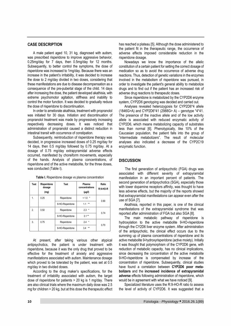
Subsequently, reintroduction of risperidone therapy was decided, in progressive increased doses of 0.25 mg/day for 14 days, then 0.5 mg/day followed by 0.75 mg/day. At a dosage of 0.75 mg/day extrapyramidal adverse effects occurred, manifested by choreiform movements, especially of the hands. Analysis of plasma concentrations, of risperidone and of the active metabolite, for the three doses, was conducted (Table I).
Table I. Risperidone dosage vs plasma concentration
Test Risperidone dosage
(mg)
Test Plasma concentrations
(µg/l)
Ratio
R:9-HO-R
1. 0.25 Risperidone < 1.0 * 0.90
9-HO-Risperidone 1.1 **
2. 0.50 Risperidone 2.3 * 0.74
9-HO-Risperidone 3.1 **
3. 0.75 Risperidone 2.4 * 0.70
9-HO-Risperidone 3.4 **
At present, after taking various other atypical antipsychotics, the patient is under treatment with risperidone, because it was the only drug that proved to be effective for the treatment of anxiety and aggressive manifestations associated with autism. Maintenance dosage which proved to be tolerated by the patient, was set at 0.5 mg/day in two divided doses.
According to the drug maker’s specifications, for the treatment of irritability associated with autism, the target dose of risperidone for patients > 20 kg is 1 mg/day. There are also clinical trials where the maximum daily dose was 2.5 mg for children > 20 kg, but at this dose the therapeutic effect
has reached a plateau [5]. Although the dose administered to the patient fit in the therapeutic range, the occurrence of adverse effects imposed considerable reduction in the risperidone dosage.
Nowadays we know the importance of the allelic constitution of a certain patient for setting the correct dosage of medication so as to avoid the occurrence of adverse drug reactions. Thus, detection of genetic variations in the enzymes involved in the metabolism of risperidone was pursued, in order to investigate the patient's general ability to metabolize drugs and to find out if the patient has an increased risk of adverse drug reactions to therapeutic doses.
Since risperidone is metabolized by the CYP2D6 enzyme system, CYP2D6 genotyping was decided and carried out.
Analyses revealed heterozygosis for CYP2D6*4 allele (1846G>A) and CYP2D6*41 (2988G> A) – genotype *4*41. The presence of the inactive allele and of the low activity allele is associated with reduced enzymatic activity of CYP2D6, which means metabolizing capacity of substrates less than normal [6]. Phenotypically, like 10% of the Caucasian population, the patient falls into the group of “intermediate metabolizers”. The result of molecular analyses also indicated a decrease of the CYP2C19 enzymatic function.
DISCUSSION
The first generation of antipsychotic (FGA) drugs was associated with different severity of extrapyramidal manifestation in an important percent of patients. The second generation of antipsychotics (SGA), especially those with lower dopamine receptors affinity, was thought to have less adverse effects, but the majority of the reports showed that extrapyramidal manifestations can appear even after the use of SGA [7].
Akathisia, reported in this paper, is one of the clinical manifestations of the extrapyramidal syndrome that was reported after administration of FGA but also SGA [8].
The main metabolic pathway of risperidone is hydroxylation to the active metabolite 9-HO-risperidone through the CY2D6 liver enzyme system. After administration of the antipsychotic, the clinical effect occurs due to the summing up of plasma concentrations of risperidone and its active metabolite 9-hydroxyrisperidone (active moiety). Initially it was thought that polymorphism of the CYP2D6 gene, with reduction of metabolic capacity, has no clinical implications, since decreasing the concentration of the active metabolite 9-HO-risperidone is compensated by increase of the concentration of risperidone. Subsequently, clinical studies have found a correlation between CYP2D6 poor meta- bolizers and the increased incidence of extrapyramidal adverse effects following administration of risperidone, which would be in agreement with what we have noticed [9].
Specialized literature uses the R:9-HO-R ratio to assess the level of activity of CYP2D6. It was suggested that a
Fiziologia - Physiology • 2016.26.1(89) 11
plasma ratio R:9-HO-R between 0.1-0.3 indicates a normal enzymatic activity, this patients being classified as extensive metabolizers, while a value >1 is associated with lack of enzymatic activity, this patients being classified as poor metabolizers [10]. For our patient, CYP2D6 intermediate metabolizer, the average ratio R:9-HO-R is 0.78, which is in trend with the things presented above.
The occurrence of these adverse effects in patients, who report high risperidone/9-HO-risperidone plasmatic ratio, characteristic to CYP2D6 deficient metabolizers, may be explained by the different pharmacodynamic profile of the two compounds. In a review, Alamo et al. analyze pharma- cological differences between risperidone and the active metabolite administered as a drug under the name of paliperidone [11]. Compared to ripseridone, 9-HO-risperidone has a higher rate of dissociation from the D2 receptors and a lower affinity to adrenergic, muscarinic and 5HT2A receptors, hence the possible differences existing in therapeutic efficacy and the profile of adverse effects between the two. Also the two compounds differ in terms of profile of the second messenger systems that they regulate [12].
Since the result of the genetic testing performed, places our patient among intermediate metabolizers, we would expected that there is an accumulation of risperidone and the active metabolite, which could explain the occurrence of adverse effects. But after determination of plasma concentrations following administration of different doses of risperidone, it appeared under-dosed, in spite of the adverse effects occurred. The analysis laboratory quotes Regenthal R. for the reference values of these analyses [13]. As far as we are concerned, we do not know about the existence in literature of data indicating exactly the optimal plasma concentration for risperidone for pediatric patients, over which the risk of adverse effects exceeds the benefits provided by the treatment.
The importance of establishing the CYP2D6 genotype in patients treated with risperidone was suggested by several studies that included cohorts of children and adolescents under treatment with risperidone [14, 15].
CONCLUSION
Thus, to implement a personalized therapy with risperidone, we sustain that CYP2D6 genotyping prior to risperidone treatment can be a useful tools in choosing the correct dosage regimen, so as to avoid the occurrence of adverse drug reactions.
Acknowledgments This work was supported by the POSDRU grant no. 159/1.5/S/136893 titled “Strategic partnership for the increase of the scientific research quality in medical universities through the award of doctoral and postdoctoral fellowships – DocMed.Net_2.0”, awarded to Dr. Andreescu Nicoleta.
REFERENCES
1. United States Food and Drug Administration. (October 2006). FDA approves the first drug to treat irritability associated with autism, risperdal. http://www.fda.gov/NewsEvents/Newsroom/.
2. Sharma A, Shaw SR. Efficacy of risperidone in managing maladaptive behaviors for children with autistic spectrum disorder: a meta-analysis. J Pediatr Health Care 2012; 26(4):291-9.
3. Ingelman-Sundberg M, Sim SC, Gomez A, et al. Influence of cytochrome P450 polymorphisms on drug therapies: Pharmacogenetic, pharmacoepigenetic and clinical aspects. Pharmacol Ther 2007; 116:496-526.
4. Rxlist.com. (2009). Risperdal indications & dosage, http://www.rxlist.com/risperdal-drug.htm
5. Risperdal (risperidone). http://www.fda.gov/Safety/ MedWatch/Safety Information/ucm175826.htm.
6. www.snpedia.com/index.php/CYPED6 7. Divac N, Prostran M, Jakovcevski I, et al. Second-Generation
Antipsychotics and Extrapyramidal Adverse Effects. Biomed Res Int. 2014; 2014: 656370.
8. Shirzadi AA, Ghaemi SN. Side effects of atypical antipsychotics: extrapyramidal symptoms and the metabolic syndrome. Harv Rev Psychiatry 2006; 14(3):152-164.
9. de Leon J, Susce MT, Pan RM, et al. The CYP2D6 poor metabolizer phenotype may be associated with risperidone adverse drug reactions and discontinuation. J Clin Psychiatry 2005; 66:15-27.
10. de Leon J, Greenlee B, Barber J, et al. Practical guidelines for the use of new generation antipsychotic drugs (except clozapine) in adult individuals with intellectual disabilities. Res Dev Disabil 2009; 30:613-669.
11. Álamo C, López-Muñoz F. The Pharmacological Role and Clinical Applications of Antipsychotics’ Active Metabolites: Paliperidone versus Risperidone. Clin Exp Pharmacol 2013; 3:117.
12. Clarke WP, Chavera TA, Silva M, et al. Signalling profile differences: paliperidone versus risperidone, Pharmacol. 2013; 170(3): 532-545.
13. Regenthal R, Koppel M, Preis R. Anestheologie und Intensivmedizin 1999; 3: 129-144.
14. Youngster I, Zachor DA, Gabis LV et al. CYP2D6 genotyping in paediatric patients with autism treated with risperidone: a preliminary cohort study. Dev Med Child Neurol 2014; 56(10): 990-4.
15. Sherwin CMT, Saldaña SN, Bies RR, et al. Population pharmacokinetic modeling of risperidone and 9-hydroxyrisperidone to estimate CYP2D6 subpopulations in children and adolescents. Ther Drug Monit 2012; 34(5): 535–544.
12 Fiziologia - Physiology • 2016.26.1(89)
DEFICITUL ENZIMATIC DE CYP2D6 ŞI EFECTELE
ADVERSE EXTRAPIRAMIDALE LA UN PACIENT CU
AUTISM TRATAT CU RISPERIDONĂ
REZUMAT Este cunoscut astăzi că variabilitatea genetică poate avea un impact major în apariția efectelor adverse ale medicamentelor antipsihotice, cum ar fi efectele adverse extrapiramidale. Din păcate, în practica clinică, testarea profilul genetic al pacienților nu se determină în mod frecvent, în vederea identificării unor astfel de riscuri. Este speranța noastră, ca în viitorul apropiat, pentru personalizarea şi buna predictibilitate a tratamentului medicamentos, identificarea genotipului pacienților și modul în care acesta ar putea contribui la apariţia efectelor adverse antipsihotice, să devenă o practică obișnuită. Această lucrare a dorit să prezinte un studiu de caz al unui pacient diagnosticat cu autism, în vârstă de 10 de ani, care ȋn urma tratamentului cu risperidonă la 0,75 mg / zi a dezvoltat efecte adverse extrapiramidale. S-a
descoperit că pacientul prezintă un deficit al citocromul P450 2D6, fiind fentotipic ȋncadrat ca metabolizor intermediar
CYP2D6, ceea ce poate explica susceptibilitatea de apariţie a efectelor secundare extrapiramidale. Deși ne-am așteptat ca şi concentraţia activă totală de rispeidonă să fie crescută, acest lucru nu a fost observat. Acest caz este un exemplu al rolului important pe care farmacogenetica ȋl poate avea, pentru a identifica riscul de a dezvolta reacții adverse
medicamentoase înainte de inițierea tratamentului, și pentru a ghida mai bine clinicienii ȋn alegerea unor doze terapeutice
personalizate pentru fiecare pacient ȋn parte, cu un minim de reacții adverse.
Cuvinte cheie: Risperidona, genotip CYP2D6, sindrom extrapiramidal
Fiziologia - Physiology • 2016.26.1(89) 13
Received January 12th 2016. Accepted March 16th 2016. Address for correspondence: Olukiran Olaoluwa Sesan, Department of Physiological Sciences, Obafemi Awolowo University, Ile-Ife, Nigeria; tel: +234 8123872009; e- mail: [email protected]
EFFECTS OF CHRONIC COPPER SULPHATE
ADMINISTRATION ON FEEDING PATTERN AND MARKERS
OF RENAL AND LIVER FUNCTIONS OF WISTAR RATS
RUFUS O. AKOMOLAFE1
, OLAOLUWA S. OLUKIRAN1
, CHRISTIAN E.
IMAFIDON1
, OLUGBENGBA A. AYANNUGA2
, JOHN A. OYEKUNLE3
,
AYOWOLE A. OLADELE4
1Department of Physiological Sciences, Faculty of Basic Medical Sciences, Obafemi Awolowo University, Ile-Ife, Nigeria 2Department of Anatomy and Cell Biology, Faculty of Basic Medical Sciences, Obafemi Awolowo University, Ile-Ife, Nigeria 3Department of Chemistry, Faculty of Science, Obafemi Awolowo University, Ile-Ife, Nigeria 4Department of Medical Laboratory Science, College of Medicine, Afe Babalola University, Ado-Ekiti, Ekiti State
ABSTRACT This study was carried out to determine the changes in feeding pattern, plasma and urine concentrations of some organic constituents which are often used in the assessment of renal function following chroni c administration of copper sulphate to Wistar rats. The changes in plasma activities of some liver enzymes were also investigated. Twenty (20) adult male Wistar rats were divided into four groups of five rats each. The control group received distilled water orally. Groups II, III and IV were administered orally with 100, 200 and 300 mg/kg/day of copper sulphate, respectively, for 35 days. Results showed significant reductions in body weight, food consumption and water intake in the experimental groups when compared with the control rats. The plasma creatinine levels of the experimental groups increased significantly when compared with the control rats. A significant reduction in urine creatinine was observed in the experimental groups when compared with the control rats. This was accompanied by significant decrease in creatinine clearance. The activities of plasma AST and ALT were significantly elevated in the experimental groups when compared with the control rats. Photomicrographs of the kidney of the experimental rats revealed dose dependent tissue degeneration, while their liver did not show any visible evidence of degeneration. It is concluded that chronic administration of copper sulphate is toxic to the kidney of rats. Copper sulphate induced liver necrosis could require a longer period of exposure to develop in rats than the time needed to cause kidney damage. Keywords: Copper sulphate, kidney, creatinine, urea, rats.
INTRODUCTION
Copper (Cu) is an essential trace element and one of the most important heavy metals capable of producing toxic effects in man and animals when ingested acutely or chronically in excess. It is used chiefly for agricultural purposes as a pesticide and in leather industry [1]. Copper is needed by the body for a number of functions, predominantly as a cofactor for a number of enzymes such as ceruloplasmin, cytochrome oxidase, dopamine β-hydroxylase, superoxide dismutase and tyrosinase. It is present in several haematinic and its salts are also used therapeutically because of their astringent and antiseptic properties but sometimes copper salts are poisonous for human organ system. Exposure of humans to copper occurs primarily from the consumption of food and drinking water [2].
Chronic copper exposure is increasingly recognized as a public health issue; its early effects remain largely unknown [3]. Ingestion of significant quantities of copper carries a risk of multi organ failure and death [1]. Initially, copper accumulates in the liver and disrupts the liver’s ability to detoxify elevated copper concentration in the body, thus it adversely affects the nervous system, reproductive system, adrenal function, connective tissue and learning ability. Copper toxicity has also been associated indirectly with a number of neurological disorders, including Alzheimer’s disease and prion diseases, such as bovine spongiform encephalopathy [4].
In a preliminary study of a two-week oral administration of 100 to 200 mg/kg of copper sulphate, we observed changes in some of the renal functions and feeding patterns of Wistar rats. We reported a pattern of anorexia
14 Fiziologia - Physiology • 2016.26.1(89)
which manifested as a significant reduction in the food and water intake of all the experimental rats. The effects of the same doses of copper on the plasma and urine levels of some markers for the assessment of kidney functions in Wistar rats revealed that their concentrations in urine reduced and increased marginally in the plasma. This was accompanied by decrease in creatinine clearance. The decrease in creatinine clearance is an indication of tissue damage, which was supposed to have been accompanied with a significant increase in plasma concentration of creatinine. The fact that the plasma level of creatinine did not rise significantly in the treated rats could be due to the acute nature of this study [5].
Most of the reported studies on the effect of copper sulphate on renal function of rats measured the plasma creatinine and urea concentrations and indicated elevation of these indices as an indication of renal tissue damage. Elevated plasma creatinine level could not be attributed to renal dysfunction only, it might be elevated as a result of other pathological conditions like muscle wasting and damage to other tissues apart from the kidney. A measure of creatinine clearance is a better assessment of renal handling of this important metabolite as well as blood flow to the kidneys. We have also reported that short term administration of Cu did not significantly alter the histology of the renal tissue of rats. We therefore decided to investigate the effects of chronic ingestion of copper sulphate on the feeding pattern and creatinine clearance and some markers for the assessment of kidney and liver functions in Wistar rats.
MATERIALS AND METHODS
Twenty (20) adult male Wistar rats weighing 120 g - 150 g obtained from the Animal House of the College of Health Sciences, Obafemi Awolowo University, Ile-Ife, were allowed to acclimatize in the laboratory for one week before the commencement of the study. They were kept under normal environmental conditions with a natural light/dark cycle and were fed on standard rodent pellet diet (Caps Feed PLC, Osogbo, Nigeria) and water ad libitum. Each rat was housed in a separate metabolic cage (Ohaus R Model; Ohaus, Pine Brook, NJ, USA) during the experiment to obtain a 24 hr urine sample. All experimental procedures carried out were in strict compliance with the principles for the care and use of laboratory animals in Biomedical Research, College of Health Sciences, Obafemi Awolowo University, Ile-Ife.
Experimental procedure The rats were divided into four groups of five rats each.
The control group received distilled water orally. Groups II, III and IV were administered orally with 100, 200 and 300 mg/kg/day of copper sulphate, respectively, for 35 days. On the 36th day, rats were sacrificed by cervical dislocation. Blood was collected into separate heparinized bottles for haematological analysis using an auto-analyzer machine (SFRI Blood Cell Counter, H18 Light, France) and then centrifuged at 4000 rpm for 15minutes at 4ºC, using a cold centrifuge (Centurium Scientific, Model 8881). The obtained plasma was collected into separate plain bottles for the assessment of organic constituents that are routinely used in the assessment of kidney function. The kidney of each rat was carefully excised and fixed inside 10% formo-saline for histopathological studies.
During the experiment, the body weight of the animals was measured once a week using a weighing balance (Camry; Zhongshan Guangdong, China) to determine the weight gain or loss in each group. Water intake and food consumption were measured with the aid of a measuring cylinder and weighing balance respectively. The daily food consumption and water intake of the rats was determined by subtracting the previous day volume of water and weight of food from the left-over.
Biochemical assays Creatinine, urea, aspartate aminotransferase, alanine
aminotransferase and total bilirubin concentrations in the plasma were assayed with the use of commercially available biochemical kits (Randox Laboratories Limited, Antrim, UK). The urine concentrations of urea and creatinine were estimated in the last samples of urine collected from the rats, using the same methods that were used in the analysis of plasma. Creatinine clearance was then calculated.
Creatinine was determined by alkaline picrate method [6]. Urea assay was carried out according to the method of Berthelot [7]. Aspartate aminotransferase and alanine aminotransferase were measured by the method of Reitman and Frankel [8]. Total bilirubin was estimated using the method of Sherlock [9].
Histological examination The fixed kidney samples were dehydrated in graded
alcohol, cleared by xylene and embedded in paraffin wax. The tissues were then cut into 7-8 μm thick sections by a microtome, fixed on the slides and stained with haematoxylin-eosin. The slides were examined under a light microscope (Olympus CH; Olympus, Tokyo, Japan) and photomicrographs were taken with a Leica DM 750 Camera at x100 and 1000 magnifications.
Fiziologia - Physiology • 2016.26.1(89) 15
Statistical analysis Statistical analysis was carried out using one-way
analysis of variance (ANOVA) followed by Tukey’s post-hoc test for multiple comparison using GraphPad 5.03 (GraphPad Software Inc., CA, USA). Differences with probability values of p < 0.05 were considered significant.
RESULTS
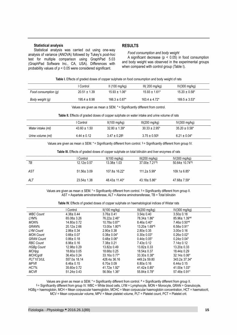
Food consumption and body weight A significant decrease (p < 0.05) in food consumption
and body weight was observed in the experimental groups when compared with control group (Table I).
Table I. Effects of graded doses of copper sulphate on food consumption and body weight of rats
I Control II (100 mg/kg) III( 200 mg/kg) IV(300 mg/kg)
Food consumption (g) 20.51 ± 1.39 15.93 ± 1.06* 15.93 ± 1.61* 15.20 ± 0.58*
Body weight (g) 195.4 ± 8.98 166.3 ± 0.87* 163.4 ± 4.72* 169.5 ± 3.53*
Values are given as mean ± SEM. * = Significantly different from control.
Table II. Effects of graded doses of copper sulphate on water intake and urine volume of rats
I Control II(100 mg/kg) III(200 mg/kg) IV(300 mg/kg)
Water intake (ml) 43.60 ± 1.50 32.80 ± 1.39* 30.33 ± 2.95* 35.20 ± 0.58*
Urine volume (ml) 4.44 ± 0.12 3.47 ± 0.28§ 3.75 ± 0.50§ 6.21 ± 0.04*
Values are given as mean ± SEM. * = Significantly different from control. § = Significantly different from group IV.
Table III. Effects of graded doses of copper sulphate on total bilirubin and liver enzymes of rats
I Control II(100 mg/kg) III(200 mg/kg) IV(300 mg/kg)
TB 12.12± 0.57 13.38± 1.03 37.00± 7.21*§ 50.64± 10.74*§
AST 51.56± 3.09 107.8± 16.22* 111.2± 5.99* 109.1± 6.85*
ALT 23.54± 1.38 48.43± 11.40* 43.18± 5.86* 47.68± 7.59*
Values are given as mean ± SEM. * = Significantly different from control. § = Significantly different from group II. AST = Aspartate aminotransferase, ALT = Alanine aminotransferase, TB = Total bilirubin
Table IV. Effects of graded doses of copper sulphate on haematological indices of Wistar rats
I Control II(100 mg/kg) III(200 mg/kg) IV(300 mg/kg)
WBC Count 4.38± 0.44 3.76± 0.41 3.54± 0.40 3.50± 0.18 LYM% 65.08± 3.26 76.22± 2.46* 78.34± 1.86* 85.96± 1.39*# MON% 14.80± 0.72 10.78± 0.87* 8.46± 0.40* 7.46± 0.50*# GRAN% 20.12± 2.66 13.00± 1.80*§ 13.20± 1.69*§ 6.58± 0.91* LYM Count 2.86± 0.34 2.90± 0.38 2.80± 0.35 3.00± 0.16 MON Count 0.66± 0.07 0.38± 0.04* 0.30± 0.03* 0.26± 0.02* GRAN Count 0.86± 0.18 0.48± 0.06* 0.44± 0.05* 0.24± 0.04* RBC Count 6.96± 0.16 7.38± 0.21 7.43± 0.12 7.14± 0.12 HGBg Count 12.98± 0.29 13.82± 0.49 13.82± 0.33 13.20± 0.33 MCHpg 18.60± 0.05 18.68± 0.25 18.54± 0.37 18.44± 0.29 MCHCg/dl 36.40± 0.24 33.16± 0.77* 33.30± 0.38* 32.14± 0.06* PLT*10*3/UL 557.0± 18.14 428.4± 36.16 449.2± 59.80 343.2± 37.34* MPVfl 6.46± 0.15 6.70± 0.09 6.80± 0.16 6.44± 0.15 HCT% 35.60± 0.72 41.72± 1.92* 41.42± 0.85* 41.00± 1.03* MCVfl 51.24± 0.43 56.56± 1.36* 55.84± 0.78* 57.48± 0.91*
Values are given as mean ± SEM. * = Significantly different from control. # = Significantly different from group II. § = Significantly different from group IV. WBC = White blood cells, LYM = Lymphocyte, MON = Monocyte, GRAN = Granulocyte,
HGBg = haemoglobin, MCH = Mean corpuscular haemoglobin, MCHC = Mean corpuscular haemoglobin concentration, HCT = haematocrit, MCV = Mean corpuscular volume, MPV = Mean platelet volume, PLT = Platelet count, PCT = Platelet crit.
16 Fiziologia - Physiology • 2016.26.1(89)
Water intake and urine volume The water intake of the experimental groups decreased
significantly (p < 0.05) when compared with the control rats (Table II). This was accompanied by a significant increase (p < 0.05) in urine volume in group IV when compared with the control and groups II and III. However, the urine volume of groups II and III was not significantly different (p > 0.05) from that of the control group (Table II).
Total bilirubin, aspartate aminotransferase and alanine aminotransferase The activities of plasma aspartate aminotransferase
(AST) and alanine aminotransferase (ALT) was significantly elevated (p < 0.05) in the experimental groups when compared with the control rats (Table III). There was no significant difference (p > 0.05) in total bilirubin level of group I when compared with the control rats. However, a significant increase (p < 0.05) in total bilirubin level was observed in groups II and III (Table III). Also, the total bilirubin level of groups II and III was significantly higher than that of group I.
Haematological indices There was no significant difference (p > 0.05) in red
blood cell (RBC) and haemoglobin concentrations of the experimental groups when compared with the control rats (Table IV). The percentage haematocrit (HCT%), mean corpuscular volume (MCV) of the experimental groups increased significantly (p < 0.05), but the mean corpuscular haemoglobin concentration (MCHC) dropped significantly (p < 0.05) in the experimental groups when compared with the control (Table IV).
The white blood cell (WBC) counts of the treated groups was not significantly different (p > 0.05) from that of the control rats (Table IV). On the other hand, a significant increase (p < 0.05) in percentage lymphocyte was observed in the experimental groups. The percentage granulocyte and monocyte dropped significantly (p < 0.05) in the treated groups when compared with the control rats. There was a significant increase (p < 0.05) in percentage lymphocyte of group IV when compared with group II, whilst the percentage monocyte dropped significantly (p < 0.05) in group IV when compared with group II.
The percentage granulocyte of group IV reduced significantly (p < 0.05) when compared with groups II and III (Table IV).
A significant reduction (p < 0.05) in platelet count and percentage platelet was seen in group IV when compared with the control rats (Table IV).
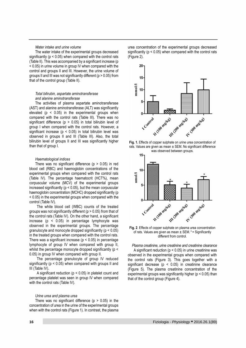
Urine urea and plasma urea There was no significant difference (p > 0.05) in the
concentration of urea in the urine of the experimental groups when with the control rats (Figure 1). In contrast, the plasma
urea concentration of the experimental groups decreased significantly (p < 0.05) when compared with the control rats (Figure 2).
Fig. 1. Effects of copper sulphate on urine urea concentration of rats. Values are given as mean ± SEM. No significant difference
was observed between groups.
Fig. 2. Effects of copper sulphate on plasma urea concentration of rats. Values are given as mean ± SEM. * = Significantly
different from control.
Plasma creatinine, urine creatinine and creatinine clearance A significant reduction (p < 0.05) in urine creatinine was
observed in the experimental groups when compared with the control rats (Figure 3). This goes together with a significant decrease (p < 0.05) in creatinine clearance (Figure 5). The plasma creatinine concentration of the experimental groups was significantly higher (p < 0.05) than that of the control group (Figure 4).
Fiziologia - Physiology • 2016.26.1(89) 17
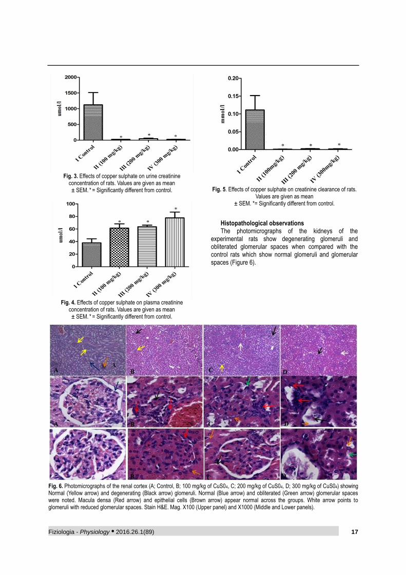
Fig. 3. Effects of copper sulphate on urine creatinine
concentration of rats. Values are given as mean ± SEM. * = Significantly different from control.
Fig. 4. Effects of copper sulphate on plasma creatinine
concentration of rats. Values are given as mean ± SEM. * = Significantly different from control.
Fig. 5. Effects of copper sulphate on creatinine clearance of rats.
Values are given as mean ± SEM. *= Significantly different from control.
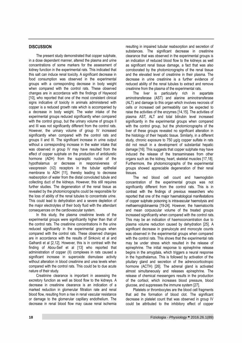
Histopathological observations The photomicrographs of the kidneys of the
experimental rats show degenerating glomeruli and obliterated glomerular spaces when compared with the control rats which show normal glomeruli and glomerular spaces (Figure 6).
Fig. 6. Photomicrographs of the renal cortex (A; Control, B; 100 mg/kg of CuS04, C; 200 mg/kg of CuS04, D; 300 mg/kg of CuS04) showing Normal (Yellow arrow) and degenerating (Black arrow) glomeruli. Normal (Blue arrow) and obliterated (Green arrow) glomerular spaces were noted. Macula densa (Red arrow) and epithelial cells (Brown arrow) appear normal across the groups. White arrow points to glomeruli with reduced glomerular spaces. Stain H&E. Mag. X100 (Upper panel) and X1000 (Middle and Lower panels).
18 Fiziologia - Physiology • 2016.26.1(89)
DISCUSSION
The present study demonstrated that copper sulphate, in a dose dependent manner, altered the plasma and urine concentrations of some markers for the assessment of kidney function in the experimental rats. This indicated that this salt can induce renal toxicity. A significant decrease in food consumption was observed in the experimental groups with a corresponding decrease in body weight when compared with the control rats. These observed changes are in accordance with the findings of Haywood [10], who reported that one of the most consistent clinical signs indicative of toxicity in animals administered with copper is a reduced growth rate which is accompanied by a decrease in body weight. The water intake of the experimental groups reduced significantly when compared with the control group, but the urinary volume of groups II and III was not significantly different from the control rats. However, the urinary volume of group IV increased significantly when compared with the control rats and groups II and III. The significant increase in urine output without a corresponding increase in the water intake that was observed in group IV may have resulted from the effect of copper sulphate on the production of antidiuretic hormone (ADH) from the supraoptic nuclei of the hypothalamus or decrease in responsiveness of vasopressin (V2) receptors in the tubular epithelial membrane to ADH [11], thereby leading to decrease reabsorption of water from the distal convoluted tubule and collecting duct of the kidneys. However, this still requires further studies. The degeneration of the renal tissue as revealed by the photomicrographs could be responsible for the loss of ability of the renal tubules to concentrate urine. This could lead to dehydration and a severe depletion of the major electrolytes of their body fluid with the attendant consequences on the cardiovascular system.
In this study, the plasma creatinine levels of the experimental groups were significantly higher than that of the control rats. The creatinine concentrations in the urine reduced significantly in the experimental groups when compared with the control rats. These observed changes are in accordance with the results of Sinkovic et al and Galhardi et al [2,12]. However, this is in contrast with the finding of Abou-Seif et al [13] who reported that administration of copper (II) complexes in rats caused a significant increase in superoxide dismutase activity without alteration in blood creatinine and urea levels when compared with the control rats. This could be to due acute nature of their study.
Creatinine clearance is important in assessing the excretory function as well as blood flow to the kidneys. A decrease in creatinine clearance is an indication of a marked reduction in glomerular filtration rate and renal blood flow, resulting from a rise in renal vascular resistance or damage to the glomerular capillary endothelium. The decrease in renal blood flow may cause renal ischemia
resulting in impaired tubular reabsorption and secretion of substances. The significant decrease in creatinine clearance that was observed in the experimental groups is an indication of reduced blood flow to the kidneys as well as significant renal tissue damage, a fact that was also corroborated by the photomicrographs of the renal tissue and the elevated level of creatinine in their plasma. The decrease in urine creatinine is a further evidence of reduced ability of the renal tubules to extract and remove creatinine from the plasma of the experimental rats.
The liver is particularly rich in aspartate aminotransferase (AST) and alanine aminotransferase (ALT) and damage to this organ which involves necrosis of cells or increased cell permeability can be expected to raise the activities of the enzymes [14,15]. The activities of plasma AST, ALT and total bilirubin level increased significantly in the experimental groups when compared with the control group, but the photomicrographs of the liver of these groups revealed no significant alteration in the histology of their hepatic tissue. Similarly, in a different study, chronic exposure to 750 μg/g copper sulfate in rats did not result in a development of substantial hepatic damage [16]. This suggests that copper sulphate may have induced the release of the transaminases from other organs such as the kidney, heart, skeletal muscles [17,18]. Furthermore, the photomicrographs of the experimental groups showed appreciable degeneration of their renal tissues.
The red blood cell count and haemoglobin concentration of the experimental groups was not significantly different from the control rats. This is in contrast with the findings of previous researchers who reported that one of the major haematological manifestations of copper sulphate poisoning is intravascular haemolysis and methaemoglobinaemia [19-24]. However, the haematocrits and mean corpuscular volume of the treated groups increased significantly when compared with the control rats. This may be an indication of haemoconcentration due to plasma volume reduction caused by dehydration [25]. A significant decrease in granulocyte and monocyte counts was observed in the experimental groups when compared with the control rats. This shows that the experimental rats may be under stress which resulted in the release of epinephrine. The initial response to epinephrine release begins in the amygdala, which triggers a neural response in the hypothalamus. This is followed by activation of the pituitary gland and secretion of the adrenocorticotropic hormone (ACTH) [26]. The adrenal gland is activated almost simultaneously and releases epinephrine. The release of chemical messengers results in the production of the cortisol, which increases blood pressure, blood glucose, and suppresses the immune system [27].
Platelets or thrombocytes are the blood cell fragments that aid the formation of blood clot. The significant decrease in platelet count that was observed in group IV could be attributed to the inhibitory effect of copper
Fiziologia - Physiology • 2016.26.1(89) 19
sulphate on the production of thrombopoietin, a circulating protein factor which facilitates megakaryocyte maturation, is produced constitutively by the kidneys [11].
CONCLUSION
From the results of this study, it is concluded that chronic administration of copper sulphate to rats induced nephrotoxicity which manifested as renal tissue damage, decreased creatinine clearance, and elevated levels of plasma and urine creatinine resulting from impaired renal function and elevation of the activities of aminotransferases in the blood.
REFERENCES
1. Saravu K, Jose J, Bhat MN, et al. Acute ingestion of copper sulphate: A review on its clinical manifestations and management. Indian J Crit Care Med 2007; 11:74-80.
2. Sinkovic A, Strdin A, Svensek F. Severe acute copper sulphate poisoning: A case report. Arh High Rada Toksikol 2008; 59: 31-5.
3. Kayacan SM, Vatansever S, Akkaya V, et al. A case of copper sulphate intoxication that is presented with prolonged hemolysis and acute renal failure. J Ist Faculty Med 2007; 70: 47-50.
4. Babaei H, Kheirandish R, Ebrahimi L. The effects of copper toxicity on histopathological and morphometrical changes of the rat testes. Asian Pac J Trop Biomed 2012; 2: S1615-S1619.
5. Akomolafe RO, Olukiran OS, Imafidon CE, et al. A study of two weeks’ administration of copper sulphate on markers of renal function and feeding pattern of Wistar rats. Afr J Biochem Res 2014; 8(9): 158-165.
6. Bonsnes RW, Taussky HH. On the colorimetric determination of creatinine by the Jaffe reaction. J Biol Chem 1945; 158: 581-91.
7. Fawcett JK, Scott EA. Rapid and precise method for the determination of urea. J Clin Pathol 1960; 13: 156-9.
8. Reitman S, Frankel SA. Colorimeter method for the determination of serum glutamic oxaloacetic and glutamic pyruvate transaminases. Am J Clin Pathol 1957; 26: 56-60.
9. Sherlock S. Circulatory studies on patients with ascites. Churchill, London 1951; 204.
10. Haywood S. The effect of the sex of weaned rats on the accumulation of dietary copper in their livers. J Comp Pathol 1978; 89: 481-486.
11. Ganong WF. Review of Medical Physiology, 23rd ed. Mc Graw Hill, New York, 2009, pp 65.
12. Galhardi CM, Diniz YS, Faine LA, et al. Toxicity of copper intake: Lipid profile, oxidative stress, and susceptibility to renal dysfunction. Food Chem Toxicol 2004; 42(12): 2053-60.
13. Abou- Seif MA, El-Naggar MM, El-Far M, et al. Prevention of biochemical changes in gamma-irradiated rats by some metal complexes. Clin Chem Lab Med 2003; 41(7): 926-33.
14. Naik SR, Panda VS. Hepatoprotective effect of Ginkgoselect phytosome in rifampicin induced liver injury in rats. Fitoterapia 2008; 79-439-45.
15. Rajesh MG, Latha MS. Preliminary evaluations of the antihepatotoxic effect of Kamilari, a polyherbal formulation. J Ethnopharmacol 2004; 91: 99-104.
16. Aburto EM. Role of selenium supplementation in the prevention of copper-induced hepatocellular damage in Fischer 344 rats [PhD dissertation]. Charlottetown, Prince Edward Island: University of Prince Edward Island, 1999, 186p.
17. Pratt DS, Kaplan MM, Laboratory tests. In: Schiff ER, Sorell MF, Maddrey WC eds. Schiff’s diseases of the liver, 8th ed. Philadelphia: Lippincott-Raven, 1999; 1: 205-44.
18. Rosen HR, Keefe EB. Evaluation of abnormal liver enzymes, use of liver tests and the serology of viral hepatitis: Liver disease, diagnosis and management, 1st ed. New York; Churchill Livingstone publishers 2000; 24-35.
19. Oldenquist G, Salem M. Parenteral copper sulfate poisoning causing acute renal failure. Nephrol Dial Transplant 1999; 14(2): 441‐3.
20. Liu J, Kashimura S, Hara K, et al. Death following cupric sulfate emesis. J Toxicol Clin Toxicol 2001; 39(2):161‐3.
21. Hantson P, Lievens M, Mahieu P. Accidental ingestion of a zinc and copper sulfate preparation. J Toxicol Clin Toxicol 1996; 34(6):725‐30.
22. Agarwal SK, Tiwari SC, Dash SG. Spectrum of poisoning requiring haemodialysis in a tertiary care hospital in India. Int J Artif Organs 1993;16 (1):20‐2.
23. Takeda T, Yukioka T, Shimazaki S. Cupric sulfate intoxication with rhabdomyolysis, treated with chelating agents and blood purification. Intern Med 2000; 39(3): 253‐5.
24. Ahasan HA, Chowdhury MA, Azhar MA, Rafiqueddin AK. Copper sulphate poisoning. Trop Doct 1994; 24(2):52‐3.
25. Wilson R, Taylor EW. The physiological responses of freshwater rainbow trout, Oncorhynchus mykiss, during acutely lethal copper exposure. J Comp Physiol 1993; 163: 38-47.
26. Margioris A, Tsatsanis, C. ACTH Action on the Adrenal. Endotext.org. Retrieved 18 April 2013.
27. Padgett D, Glaser R. How stress influences the immune response. Trends in Immunology. 2003; 24 (8): 444-448.
20 Fiziologia - Physiology • 2016.26.1(89)
Received February 5th 2016. Accepted March 20th 2016. Address for correspondence: Costela Serban, Department of Functional Sciences, “Victor Babes” University of Medicine and Pharmacy Timisoara, 2A Eftimie Murgu Square, Timisoara, RO-300041; phone: +0256490507, e-mail: [email protected]
CONTRIBUTION OF SELF-PERCEIVED STRESS TO
POSTSURGICAL LIFE QUALITY CHANGE OF PATIENTS
UNDERGOING CATHARACT SURGERY
CIOBOTEA DANA1
, ȘERBAN COSTELA2
, PUTNOKY SALOMEIA1
,
FIRA MLADINESCU CORNELUȚA1
, TUȚĂ SAS IOANA1
,
BĂCEAN MILOICOV CODRUȚA1
, VLAICU BRIGITHA1
1Departament 14 - Microbiology, Discipline of Hygiene, ”Victor Babeș” University of Medicine and Pharmacy Timișoara 2Departament 3 - Functional science, Discipline of Informatics and Biostatistics, ”Victor Babeș” University of Medicine and
Pharmacy Timișoara
ABSTRACT The research aimed to investigate pre- and postsurgical self-perceived stress and life quality of patients programmed to receive crystalline implants in relation to demographics and clinical status, but also to assess the contribution of modified stress to the improvement of life quality in between assessments. The study included 53 patients with catharact in whom the procedure of phacoemulsification was used for crystalline replacement. Presurgically, demographic and clinical variables do not influence the size of the two measures, which have 20% common variance. Postsurgically, the stres level decreased and life quality improved. The reduction of stress level and the increase of sleep duration explain 38% of life quality variance.
INTRODUCTION
Anxiety and stress in patients before surgical interventions represent a global problem which meay lead to profound effects. Among these effects the risk of increased postsurgical pain, the risk of infection, and the decreased healing rate may be mentioned. There are multiple factors affecting the level of presurgical anxiety, including the need for surgery, the perceived loss of control, the fear of postsurgical pain and the alteration of body image [1].
Numerous studies demonstrated that the improvement in visual acuity induced by catharact surgery leads to considerable gain in real life activities, in emotional and social life components [2].
In the present study we intended to investigate the self-perceived stress and life quality in patients scheduled to receive crystalline implants, the size of changes in the two measurements as a consequence of catharact surgery in relation with the demographic and clinical situations and the contribution of changed stress levels to a changed life quality in between assessments.
MATERIAL AND METHOD
Material The study included 53 catharact patients who adressed
the Clinic of Ophthalmology in Timisoara between January and April 2015, with the following demographic characteristics: 49.1% of patients belong to the masculine gender, with an age median value of 70 years and age interval between 50 and 84 years.
Presurgically, for the eye with the best results there were 69.8% patients without visual deficiency or with minor visual deficiencies, 20.8% with moderate visual deficiency and 9.4% with severe deficiencies or cecity. For the eye with the worst results, we found 20.8% patients without visual deficiencies or with minor deficiencies, 24.5% with moderate visual deficiencies and 54.7% with severe deficiencies or with cecity. One month after the surgical intervention, visual acuity was classified as no visual deficit in all patients, with 83% being recorded with maximum values.
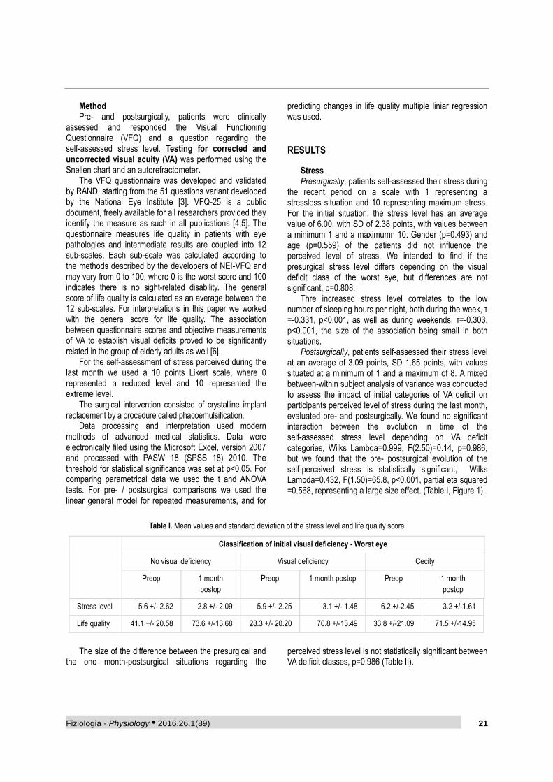
Fiziologia - Physiology • 2016.26.1(89) 21
Method Pre- and postsurgically, patients were clinically
assessed and responded the Visual Functioning Questionnaire (VFQ) and a question regarding the self-assessed stress level. Testing for corrected and uncorrected visual acuity (VA) was performed using the Snellen chart and an autorefractometer.
The VFQ questionnaire was developed and validated by RAND, starting from the 51 questions variant developed by the National Eye Institute [3]. VFQ-25 is a public document, freely available for all researchers provided they identify the measure as such in all publications [4,5]. The questionnaire measures life quality in patients with eye pathologies and intermediate results are coupled into 12 sub-scales. Each sub-scale was calculated according to the methods described by the developers of NEI-VFQ and may vary from 0 to 100, where 0 is the worst score and 100 indicates there is no sight-related disability. The general score of life quality is calculated as an average between the 12 sub-scales. For interpretations in this paper we worked with the general score for life quality. The association between questionnaire scores and objective measurements of VA to establish visual deficits proved to be significantly related in the group of elderly adults as well [6].
For the self-assessment of stress perceived during the last month we used a 10 points Likert scale, where 0 represented a reduced level and 10 represented the extreme level.
The surgical intervention consisted of crystalline implant replacement by a procedure called phacoemulsification.
Data processing and interpretation used modern methods of advanced medical statistics. Data were electronically filed using the Microsoft Excel, version 2007 and processed with PASW 18 (SPSS 18) 2010. The threshold for statistical significance was set at p<0.05. For comparing parametrical data we used the t and ANOVA tests. For pre- / postsurgical comparisons we used the linear general model for repeated measurements, and for
predicting changes in life quality multiple liniar regression was used.
RESULTS
Stress Presurgically, patients self-assessed their stress during
the recent period on a scale with 1 representing a stressless situation and 10 representing maximum stress. For the initial situation, the stress level has an average value of 6.00, with SD of 2.38 points, with values between a minimum 1 and a maximumn 10. Gender (p=0.493) and age (p=0.559) of the patients did not influence the perceived level of stress. We intended to find if the presurgical stress level differs depending on the visual deficit class of the worst eye, but differences are not significant, p=0.808.
Thre increased stress level correlates to the low number of sleeping hours per night, both during the week, τ =-0.331, p<0.001, as well as during weekends, τ=-0.303, p<0.001, the size of the association being small in both situations.
Postsurgically, patients self-assessed their stress level at an average of 3.09 points, SD 1.65 points, with values situated at a minimum of 1 and a maximum of 8. A mixed between-within subject analysis of variance was conducted to assess the impact of initial categories of VA deficit on participants perceived level of stress during the last month, evaluated pre- and postsurgically. We found no significant interaction between the evolution in time of the self-assessed stress level depending on VA deficit categories, Wilks Lambda=0.999, F(2.50)=0.14, p=0.986, but we found that the pre- postsurgical evolution of the self-perceived stress is statistically significant, Wilks Lambda=0.432, F(1.50)=65.8, p<0.001, partial eta squared =0.568, representing a large size effect. (Table I, Figure 1).
Table I. Mean values and standard deviation of the stress level and life quality score
Classification of initial visual deficiency - Worst eye
No visual deficiency Visual deficiency Cecity
Preop 1 month
postop
Preop 1 month postop Preop 1 month
postop
Stress level 5.6 +/- 2.62 2.8 +/- 2.09 5.9 +/- 2.25 3.1 +/- 1.48 6.2 +/-2.45 3.2 +/-1.61
Life quality 41.1 +/- 20.58 73.6 +/-13.68 28.3 +/- 20.20 70.8 +/-13.49 33.8 +/-21.09 71.5 +/-14.95
The size of the difference between the presurgical and
the one month-postsurgical situations regarding the perceived stress level is not statistically significant between VA deificit classes, p=0.986 (Table II).
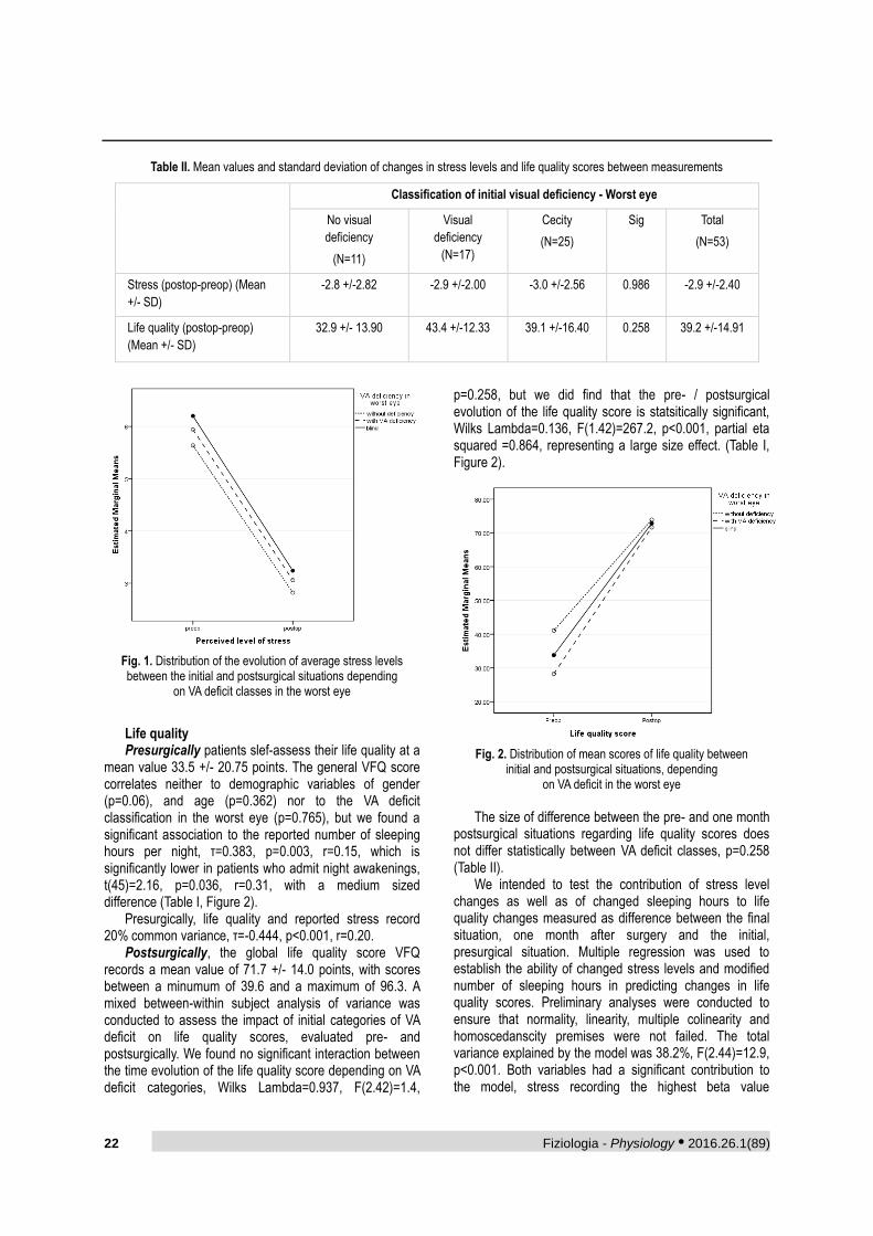
22 Fiziologia - Physiology • 2016.26.1(89)
Table II. Mean values and standard deviation of changes in stress levels and life quality scores between measurements
Classification of initial visual deficiency - Worst eye
No visual
deficiency
(N=11)
Visual
deficiency
(N=17)
Cecity
(N=25)
Sig Total
(N=53)
Stress (postop-preop) (Mean
+/- SD)
-2.8 +/-2.82 -2.9 +/-2.00 -3.0 +/-2.56 0.986 -2.9 +/-2.40
Life quality (postop-preop)
(Mean +/- SD)
32.9 +/- 13.90 43.4 +/-12.33 39.1 +/-16.40 0.258 39.2 +/-14.91
Fig. 1. Distribution of the evolution of average stress levels between the initial and postsurgical situations depending
on VA deficit classes in the worst eye
Life quality Presurgically patients slef-assess their life quality at a
mean value 33.5 +/- 20.75 points. The general VFQ score correlates neither to demographic variables of gender (p=0.06), and age (p=0.362) nor to the VA deficit classification in the worst eye (p=0.765), but we found a significant association to the reported number of sleeping hours per night, τ=0.383, p=0.003, r=0.15, which is significantly lower in patients who admit night awakenings, t(45)=2.16, p=0.036, r=0.31, with a medium sized difference (Table I, Figure 2).
Presurgically, life quality and reported stress record 20% common variance, τ=-0.444, p<0.001, r=0.20.
Postsurgically, the global life quality score VFQ records a mean value of 71.7 +/- 14.0 points, with scores between a minumum of 39.6 and a maximum of 96.3. A mixed between-within subject analysis of variance was conducted to assess the impact of initial categories of VA deficit on life quality scores, evaluated pre- and postsurgically. We found no significant interaction between the time evolution of the life quality score depending on VA deficit categories, Wilks Lambda=0.937, F(2.42)=1.4,
p=0.258, but we did find that the pre- / postsurgical evolution of the life quality score is statsitically significant, Wilks Lambda=0.136, F(1.42)=267.2, p<0.001, partial eta squared =0.864, representing a large size effect. (Table I, Figure 2).
Fig. 2. Distribution of mean scores of life quality between
initial and postsurgical situations, depending on VA deficit in the worst eye
The size of difference between the pre- and one month
postsurgical situations regarding life quality scores does not differ statistically between VA deficit classes, p=0.258 (Table II).
We intended to test the contribution of stress level changes as well as of changed sleeping hours to life quality changes measured as difference between the final situation, one month after surgery and the initial, presurgical situation. Multiple regression was used to establish the ability of changed stress levels and modified number of sleeping hours in predicting changes in life quality scores. Preliminary analyses were conducted to ensure that normality, linearity, multiple colinearity and homoscedanscity premises were not failed. The total variance explained by the model was 38.2%, F(2.44)=12.9, p<0.001. Both variables had a significant contribution to the model, stress recording the highest beta value
Fiziologia - Physiology • 2016.26.1(89) 23
(beta=0.418, p=0.003), and for additional sleeping hours beta=0.326, p=0.016. The changed stress level has the highest unique contribution of 15% to the model, the
unique contribution of additional sleeping hours being 9.1%, the remaining 14.1% variance being common to the two measures (Table III).
Table III. Coefficients for predicting changed life quality scores in between measurements
Model Unstandardized
Coefficients
Standardized
Coefficients
t Sig. 95.0% Confidence Interval
for B
B Std. Error Beta Lower
Bound
Upper
Bound
(Constant) 20.672 4.212 4.908 .000 12.171 29.173
dif_hours_week 5.004 2.002 .326 2.500 .016 .964 9.043
dif_stress 2.855 .893 .418 3.197 .003 1.053 4.658
a. Dependent Variable: dif_VFQ
DISCUSSIONS
Demographic and clinical variables did not influence the perceived stress level or life quality, as assessed for the month prior to catharact surgery. Other authors [7] found in patients with eye pathology inducing decreased sight that the state of mind and anxiety were associated with objective measures of the visual function. Kroll [8] found that specific situation coping was a mediator between the patient's personality type and adaptation criteria to the stress level as measured before catharact surgery. Pruessner et al., using functional magnetic resonance (fMRI) showed that stress exposure and the activation of the hypothalamus-hypophysis-adrenal axis are associated with reduced activities in some major portions of the limbic system allowing them to speculate on the effects of stress upon cognitive and emotional brain regulation.
We found that increased stress levels correlate to the decreased number of sleeping hours per night and to reduced life quality scores. Within the controled environment of the somnology laboratory, in a case-control study, Mikel et al [9] showed that sleep deprivation decreases the psychological threshold of stress perception manipulated by cognitive tests, time pressure and performance feedback.
In our study we showed that the postsurgical reduction of the stress level and the increase in life quality levels are significant, the modification size being similar in all 3 VA classes.
A recent study [10] demonstrated that among catharact patients assessed by IDATE-T (Anxiety Scale) and WHOQOL (WHO Quality of Life Questionnaire) there is a high psychological burden. The authors showed that patients' life quality is inversely related to the anxiety level. This finding confirms previous studies which demonstrated reduced life quality scores in catharact patients [11] and a
negative impact of anxiety disorders on the life quality of patients who underwent catharact surgery [12].
The increase in life quality may be explained in over 38% of cases by the decrease in stress levels and increase in the number of hours of sleep. These results support our recommendation to patients who postpone catharact surgery or who present counterindications for catharact surgery to undergo stress reduction measures by specific psychotherapy and blue light therapy for decreasing anxiety and depression and improve sleep duration [13,14].
CONCLUSIONS
‐ Presurgically, the stress level and life quality do not depend on demographic variables or on the objective level of visual deficit
‐ Postsurgically, one month after phacoemulsification, the stress level and life quality improved in a liniary relation, correlated neither to the initial clinical status nor to the clinical level of improvement in the operated eye
‐ The size of life quality change may be 38.2% justified by changes in stress levels and sleep duration, as recorded before and after catharact surgery
‐ We think that presurgical adjuvant methods such as blue light exposure to reduce anxiety and to improve sleep, but also psychological therapy are beneficial in coping with stress and anxiety, depending on the type of patient personality.
REFERENCES 1. Bailey L. Continuing Education Strategies for Decreasing
Patient Anxiety in the Perioperative Setting. AORN Journal. 2010;92(4):445–460
24 Fiziologia - Physiology • 2016.26.1(89)
2. Lamoureux E, Fenwick E, Pesudovs K, Tan D. The impact of cataract surgery on quality of life. Curr Opin Ophthalmol. 2011; 22 (1): 19-27.
3. http://www.rand.org/health/surveys_tools/vfq.html 4. Mangione CM, Lee PP, Gutierrez PR, et al. Development of
the 25-item National Eye Institute Visual Function Questionnaire (VFQ-25). Arch Ophthalmol. 2001;119: 1050-1058.
5. World Health Organization International Statistical Classification of Diseases and Related Health Problems 10th revision Current version Version for 2003 Chapter VII H54 Blindness and low vision. http://www.who.int/classifications/icd/en/
6. Owen CG, Rudnicka AR, Smeeth L, Evans JR, Wormald RP, Fletcher AE. Is the NEI-VFQ-25 a useful tool in identifying visual impairment in an elderly population? BMC Ophthalmol. 2006, 9;6:24.
7. Eramudugolla R, Wood J, Anstey KJ. Co-morbidity of depression and anxiety in common age-related eye diseases: a population-based study of 662 adults. Front Aging Neurosci. 2013; 5: 56.
8. Knoll N, Rieckmann N, Schwarzer R. Coping as a mediator between personality and stress outcomes: a longitudinal study with cataract surgery patients. Eur J Pers. 2005, 19: 229-247.
9. Minkel JD, Banks S, Htaik O, Moreta MC, Jones CW, McGlinchey EL, Simpson NS, Dinges DF. Sleep deprivation and stressors: evidence for elevated negative affect in response to mild stressors when sleep deprived. Emotion, 2012; 12(5): 1015-20.
10. Fazzi L, Dobrianskyj FM, Abreu Reggi JR, et al. The influence of anxiety on quality of life among patients with an indication for cataract surgery. Sao Paulo Med. J. 2015; 133(2)
11. Skiadaresi E, McAlinden C, Pesudovs K, et al. Subjective Quality of Vision Before and After Cataract Surgery. Arch Ophthalmol. 2012;130(11):1377-1382.
12. Yokoi T, Moriyama M, Hayashi K, et al. Predictive factors for comorbid psychiatric disorders and their impact on vision-related quality of life in patients with high myopia. Int Ophthalmol. 2014; 34(2): 171-83.
13. Lieverse R, Nielen MM, Veltman DJ, et al. Bright light in elderly subjects with nonseasonal major depressive disorder: a double blind randomised clinical trial using early morning bright blue light comparing dim red light treatment. Trials 2008, 9: 48.
14. Lieverse R, Van Someren EJ, Nielen MM, et al. Bright light treatment in elderly patients with nonseasonal major depressive disorder: a randomized placebo-controlled trial. Arch Gen Psychiatry 2011, 68: 61-70.
CONTRIBUȚIA STRESULUI AUTOPERCEPUT LA
MODIFICAREA POSTOPERATORIE A CALITĂȚII VIEȚII
LA PACIENȚI SUPUȘI OPERAȚIEI DE CATARACTĂ
REZUMAT Cercetarea și-a propus investigarea pre și postoperatorie a stresului autoperceput și a calității vieții la pacienți programați pentru implant de cataractă în relație cu statusul demografic și clinic, dar și contribuția modificării stresului la ameliorarea calității vieții între evaluări. Au fost incluși 53 pacienți cu diagnostic de cataractă, la care s-a folosit procedeul de facoemulsificare pentru înlocuirea cristalinului. Preoperator, variabilele demografice și clinice nu influențează mărimea celor două măsuri, ele având 20% varianță comună. Postoperator nivelul de stres s-a redus, iar calitatea vieții s-a îmbunătățit. Reducerea nivelului de stres și creșterea duratei somnului explică 38% din varianța calității vieții.
Fiziologia - Physiology • 2016.26.1(89) 25
Received February 12th 2016. Accepted March 6th 2016. Address for correspondence: Roxana Voisan, UPU-SMURD, “Pius Brinzeu” Emergency Clinical County Hospital Timisoara, Liviu Rebreanu 156 Street, Timisoara, RO-300723; phone: +40356433111, e-mail: [email protected]
BIOCOMPOSITE PROCESSING TECHNOLOGY
AND MEDICAL APPLICATIONS
ROXANA VOISAN1
, OANA GINGU2
1UPU-SMURD SCJU Timisoara 2Faculty of Mechanics, University of Craiova
ABSTRACT Porous biocomposite materials - hydroxyapatite reinforced by titanium -made by powder metallurgy technology, are qualitatively assessed from the point of view of their behavior along the wear tests against metallic and ceramic counterparts. This assessment is monitored through some representative parameters of wear tests: friction coefficient, wear rate and wear track area. The experimental results point out the distinction of the wear behavior between the biocomposite materials which are elaborated by two different processes: spark plasma sintering and two step sintering. The wear results, related to the structural properties of the biocomposite materials, could recommend each of the processing technologies for specific bone grafts manufacturing, namely for the cortical or trabecular hard tissue. Because of some disadvantages reported up to now (mainly due to the difference between the mechanical characteristics of the human bone and the metallic implant), the biocomposite become an interesting solution to improve the hard tissue implants for the human body. Keywords: biomaterials, hydroxyapatite, titanium, bone
INTRODUCTION
One of the most important targets of the science is to develop new materials for bone implants. Titanium and Ti-based alloys have been usually used as artificial bone materials because of their great mechanical properties and they show non-toxic, i.e., when these materials are utilized for bone implants they are considered bioinert [1].
Titanium alloys present the greatest biocompatibility among metallic materials for biomedical applications because of its high chemical stability in the human body, relative low module of elasticity, low density, high strength and corrosion resistance. However, even if they are considered into bioinert materials and its biocompatibility is inferior to that of hydroxyapatite (HAP: Ca(PO4)3OH), which is grouped into bioactive materials [2]. Due to the reported geometrical instability, its reinforcing by some form of stable osteosynthesis, preferably with an angularly stable plate is recommended [3]. Much better biomaterials for hard tissue replacement implants may be acquired by the preparation of titanium composites. The most commonly used materials are ceramics such as hydroxyapatites, bioglasses or silicas.
Therefore, the connection of excellent biocompatibility and bioactivity of ceramics with good mechanical
properties of titanium is considered to be a promising approach for fabrication of more perfect hard tissue replacement implants. There are two creative ways to meet this idea: use of ceramics as a coating or as a reinforcing phase in metallic-ceramic composites [4].
Direct HAP deposition methods on titanium substrate are, for example: ion plating [5], plasma spray method [6], pulse laser deposition method [7,8], RF magnetron sputtering [9], super plastic joining method [10], ion beam dynamic mixing method [11], processing of nanostructured biomaterials [12, 13], obtaining of an intermediate layer at coating/implant interface [14, 15].
The most approached processing technologies for the HAP/Ti advanced biomaterials are: powder metallurgy [16], pulsed laser deposition [17,18], microarc oxidation [19], microwave sintering [20], radio-frequency thermal plasma spraying (RF-TPS) technology [21], electrochemical treatment [22, 23].
In order to obtain nanostructured sintered biocomposites, two steps sintering (TSS) and spark plasma sintering (SPS) are developed at different technological parameters in this research.
One of the challenges of tissue engineering field is to control the friction respectively wear phenomena occurring during the implants / grafts long life. The medical studies
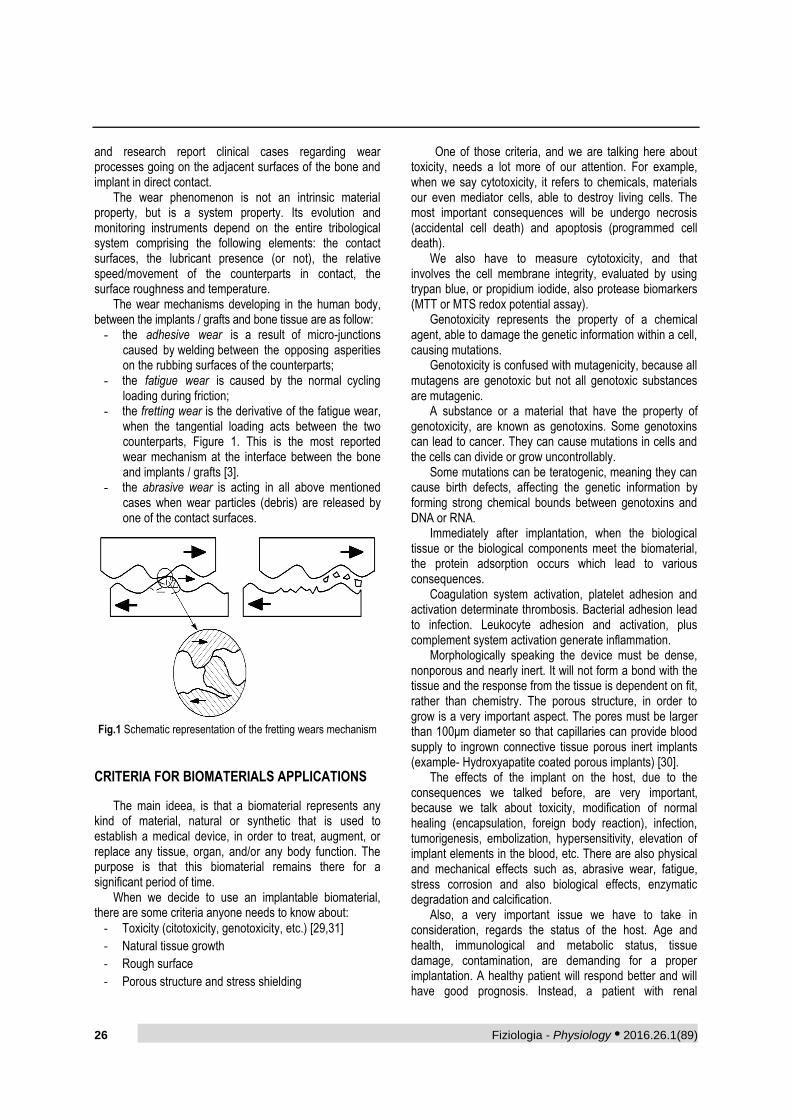
26 Fiziologia - Physiology • 2016.26.1(89)
and research report clinical cases regarding wear processes going on the adjacent surfaces of the bone and implant in direct contact.
The wear phenomenon is not an intrinsic material property, but is a system property. Its evolution and monitoring instruments depend on the entire tribological system comprising the following elements: the contact surfaces, the lubricant presence (or not), the relative speed/movement of the counterparts in contact, the surface roughness and temperature.
The wear mechanisms developing in the human body, between the implants / grafts and bone tissue are as follow:
- the adhesive wear is a result of micro-junctions caused by welding between the opposing asperities on the rubbing surfaces of the counterparts;
- the fatigue wear is caused by the normal cycling loading during friction;
- the fretting wear is the derivative of the fatigue wear, when the tangential loading acts between the two counterparts, Figure 1. This is the most reported wear mechanism at the interface between the bone and implants / grafts [3].
- the abrasive wear is acting in all above mentioned cases when wear particles (debris) are released by one of the contact surfaces.
Fig.1 Schematic representation of the fretting wears mechanism
CRITERIA FOR BIOMATERIALS APPLICATIONS
The main ideea, is that a biomaterial represents any kind of material, natural or synthetic that is used to establish a medical device, in order to treat, augment, or replace any tissue, organ, and/or any body function. The purpose is that this biomaterial remains there for a significant period of time.
When we decide to use an implantable biomaterial, there are some criteria anyone needs to know about:
‐ Toxicity (citotoxicity, genotoxicity, etc.) [29,31]
‐ Natural tissue growth
‐ Rough surface
‐ Porous structure and stress shielding
One of those criteria, and we are talking here about toxicity, needs a lot more of our attention. For example, when we say cytotoxicity, it refers to chemicals, materials our even mediator cells, able to destroy living cells. The most important consequences will be undergo necrosis (accidental cell death) and apoptosis (programmed cell death).
We also have to measure cytotoxicity, and that involves the cell membrane integrity, evaluated by using trypan blue, or propidium iodide, also protease biomarkers (MTT or MTS redox potential assay).
Genotoxicity represents the property of a chemical agent, able to damage the genetic information within a cell, causing mutations.
Genotoxicity is confused with mutagenicity, because all mutagens are genotoxic but not all genotoxic substances are mutagenic.
A substance or a material that have the property of genotoxicity, are known as genotoxins. Some genotoxins can lead to cancer. They can cause mutations in cells and the cells can divide or grow uncontrollably.
Some mutations can be teratogenic, meaning they can cause birth defects, affecting the genetic information by forming strong chemical bounds between genotoxins and DNA or RNA.
Immediately after implantation, when the biological tissue or the biological components meet the biomaterial, the protein adsorption occurs which lead to various consequences.
Coagulation system activation, platelet adhesion and activation determinate thrombosis. Bacterial adhesion lead to infection. Leukocyte adhesion and activation, plus complement system activation generate inflammation.
Morphologically speaking the device must be dense, nonporous and nearly inert. It will not form a bond with the tissue and the response from the tissue is dependent on fit, rather than chemistry. The porous structure, in order to grow is a very important aspect. The pores must be larger than 100μm diameter so that capillaries can provide blood supply to ingrown connective tissue porous inert implants (example- Hydroxyapatite coated porous implants) [30].
The effects of the implant on the host, due to the consequences we talked before, are very important, because we talk about toxicity, modification of normal healing (encapsulation, foreign body reaction), infection, tumorigenesis, embolization, hypersensitivity, elevation of implant elements in the blood, etc. There are also physical and mechanical effects such as, abrasive wear, fatigue, stress corrosion and also biological effects, enzymatic degradation and calcification.
Also, a very important issue we have to take in consideration, regards the status of the host. Age and health, immunological and metabolic status, tissue damage, contamination, are demanding for a proper implantation. A healthy patient will respond better and will have good prognosis. Instead, a patient with renal
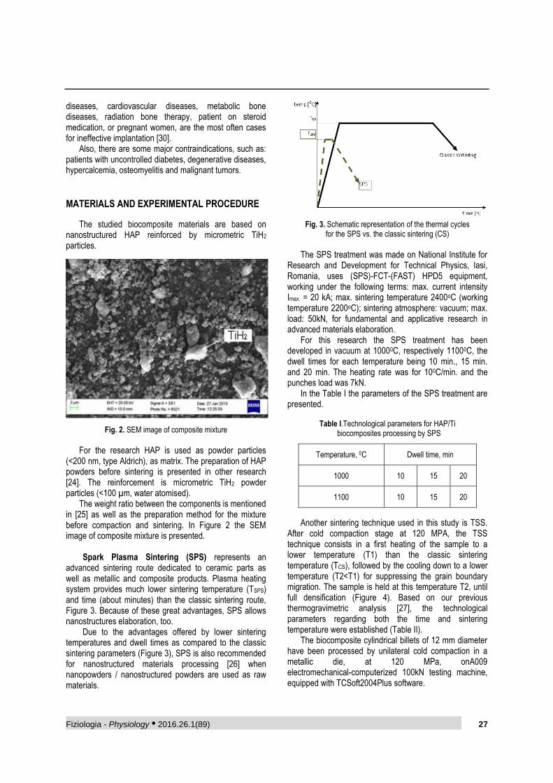
Fiziologia - Physiology • 2016.26.1(89) 27
diseases, cardiovascular diseases, metabolic bone diseases, radiation bone therapy, patient on steroid medication, or pregnant women, are the most often cases for ineffective implantation [30].
Also, there are some major contraindications, such as: patients with uncontrolled diabetes, degenerative diseases, hypercalcemia, osteomyelitis and malignant tumors.
MATERIALS AND EXPERIMENTAL PROCEDURE
The studied biocomposite materials are based on nanostructured HAP reinforced by micrometric TiH2 particles.
Fig. 2. SEM image of composite mixture
For the research HAP is used as powder particles
(<200 nm, type Aldrich), as matrix. The preparation of HAP powders before sintering is presented in other research [24]. The reinforcement is micrometric TiH2 powder particles (<100 µm, water atomised).
The weight ratio between the components is mentioned in [25] as well as the preparation method for the mixture before compaction and sintering. In Figure 2 the SEM image of composite mixture is presented.
Spark Plasma Sintering (SPS) represents an advanced sintering route dedicated to ceramic parts as well as metallic and composite products. Plasma heating system provides much lower sintering temperature (TSPS) and time (about minutes) than the classic sintering route, Figure 3. Because of these great advantages, SPS allows nanostructures elaboration, too.
Due to the advantages offered by lower sintering temperatures and dwell times as compared to the classic sintering parameters (Figure 3), SPS is also recommended for nanostructured materials processing [26] when nanopowders / nanostructured powders are used as raw materials.
Fig. 3. Schematic representation of the thermal cycles
for the SPS vs. the classic sintering (CS)
The SPS treatment was made on National Institute for
Research and Development for Technical Physics, Iasi, Romania, uses (SPS)-FCT-(FAST) HPD5 equipment, working under the following terms: max. current intensity Imax. = 20 kA; max. sintering temperature 2400oC (working temperature 2200oC); sintering atmosphere: vacuum; max. load: 50kN, for fundamental and applicative research in advanced materials elaboration.
For this research the SPS treatment has been developed in vacuum at 10000C, respectively 11000C, the dwell times for each temperature being 10 min., 15 min. and 20 min. The heating rate was for 100C/min. and the punches load was 7kN.
In the Table I the parameters of the SPS treatment are presented.
Table I.Technological parameters for HAP/Ti biocomposites processing by SPS
Temperature, 0C Dwell time, min
1000 10 15 20
1100 10 15 20
Another sintering technique used in this study is TSS. After cold compaction stage at 120 MPA, the TSS technique consists in a first heating of the sample to a lower temperature (T1) than the classic sintering temperature (TCS), followed by the cooling down to a lower temperature (T2<T1) for suppressing the grain boundary migration. The sample is held at this temperature T2, until full densification (Figure 4). Based on our previous thermogravimetric analysis [27], the technological parameters regarding both the time and sintering temperature were established (Table II).
The biocomposite cylindrical billets of 12 mm diameter have been processed by unilateral cold compaction in a metallic die, at 120 MPa, onA009 electromechanical-computerized 100kN testing machine, equipped with TCSoft2004Plus software.
HAP
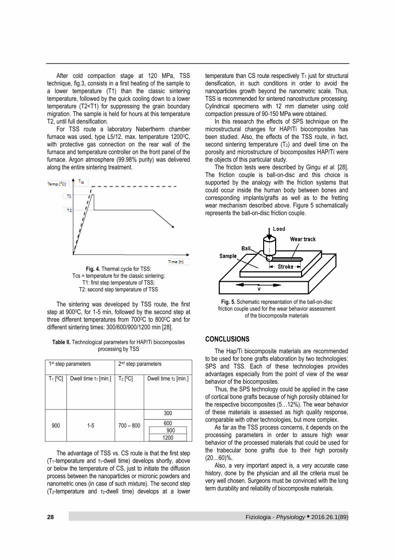
28 Fiziologia - Physiology • 2016.26.1(89)
After cold compaction stage at 120 MPa, TSS technique, fig.3, consists in a first heating of the sample to a lower temperature (T1) than the classic sintering temperature, followed by the quick cooling down to a lower temperature (T2<T1) for suppressing the grain boundary migration. The sample is held for hours at this temperature T2, until full densification.
For TSS route a laboratory Nabertherm chamber furnace was used, type L5/12, max. temperature 12000C, with protective gas connection on the rear wall of the furnace and temperature controller on the front panel of the furnace. Argon atmosphere (99.98% purity) was delivered along the entire sintering treatment.
Fig. 4. Thermal cycle for TSS:
Tcs = temperature for the classic sintering; T1: first step temperature of TSS;
T2: second step temperature of TSS
The sintering was developed by TSS route, the first
step at 9000C, for 1-5 min, followed by the second step at three different temperatures from 7000C to 8000C and for different sintering times: 300/600/900/1200 min [28].
Table II. Technological parameters for HAP/Ti biocomposites processing by TSS
1st step parameters 2nd step parameters
T1 [0C] Dwell time τ1 [min.] T2 [0C] Dwell time τ2 [min.]
900 1-5 700 – 800
300
600
900
1200
The advantage of TSS vs. CS route is that the first step
(T1-temperature and τ1-dwell time) develops shortly, above or below the temperature of CS, just to initiate the diffusion process between the nanoparticles or micronic powders and nanometric ones (in case of such mixture). The second step (T2-temperature and τ2-dwell time) develops at a lower
temperature than CS route respectively T1 just for structural densification, in such conditions in order to avoid the nanoparticles growth beyond the nanometric scale. Thus, TSS is recommended for sintered nanostructure processing. Cylindrical specimens with 12 mm diameter using cold compaction pressure of 90-150 MPa were obtained.
In this research the effects of SPS technique on the microstructural changes for HAP/Ti biocomposites has been studied. Also, the effects of the TSS route, in fact, second sintering temperature (T2) and dwell time on the porosity and microstructure of biocomposites HAP/Ti were the objects of this particular study.
The friction tests were described by Gingu et al. [28]. The friction couple is ball-on-disc and this choice is supported by the analogy with the friction systems that could occur inside the human body between bones and corresponding implants/grafts as well as to the fretting wear mechanism described above. Figure 5 schematically represents the ball-on-disc friction couple.
Fig. 5. Schematic representation of the ball-on-disc
friction couple used for the wear behavior assessment of the biocomposite materials
CONCLUSIONS
The Hap/Ti biocomposite materials are recommended to be used for bone grafts elaboration by two technologies: SPS and TSS. Each of these technologies provides advantages especially from the point of view of the wear behavior of the biocomposites.
Thus, the SPS technology could be applied in the case of cortical bone grafts because of high porosity obtained for the respective biocomposites (5…12%). The wear behavior of these materials is assessed as high quality response, comparable with other technologies, but more complex.
As far as the TSS process concerns, it depends on the processing parameters in order to assure high wear behavior of the processed materials that could be used for the trabecular bone grafts due to their high porosity (20…60)%.
Also, a very important aspect is, a very accurate case history, done by the physician and all the criteria must be very well chosen. Surgeons must be convinced with the long term durability and reliability of biocomposite materials.
Fiziologia - Physiology • 2016.26.1(89) 29
Cellular, molecular, and genetic level research is producing exciting new information that has improved understanding of the host/biomaterials interactions.
That is why the development of new generations of biomaterials and implant devices and also the improved strategies for their evaluation and use should be the number one priority. ACKNOWLEDGMENTS We hereby acknowledge the research project PN-II-PT-PCCA-2013-4-2094, title: ''Research on the bone substitution with biocomposite materials processed by powder metallurgy specific techniques'', acronym BONY, for the financial support.
REFERENCES
1. Rakngarm A, Miyashita Y, Mutoh Y. Formation of hydroxyapatite layer on bioactive Ti and Ti–6Al–4V by simple chemical technique. J Mater Sci Mater Med. 2008;19(5):1953-61.
2. Niinomi M. Recent research and development in titanium alloys for biomedical applications and healthcare goods. Science and Technology of Advanced Materials, 2003; 4: 445-454.
3. Ahmed AR, Watanabe H, Takagishi K. Reconstruction with autologous pasteurized whole knee joint I: experimental study in a rabbit model. J Orthop Sci. 2003; 8: 170-179.
4. Hench LL. Biomaterials: A forecast for the future. Biomaterials, 1998; 199: 1419-1423.
5. Niinomi M, Okabe T, Taleff EM, Lesuer DR, Lippard HF, Eds. of Structural Biomaterials for the 21st Century, TMS, USA, 2001; 25-34.
6. Hanumantharaju HG, et al. Study on hydroxyapatite coating on biomaterials by plasma spray method. Int. J. of Eng. Science and Technology (IJEST), 2012; 4(9): 4152-4159.
7. Rajesh P, Muraleedharan CV, Komath M, Varma H. Pulsed laser deposition of hydroxyapatite on titanium substrate with titania interlayer. J. Mater. Sci. Mater Med. 2011; 22(3):497-505.
8. Fischer D, de la Fuente GF, Jansen M. A new pulsed laser deposition technique: scanning multi-component pulsed laser deposition method. Rev. Sci. Instrum. 2012; 83(4): 043901.
9. Cormier PA, Balhamri A, Thomann AL, Dussart R, Semmar N, Mathias J, Snyders R, Konstantinidis S. Measuring the energy flux at the substrate position during magnetron sputter deposition processes. J. Appl. Phys. 2013; 113.
10. Nonami T, Naganuma K, Kameyama T. Superplastic bonding of biomaterial-apatite and Ti-alloy. Mater. Jpn, 1998; 37: 856-861.
11. Choi JM, Kim HE, Lee In-S. Ion-beam-assisted deposition (IBAD) of hydroxyapatite coating layer on Ti-based metal substrate. Biomaterials 2000; 21: 469-473.
12. Niespodziana K, et al. Synthesis of titanium alloys for biomedical applications. Rev. Adv. Mater. Sci. 2008; 18: 236-240.
13. Hari Singh N. Handbook of nanostructured biomaterials and their applications in nanobiotechnology, ed. by American Scientific Publishers, 2 vol, New York, 2005.
14. Lin C-M, Yen S-K. Characterization and bond strength of electrolytic HA/TiO2 double layers for orthopaedic applications. J. Mater. Sci. Med. 2005; 16: 889-897.
15. Valanezhad A, Tsuru K, Maruta M, Kawachi G, Matsuya S, Ishikawa K. Novel Ceramic Coating on Titanium with High Mechanical Properties. Bioceramics Dev. and Appls. 2011; 1: 27-30.
16. Marceloa TM, Livramentoa V, de Oliveirab MV, Carvalho MH. Microstructural Characterization and Interactions in Ti and TiH2 -Hydroxyapatite Vacuum Sintered Composites. Materials Res, 2006; 9(1):65-71.
17. Tanaskovic D, Jokic B, Socol G, Popescu A, Mihailescu IN, Petrovic R, Janackovic DJ. Synthesis of functionally graded bioactive glass-apatite multistructures on Ti substrates by pulsed laser deposition, Appl. Surf. Sci. 2007; 254(5): 1279-1282.
18. Hashimoto Y, et al. Cytocompatibility of calcium phosphate coatings deposited by an ArF pulsed laser. J. Mater. Sci. Med. 2008; 19: 327-333.
19. Liu F, et al. Formation characterization of hydroxyapatite on titanium by microarc oxidation and hydrothermal treatment. J. Biosci. Bioeng. 2005; 100(N1): 100-104.
20. Nath S, et al. A comparative study of conventional sintering with microwave sintering of hydroxyapatite synthesized by chemical route. Trends Biomater. Artif. Organs, 2006; 19: 93-98.
21. Karlis A, Gross AB, Muller D, Lucas H, Haynes DR. Osteoclast resorption of thermal spray hydoxyapatite coatings is influenced by surface topography. Acta Biomat. 2012; 8: 1948-1956.
22. Kawashita M, et al. Apatite formation on titanium substrates by electrochemical deposition in metastable calcium phosphate solution. J. Mater. Sci. Med. 2008; 19: 137-142.
23. Xiao X, Liu R, Huang Q. Preparation and characterization of nano-hydroxyapatite/polymer composite scaffolds. J. Mater. Sci. Mater. Med. 2008; 19(11): 3429-3435.
24. Pascu I, Gingu O, Ciupitu I, Rotaru P. Patent RO, no. A/00317/2010.
25. Gingu O, Pascu I, Lupu N, Benga G. Patent RO, no. A/00318/2010.
26. Maiwa H. Structure and properties of Ba(Zr0.2Ti0.8)O3 ceramics prepared by spark plasma sintering. J. Mater. Sci. 2008; 43: 6385-6390.
27. Gingu O, et al. Spark plasma sintering behavior of ceramic biocomposites based on hydroxyapatite nanopowders. In Proc. International Congress and Exhibition on Powder Metallurgy, EUROPM 2009, Coperhagen, Denmark, 12-14 Oct. 2009.
28. Gingu O, et al. Wear behaviour of ceramic biocomposites based on hydroxyapatite nanopowders. J. Proc. Mech. Eng. 2011; 225: 62-70.
29. Kim HR, Park YJ, Shin DY, Oh SM, Chung KH. Methods for Genotoxicity Testing of Silver Nanoparticles. Environmental Health and Toxicology, 2013; 28: e2013003.
30. Wang X. Overview on Biocompatibilities of Implantable Biomaterials. In Advances in Biomaterials Science and Biomedical Applications, R. Pignatello, Ed. InTech, 2013.
31. Leggat PA, Smith DR, Kedjarune U. Surgical Applications of Cyanoacrylate Adhesives: A Review of Toxicity. ANZ Journal of
Surgery, 2007; 77(4): 209-213.
30 Fiziologia - Physiology • 2016.26.1(89)
TEHNOLOGIA PROCESARII MATERIALELOR
BIOCOMPOZITE SI APLICABILITATEA
IN DOMENIUL MEDICAL
REZUMAT Materiale biocompozite poroase, pe baza de hidroxiapatita ranforsata cu particule de titan, au fost obtinute prin tehnologia metalurgiei pulberilor. In cadrul acestei lucrari biocompositele elaborate sunt evaluate din punct de vedere calitativ in ceea ce priveste comportamentul la uzare, avand ca si piesa conjugata produse metalice si ceramice. Parametrii de evaluare calitativa sunt: coeficientul de frecare, rata uzarii si suprafata urmei uzata. Rezultatele experimentale evidentiaza diferenta privind comportamentul la uzare al biocompozitelor obtinute prin doua metode diferite: sinterizarea cu descarcare in plasma (SPS) respectiv sinterizarea in doi pasi (TSS). Valorile experimentale obtinute, coroborate cu proprietatile structurale ale biocompozitelor studiate, pot recomanda fiecare dintre tehnologiile mentionate pentru fabricarea grafoanelor osoase respectiv SPS pentru tesut osos cortical, iar TSS pentru tesut trabecular. Din cauza unor dezavantaje raportate pana in prezent, (mai ales provocate de diferentele din structura osoasa umana si implantul metalic), biocompositele au inceput sa reprezinte o solutie interesanta in ceea ce priveste cresterea capacitatii de integrare a implantului in organismul uman. Cuvinte cheie: biomateriale, biocompozite tip hidroxiapatita /titan, structura osoasa.
Fiziologia - Physiology • 2016.26.1(89) 31
Received February 24th 2016. Accepted March 4th 2016. Address for correspondence: Hogea Lavinia, Psychology Discipline, Neurosciences Department, “Victor Babes” University of Medicine and Pharmacy Timisoara, Eftimie Murgu Square No. 2A, Timisoara, RO-300041, e-mail: [email protected] *The first two authors should be considered as first authors due to the equal contribution to this study
ASSESSMENT OF OBSESSIVE-COMPULSIVE BEHAVIOR
IN PATIENTS WITH HAEMOPHILIA
LAVINIA HOGEA*1
, LAURA NUSSBAUM*2
, RALUCA DUMACHE3
1Psychology Discipline, Neurosciences Department, “Victor Babes” University of Medicine and Pharmacy Timișoara, Romania 2Pedopsychiatry Discipline, Neurosciences Department, “Victor Babes” University of Medicine and Pharmacy Timisoara, Romania 3Department of Forensic Medicine, Bioethics, Deontology and Medical Law, Neurosciences Department, “Victor Babes” University of Medicine and Pharmacy Timisoara, Romania
ABSTRACT Introduction: Hemophilia, like other rare disease, is a chronic disease, debilitating or crippling threatening life and the quality of life it requires a special effort and support from the community. Permanent disability to which a hemophiliac body is prone to leads to an alteration of the psyche, with repercussions on the whole personality of the individual. Intense and permanent headaches with witch the patient is facing are causing an increase in psychic tension and internalization of one's personality, even more so, the hemorrhagic accidents are more frequent and more intense. Material and methods: in the study there were 200 subjects grouped into two equal groups (patients with hemophilia A and B and healthy population) aged between 16 and 45 years. Results: statistically significant differences exist between the hemophiliacs and non-hemophiliacs, hemophiliacs have a higher level of obsessive compulsive behavior, compared to non-hemophiliacs. Conclusions: Patients with chronic conditions, pose a twice as high risk for the occurrence of psychiatric complications, compared to their healthy peers. Key words: hemophilia, obsessive-compulsive behavior
INTRODUCTION
Hemophiliac patients must go through periods of disability, must suffer lengthy treatments and in the meantime they must deal with various experiences and feelings, such as fear and uncertainty [1]. The process of adapting to the disease is a difficult one and can lead over time to the appearance of psychological dysfunctional symptoms, which can be associated with psychiatric morbidity, both to the patient and members of his family [1]. Hemophilia patient's mental disorders are much more numerous than those presented to the doctor, because both the patients and their parents often conceal their existence. Fear, embarrassment, anxiety or mistrust may be some of the reasons why the patient does not communicate the existence of psychological problems.
The type of mental disorders encountered more often at hemophiliac patients are: mentally retarded, anxiety, nocturnal psychogenic enuresis, retardation in language acquisitions, anxious-phobious neurosis, affective mood disorder, Tourette Syndrome, ADHD, conduct disorder, stuttering, sociopathic behavior disorder, separation anxiety,
night pavor, opioid drug abuse, alcohol abuse, depressive reaction, self-harming, suicidal tendency, focus and attention disorders, temporary depression moods, headaches, insomnia.
PURPOSE
There are statistically significant differences regarding the level of obsessive-compulsive behaviors in hemophiliac patients compared to the control group.
MATERIAL AND METHODS
The study was conducted on a sample of 200 subjects grouped into two groups of equal size. The clinical batch consists of 100 people with hemophilia A and B (50%), with ages between 16 and 45 years, while the control group is made up of 100 people without hemophilia (50%) (Figure 1).
Inclusion criteria in the study are: diagnosis of hemophilia A or hemophilia B; patients aged between 16 and 45 years; intelligence quotient > 100.
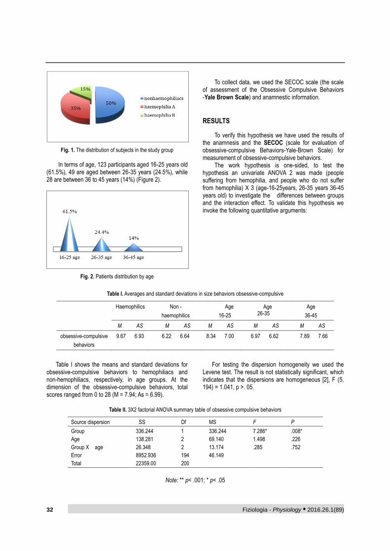
32 Fiziologia - Physiology • 2016.26.1(89)
Fig. 1. The distribution of subjects in the study group
In terms of age, 123 participants aged 16-25 years old
(61.5%), 49 are aged between 26-35 years (24.5%), while 28 are between 36 to 45 years (14%) (Figure 2).
Fig. 2. Patients distribution by age
To collect data, we used the SECOC scale (the scale
of assessment of the Obsessive Compulsive Behaviors -Yale Brown Scale) and anamnestic information.
RESULTS
To verify this hypothesis we have used the results of the anamnesis and the SECOC (scale for evaluation of obsessive-compulsive Behaviors-Yale-Brown Scale) for measurement of obsessive-compulsive behaviors.
The work hypothesis is one-sided, to test the hypothesis an univariate ANOVA 2 was made (people suffering from hemophilia, and people who do not suffer from hemophilia) X 3 (age-16-25years, 26-35 years 36-45 years old) to investigate the differences between groups and the interaction effect. To validate this hypothesis we invoke the following quantitative arguments:
Table I. Averages and standard deviations in size behaviors obsessive-compulsive
Haemophilics Non -
haemophilics
Age
16-25
Age 26-35
Age
36-45
M AS M AS M AS M AS M AS
obsessive-compulsive
behaviors
9.67 6.93 6.22 6.64
8.34 7.00
6.97 6.62
7.89 7.66
Table I shows the means and standard deviations for
obsessive-compulsive behaviors to hemophiliacs and non-hemophiliacs, respectively, in age groups. At the dimension of the obsessive-compulsive behaviors, total scores ranged from 0 to 28 (M = 7.94; As = 6.99).
For testing the dispersion homogeneity we used the Levene test. The result is not statistically significant, which indicates that the dispersions are homogeneous [2], F (5, 194) = 1.041, p >. 05.
Table II. 3X2 factorial ANOVA summary table of obsessive compulsive behaviors
Source dispersion SS Df MS F P
Group
Age
Group X age
Error
Total
336.244
138.281
26.348
8952.936
22359.00
1
2
2
194
200
336.244
69.140
13.174
46.149
7.286*
1.498
.285
.008*
.226
.752
Note: ** p< .001; * p< .05
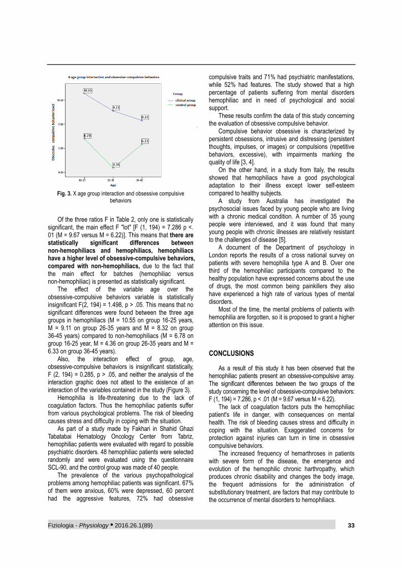
Fiziologia - Physiology • 2016.26.1(89) 33
Fig. 3. X age group interaction and obsessive compulsive behaviors
Of the three ratios F in Table 2, only one is statistically
significant, the main effect F "lot" [F (1, 194) = 7.286 p <. 01 (M = 9.67 versus M = 6.22)]. This means that there are statistically significant differences between non-hemophiliacs and hemophiliacs, hemophiliacs have a higher level of obsessive-compulsive behaviors, compared with non-hemophiliacs, due to the fact that the main effect for batches (hemophiliac versus non-hemophiliac) is presented as statistically significant.
The effect of the variable age over the obsessive-compulsive behaviors variable is statistically insignificant F(2, 194) = 1.498, p > .05. This means that no significant differences were found between the three age groups in hemophiliacs (M = 10.55 on group 16-25 years, M = 9.11 on group 26-35 years and M = 8.32 on group 36-45 years) compared to non-hemophiliacs (M = 6.78 on group 16-25 year, M = 4.36 on group 26-35 years and M = 6.33 on group 36-45 years).
Also, the interaction effect of group, age, obsessive-compulsive behaviors is insignificant statistically, F (2, 194) = 0.285, p > .05, and neither the analysis of the interaction graphic does not attest to the existence of an interaction of the variables contained in the study (Figure 3).
Hemophilia is life-threatening due to the lack of coagulation factors. Thus the hemophiliac patients suffer from various psychological problems. The risk of bleeding causes stress and difficulty in coping with the situation.
As part of a study made by Fakhari in Shahid Ghazi Tabatabai Hematology Oncology Center from Tabriz, hemophiliac patients were evaluated with regard to possible psychiatric disorders. 48 hemophiliac patients were selected randomly and were evaluated using the questionnaire SCL-90, and the control group was made of 40 people.
The prevalence of the various psychopathological problems among hemophiliac patients was significant. 67% of them were anxious, 60% were depressed, 60 percent had the aggressive features, 72% had obsessive
compulsive traits and 71% had psychiatric manifestations, while 52% had features. The study showed that a high percentage of patients suffering from mental disorders hemophiliac and in need of psychological and social support.
These results confirm the data of this study concerning the evaluation of obsessive compulsive behavior.
Compulsive behavior obsessive is characterized by persistent obsessions, intrusive and distressing (persistent thoughts, impulses, or images) or compulsions (repetitive behaviors, excessive), with impairments marking the quality of life [3, 4].
On the other hand, in a study from Italy, the results showed that hemophiliacs have a good psychological adaptation to their illness except lower self-esteem compared to healthy subjects.
A study from Australia has investigated the psychosocial issues faced by young people who are living with a chronic medical condition. A number of 35 young people were interviewed, and it was found that many young people with chronic illnesses are relatively resistant to the challenges of disease [5].
A document of the Department of psychology in London reports the results of a cross national survey on patients with severe hemophilia type A and B. Over one third of the hemophiliac participants compared to the healthy population have expressed concerns about the use of drugs, the most common being painkillers they also have experienced a high rate of various types of mental disorders.
Most of the time, the mental problems of patients with hemophilia are forgotten, so it is proposed to grant a higher attention on this issue.
CONCLUSIONS
As a result of this study it has been observed that the hemophiliac patients present an obsessive-compulsive array. The significant differences between the two groups of the study concerning the level of obsessive-compulsive behaviors: F (1, 194) = 7.286, p < .01 (M = 9.67 versus M = 6.22).
The lack of coagulation factors puts the hemophiliac patient's life in danger, with consequences on mental health. The risk of bleeding causes stress and difficulty in coping with the situation. Exaggerated concerns for protection against injuries can turn in time in obsessive compulsive behaviors.
The increased frequency of hemarthroses in patients with severe form of the disease, the emergence and evolution of the hemophilic chronic harthropathy, which produces chronic disability and changes the body image, the frequent admissions for the administration of substitutionary treatment, are factors that may contribute to the occurrence of mental disorders to hemophiliacs.
34 Fiziologia - Physiology • 2016.26.1(89)
REFERENCES
1. Cosma A. Hemofilicul îndrumător practic pentru bolnavi, familiile lor şi educatori. Ed. Medicală, Bucureşti, 1983.
2. Sava FA. Analiza datelor in cercetarea psihologica. Metode statistice complementare, ASCR. Cluj-Napoca, 2004.
3. Eisen JL, Rasmussen SA. Obsessive-compulsive disorder with psychotic features. J Clin Psychiatr, 1993; 54: 373-379.
4. Eisen JL, Phillips KA, Coles ME, Rasmussen SA. Insight in
obsessive compulsive disorder and body dysmorphic disorder. Compr Psychiatry, 2004; 45: 10-15.
5. Olsson CA, Bond L, Johnson MW, Forer DL, Boyce MF, Sawyer SM. Adolescent chronic illness: a qualitative study of psychosocial adjustment. Ann Acad Med Singapore, 2003; 32(1): 43-50.
6. Cassis FRMY. Psychosocial Care for People with Hemophilia. Treatement of hemophilia, Sao Paulo, Brazil, 2007; 44.
EVALUAREA COMPORTAMENTULUI
OBSESIV-COMPULSIV LA PACIENȚII CU HEMOFILIE
REZUMAT Introducere: Hemofilia, asemenea celorlalte boli rare, este o boală cronică debilitantă sau handicapantă, ameninţătoare de viaţă şi de calitate a vieţii, necesită efort şi susţinere specială din partea comunităţii. Invaliditatea permanentă la care este supus un organism tarat hemofilic antrenează o alterare a psihicului, cu repercusiuni asupra întregii personalităţii individuale. Durerile intense şi permanente cu care se confruntă provoacă o creştere a tensiunii psihice şi o interiorizare a personalităţii, cu atât mai mult, cu cât accidentele hemoragice sunt mai frecvente şi mai intense. Material si metode: Din studiu au făcut parte 200 de subiecţi grupaţi în două loturi egale (pacienți cu hemofilie A și B și populație sănătoasă), cu vârste cuprinse între 16 şi 45 de ani. Rezultate: Există diferenţe semnificative statistic între hemofilici şi nonhemofilici, hemofilicii au un nivel mai mare al comportamentelor obsesiv-compulsive, comparativ cu nonhemofilicii. Concluzii: Pacienții cu afecţiuni cronice, prezintă un risc dublu în apariţia complicaţiilor psihice faţă de semenii lor sănătoşi. Cuvinte cheie: hemofilie, comportament obsesiv-compulsiv
Fiziologia - Physiology • 2016.26.1(89) 35
Received 12th of January 2016.Accepted 15th of March 2016. Address for correspondence: Danina Muntean, Department of Pathophysiology, “Victor Babeș” University of Medicine and Pharmacy of Timişoara; 14 Tudor Vladimirescu st., RO-300173 Timisoara,Tel/Fax: +40-256-493085; e-mail: [email protected]
CHARACTERIZATION OF THE EFFECTS OF SARCKATP
AND MITOKATP MODULATORS ON REPERFUSION-INDUCED
ARRHYTHMIAS IN ISOLATED RAT HEARTS
ALEXANDRA PETRUȘ1
, ADRIAN STURZA1,2
, OANA DUICU1,2
,
NORBERT JOST1,2
, DANINA MUNTEAN1,2
, ISTVAN BACZKO1
1Department of Pathophysiology, 2Center for Translational Research and Systems Medicine, “Victor Babeș” University of Medicine and Pharmacy, Timișoara, Romania
ABSTRACT Malignant tachyarrhythmias, ventricular fibrillation (VF) and tachycardia (VT), associated with ischemia/reperfusion (I/R) injury represent a major cause of sudden cardiac death worldwide. Pharmacological modulation of the cardiac ATP-sensitive potassium channels, either sarcolemmal (sarcKATP) or mitochondrial (mitoKATP), has been reported by many (but not all) studies to be cardioprotective against the deleterious effects of postischemic reperfusion. The aim of the present study was to assess the effects of HMR-1098 (10 μΜ), a cardioselective sarcKATP inhibitor and of diazoxide (DZX, 50 μΜ), a mitoKATP opener, given either independently or together, on I/R-related arrhythmias. A brief episode of regional ischemia was elicited by ligation of the left anterior descending coronary artery (LAD) in Langendorff-perfused rat hearts (constant flow mode) and the compounds were given prior to the induction of ischemia. In separate administration, HMR-1098 and DZX significantly reduced the duration of reperfusion-related VF (but not of VT) as compared to the control group. Also, the incidence of both tachyarrhythmias was diminished (albeit not significantly) by the individual administration of these compounds. Intriguingly, neither isolated nor combined administration of HMR-1098 and DZX were able to significantly decrease the duration of ventricular tachycardia (VT). Moreover, in the presence of both compounds the beneficial effect on VF duration, recorded with each of the drug when given solely, was lost. In isolated rat hearts, the independent administration of a sarcKATP inhibitor and a mitoKATP opener mitigated the duration of reperfusion-induced ventricular fibrillation, whereas the association of these compounds did not influence the incidence and duration of malignant tachyarrhythmias. Keywords: isolated rat heart, sarcolemmal and mitochondrial ATP-sensitive potassium channels, ischemia-related malignant arrhythmias
INTRODUCTION
Ischemia-related ventricular tachyarrhythmias are currently the major cause of sudden cardiac death worldwide [1]. Preventing malignant arrhythmias in the setting of myocardial ischemia represents an unmet therapeutic target, ultimately aimed at decreasing mortality due to coronary artery disease [2]. The efficiency of current antiarrhythmic therapy is still under debate and a couple of studies have shown that several antiarrhythmic drugs can rather increase the risk of lethal arrhythmias in patients recovering after myocardial infarction [3]. The major mechanism underlying ventricular arrhythmias is represented by reentry, classically promoted by the heterogeneity in action potential duration (APD) and tissue repolarization, respectively. The duration of action potential
can be influenced, among others, by the activity of the ATP-sensitive potassium channels (KATP) that couple membrane excitability with cellular energetics [4]. These channels were firstly described by Noma [5] in the sarcolemmal membrane of cardiomyocytes (sarcKATP). A decade later, Inoue et al. [6] described a second type of KATP channels located in the inner mitochondrial membrane (mitoKATP). The ATP-dependent potassium channels are closed during normoxic conditions, when the cellular level of ATP is high, and open when ATP concentration drops during myocardial ischemia, as a result of the accumulation of ischemic metabolites ADP, lactate and H+ [2].
Paradoxically, both activation and inhibition of sarcKATP channels have been associated with antiarrhythmic effects [7]. In particular, opening the sarcKATP channels has been considered responsible for a significant potassium efflux
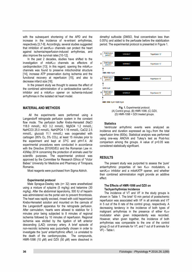
36 Fiziologia - Physiology • 2016.26.1(89)
with the subsequent shortening of the APD and the increase in the incidence of re-entrant arrhythmias, respectively [3,7-9]. Accordingly, several studies suggested that inhibition of sarcKATP channels can protect the heart against ischemia/reperfusion-induced arrhythmias, and also improve the survival rates [10-12].
In the past 2 decades, studies have shifted to the investigation of mitoKATP channels as effectors of cardioprotection [13]. In this regard, opening the mitoKATP channels was found to preserve mitochondrial structure [14], increase ATP preservation during ischemia and the functional recovery at reperfusion [15], and also to decrease infarct size [16].
In the present study we thought to assess the effect of the combined administration of a cardioselective sarcKATP inhibitor and a mitoKATP opener on ischemia-induced arrhythmias in the isolated rat heart model.
MATERIAL AND METHODS
All the experiments were performed using a Langendorff retrograde perfusion system in the constant flow mode. The perfusion buffer Krebs-Henseleit (NaCl 118.0 mmol/L; KCl 3.2 mmol/L; MgSO4 1.2 mmol/L; NaHCO3 25.0 mmol/L; NaH2PO4 1.18 mmol/L; CaCl2 2.5 mmol/L; glucoză 11.1 mmol/L) was oxygenated with carbogen (95% O2, 5% CO2), at least 15 minutes prior to the experiment and kept at 370C and pH=7.4. All experimental procedures were conducted in accordance with the Directive 2010/63/EU and the Romanian Law nr. 43/May 2014 concerning the protection of animals used for scientific purposes. The experimental protocol was approved by the Committee for Research Ethics of “Victor Babes” University for Medicine and Pharmacy of Timişoara, Romania.
Most reagents were purchased from Sigma Aldrich.
Experimental protocol Male Sprague-Dawley rats (n= 32) were anesthetized
using a mixture of xylazine (5 mg/kg) and ketamine (30 mg/kg). After the abdominal laparotomy, 500 IU of heparin was administered via the portal vein to prevent thrombosis. The heart was rapidly excised, rinsed with cold heparinized Krebs-Henseleit solution and mounted on the cannula of the Langendorff apparatus for the retrograde perfusion. After cannulation, hearts were allowed to stabilize for 3 minutes prior being subjected to 8 minutes of regional ischemia followed by 10 minutes of reperfusion. Regional ischemia was elicited by the ligation of left anterior descending (LAD) coronary artery. A brief episode of non-necrotic ischemia was purportedly chosen in order to investigate the 'pure' antiarrhythmic effect, i.e unrelated to the death of the cardiomyocytes. The compounds, HMR-1098 (10 μΜ) and DZX (50 μΜ) were dissolved in
dimethyl sulfoxide (DMSO, final concentration less than 0.05%) and added to the perfusate before the stabilization period. The experimental protocol is presented in Figure 1.
Fig. 1. Experimental protocol. (A) Control group, (B) HMR-1098, (C) DZX,
(D) HMR-1098 + DZX treated groups.
Statistics Ventricular arrhythmic events were analyzed as
incidence and duration expressed as log10 from the total reperfusion time (600s). Statistical analysis was performed using one-way ANOVA and Tukey’s test as Post-hoc comparison among the groups. A value of p<0.05 was considered statistically significant.
RESULTS
The present study was purported to assess the 'pure' antiarrhythmic properties of two KATP modulators, a sarcKATP inhibitor and a mitoKATP opener, and whether their combined administration might provide an additive effect.
The Effects of HMR-1098 and DZX on Tachyarrhythmias Incidence The incidence of VT and VF in the study groups is
shown in Table 1. The brief 10 min period of postischemic reperfusion was associated with VF in all animals and VT in 5 out of the 8 rats of the control group, respectively. A decreasing tendency in the incidence of both types of malignant arrhythmias in the presence of either KATP modulator when given independently was recorded. However, when given together, the incidence of both arrhythmias was comparable to the one of the control group (5 out of 8 animals for VT, and 7 out of 8 animals for VF) - Table I.
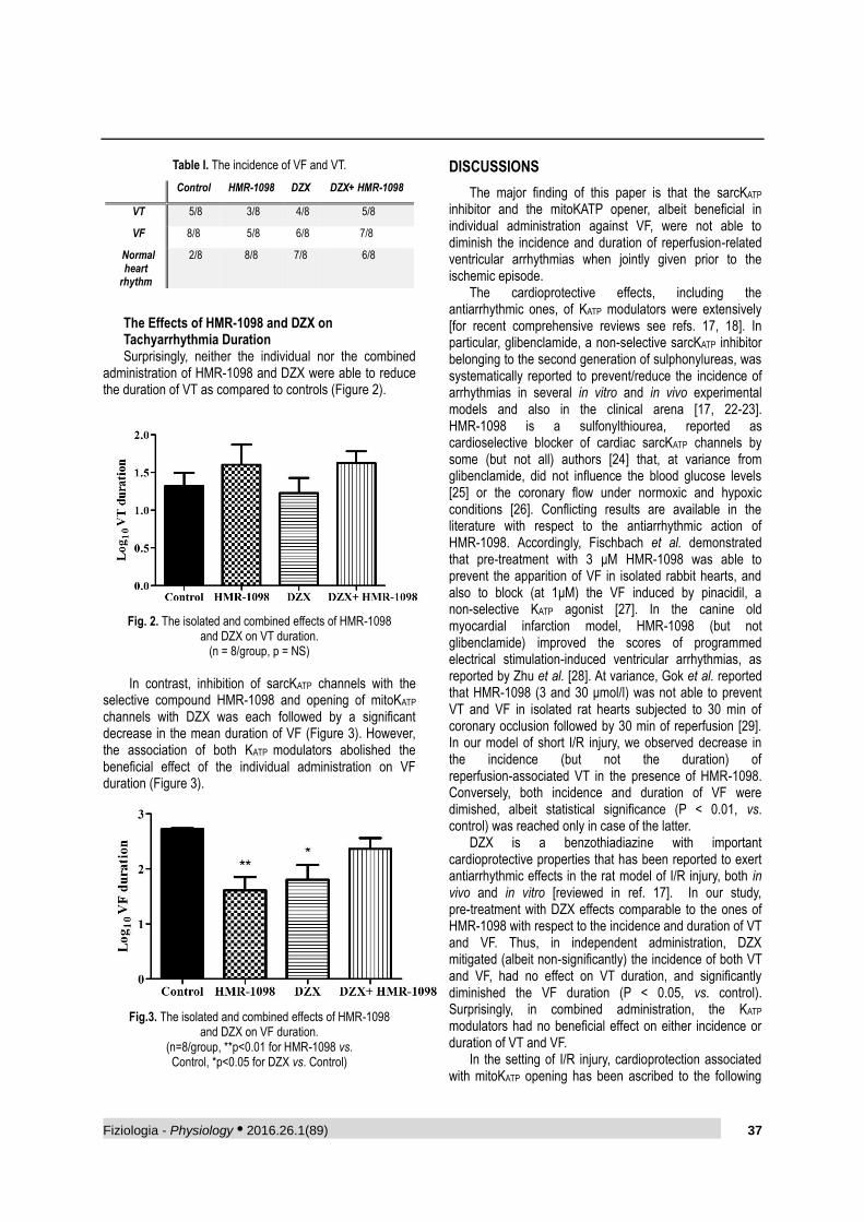
Fiziologia - Physiology • 2016.26.1(89) 37
Table I. The incidence of VF and VT.
Control HMR-1098 DZX DZX+ HMR-1098
VT 5/8 3/8 4/8 5/8
VF 8/8 5/8 6/8 7/8
Normal heart
rhythm
2/8 8/8 7/8 6/8
The Effects of HMR-1098 and DZX on Tachyarrhythmia Duration Surprisingly, neither the individual nor the combined
administration of HMR-1098 and DZX were able to reduce the duration of VT as compared to controls (Figure 2).
Fig. 2. The isolated and combined effects of HMR-1098
and DZX on VT duration. (n = 8/group, p = NS)
In contrast, inhibition of sarcKATP channels with the
selective compound HMR-1098 and opening of mitoKATP channels with DZX was each followed by a significant decrease in the mean duration of VF (Figure 3). However, the association of both KATP modulators abolished the beneficial effect of the individual administration on VF duration (Figure 3).
Fig.3. The isolated and combined effects of HMR-1098
and DZX on VF duration. (n=8/group, **p<0.01 for HMR-1098 vs. Control, *p<0.05 for DZX vs. Control)
DISCUSSIONS
The major finding of this paper is that the sarcKATP inhibitor and the mitoKATP opener, albeit beneficial in individual administration against VF, were not able to diminish the incidence and duration of reperfusion-related ventricular arrhythmias when jointly given prior to the ischemic episode.
The cardioprotective effects, including the antiarrhythmic ones, of KATP modulators were extensively [for recent comprehensive reviews see refs. 17, 18]. In particular, glibenclamide, a non-selective sarcKATP inhibitor belonging to the second generation of sulphonylureas, was systematically reported to prevent/reduce the incidence of arrhythmias in several in vitro and in vivo experimental models and also in the clinical arena [17, 22-23]. HMR-1098 is a sulfonylthiourea, reported as cardioselective blocker of cardiac sarcKATP channels by some (but not all) authors [24] that, at variance from glibenclamide, did not influence the blood glucose levels [25] or the coronary flow under normoxic and hypoxic conditions [26]. Conflicting results are available in the literature with respect to the antiarrhythmic action of HMR-1098. Accordingly, Fischbach et al. demonstrated that pre-treatment with 3 μΜ HMR-1098 was able to prevent the apparition of VF in isolated rabbit hearts, and also to block (at 1μΜ) the VF induced by pinacidil, a non-selective KATP agonist [27]. In the canine old myocardial infarction model, HMR-1098 (but not glibenclamide) improved the scores of programmed electrical stimulation-induced ventricular arrhythmias, as reported by Zhu et al. [28]. At variance, Gok et al. reported that HMR-1098 (3 and 30 μmol/l) was not able to prevent VT and VF in isolated rat hearts subjected to 30 min of coronary occlusion followed by 30 min of reperfusion [29]. In our model of short I/R injury, we observed decrease in the incidence (but not the duration) of reperfusion-associated VT in the presence of HMR-1098. Conversely, both incidence and duration of VF were dimished, albeit statistical significance (P < 0.01, vs. control) was reached only in case of the latter.
DZX is a benzothiadiazine with important cardioprotective properties that has been reported to exert antiarrhythmic effects in the rat model of I/R injury, both in vivo and in vitro [reviewed in ref. 17]. In our study, pre-treatment with DZX effects comparable to the ones of HMR-1098 with respect to the incidence and duration of VT and VF. Thus, in independent administration, DZX mitigated (albeit non-significantly) the incidence of both VT and VF, had no effect on VT duration, and significantly diminished the VF duration (P < 0.05, vs. control). Surprisingly, in combined administration, the KATP modulators had no beneficial effect on either incidence or duration of VT and VF.
In the setting of I/R injury, cardioprotection associated with mitoKATP opening has been ascribed to the following
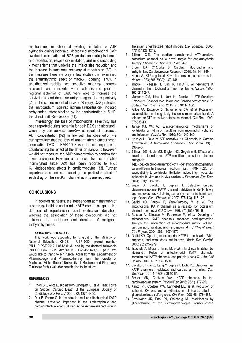
38 Fiziologia - Physiology • 2016.26.1(89)
mechanisms: mitochondrial swelling, inhibition of ATP synthesis during ischemia, decreased mitochondrial Ca2+ overload, modulation of ROS production during ischemia and reperfusion, respiratory inhibition, and mild uncoupling - mechanisms that underlie the infarct size reduction and the increase in functional recovery at reperfusion [30]. In the literature there are only a few studies that examined the antiarrhythmic effect of mitoKATP opening. Thus, in anesthetized rabbits, two selective mitoKATP openers, nicorandil and minoxidil, when administered prior to regional ischemia of LAD, were able to increase the survival rate and decrease arrhythmogenesis, respectively [2]. In the canine model of in vivo I/R injury, DZX protected the myocardium against ischemia/reperfusion- induced arrhythmias, effect blocked by the administration of 5-HD, the classic mitoKATP blocker [31].
Interestingly, the loss of mitochondrial selectivity has been reported during ischemia for both DZX and nicorandil, when they can activate sarcKATP as result of increased ADP concentration [32]. In line with this observation we can speculate that the loss of antiarrhythmic effects when associating DZX to HMR-1098 was the consequence of counteracting the effect of the latter on sarcKATP; however, we did not measure the ADP concentration to confirm that it was decreased. However, other mechanisms can be also incriminated since DZX has been reported to elicit KATP-independent effects in cardiomyocytes [33]. Further experiments aimed at assessing the particular effect of each drug on the sarcKATP channel activity are required.
CONCLUSIONS
In isolated rat hearts, the independent administration of a sarcKATP inhibitor and a mitoKATP opener mitigated the duration of reperfusion-induced ventricular fibrillation, whereas the association of these compounds did not influence the incidence and duration of malignant tachyarrhythmias.
ACKNOWLEDGEMENTS This work was supported by a grant of the Ministry of
National Education, CNCS – UEFISCDI, project number PN-II-ID-PCE-2012-4-0512 (N.J.) and by the doctoral fellowship POSDRU no. 159/1.5/S/136893 – DocMed.Net_2.0. (A.P.) We would like to thank to Mr. Karoly Acsai from the Department of Pharmacology and Pharmacotherapy from the Faculty of Medicine, “Victor Babes” University of Medicine and Pharmacy, Timisoara for his valuable contribution to the study.
REFERENCES
1. Priori SG, Aliot E, Blomstrom-Lundqvist C, et al. Task Force on Sudden Cardiac Death of the European Society of Cardiology. Eur Heart J. 2001; 22: 1374-1450.
2. Das B, Sarkar C. Is the sarcolemmal or mitochondrial KATP channel activation important in the antiarrhythmic and cardioprotective effects during acute ischemia/reperfusion in
the intact anesthetized rabbit model? Life Sciences. 2005; 77(11):1226-1248.
3. Billman G.E. The cardiac sarcolemmal ATP-sensitive potassium channel as a novel target for anti-arrhythmic therapy. Pharmacol Ther. 2008; 120: 54-70.
4. Brown DA, O’Rourke B. Cardiac mitochondria and arrhythmias. Cardiovascular Research. 2010; 88: 241-249.
5. Noma A. ATP-regulated K + channels in cardiac muscle. Nature. 1983; 305(5930): 147–148.
6. Innoue I, Nagase H, Kishi K, Higuti T. ATP-sensitive K channel in the mitochondrial inner membrane. Nature. 1990; 352: 244-247.
7. Muntean DM, Kiss L, Jost N, Baczkó I. ATP-Sensitive Potassium Channel Modulators and Cardiac Arrhythmias: An Update. Curr Pharm Des. 2015; 21: 1091-1102.
8. Wilde AA, Escande D, Schumacher CA, et al. Potassium accumulation in the globally ischemic mammalian heart. A role for the ATP-sensitive potassium channel. Circ Res. 1990; 67: 835-43.
9. Janse MJ, Wit AL. Electrophysiological mechanisms of ventricular arrhythmias resulting from myocardial ischemia and infarction. Physiol Rev. 1989; 69: 1049-169.
10. Nakaya H. Role of ATP-sensitive K+ Channels in Cardiac Arrhythmias. J Cardiovasc Pharmacol Ther. 2014; 19(3): 237-243.
11. Billman GE, Houle MS, Englert HC, Gogelein H. Effects of a novel cardioprotective ATP-sensitive potassium channel antagonist, 1-[[5-[2-(5-chloro-o-anisamido)ethyl]-b-methoxyethoxyphenyl]sulfonyl]-3-methylthiourea, sodium salt (HMR1402), on susceptibility to ventricular fibrillation induced by myocardial ischemia: in vitro and in vivo studies. J Pharmacol Exp Ther. 2004; 309(1):182-192.
12. Vajda S, Baczko I, Lepran I. Selective cardiac plasma-membrane KATP channel inhibition is defibrillatory and improves survival during acute myocardial ischemia and reperfusion. Eur J Pharmacol. 2007; 577(1-3): 115-123.
13. Garlid KD, Paucek P, Yarov-Yarovoy V, et al. The mitochondrial KATP channel as a receptor for potassium channel openers. J Biol Chem. 1996; 271(15):8796–9.
14. Rousou A, Ericsson M, Federman M, et al. Opening of mitochondrial KATP channels enhances cardioprotection through the modulation of mitochondrial matrix volume, calcium accumulation, and respiration. Am J Physiol Heart Circ Physiol. 2004; 287: 1967-1976.
15. Garlid KD. Opening mitochondrial KATP in the heart - What happens, and what does not happen. Basic Res Cardiol. 2000; 95: 275–279.
16. Tsuchida A, Miura T, Tanno M, et al. Infarct size limitation by nicorandil: Roles of mitochondrial KATP channels, sarcolemmal KATP channels, and protein kinase C. J Am Coll Cardiol. 2002; 40: 1523–1530.
17. Baczko I, Husti Z, Lang V, Lepran I, Light PE. Sarcolemmal KATP channels modulatos and cardiac arrhythmias. Curr Med Chem. 2011; 18(24): 3640-61.
18. Foster MN, Coetzee WA. KATP channels in the cardiovascular system. Physiol Rev 2016; 96(1): 177-252.
19. Kantor PF, Coetzee WA, Carmeliet EE, et al. Reduction of ischemic K+ loss and arrhythmias in rat hearts: effect of glibenclamide, a sulfonylurea. Circ Res. 1998; 66: 478−485.
20. Smallwood JK, Ertel PJ, Steinberg MI. Modification by glibenclamide of the electrophysiological consequences
Fiziologia - Physiology • 2016.26.1(89) 39
ofmyocardial ischaemia in dogs and rabbits. Naunyn Schmiedebergs Arch Pharmacol. 1990; 342: 214−220.
21. Benndorf K, Friedrich M, Hirche H. Anoxia opens ATP regulated K channels in isolated heart cells of the guinea pig. Pflugers Archive. 1991; 419: 108−110.
22. Dhein S, Pejman P, Krusemann K. Effects of the IK.ATP blockers glibenclamide and HMR1883 on cardiac electrophysiology during ischemia and reperfusion. Eur J Pharmacol. 2000; 398: 273−284.
23. Bril A, Laville M.P, Gout B. Effects of glibenclamide on ventricular arrhythmias and cardiac function in ischaemia and reperfusion in isolated rat heart. Cardiovasc Res. 1992; 26: 1069−1076.
24. Manning Fox J, Kanji H, French R, et al. Cardioselectivity of the sulphonylurea HMR 1098: studies on native and recombinant cardiac and pancreatic KATP channels. Br J Pharmacol. 2002; 135: 480-488.
25. Billman G. E, Englert H.C, Schoelkens B.A. HMR 1883, a novel cardioselective inhibitor of the ATP- sensitive potassium channel; Part II: effects on susceptibility to ventricular fibrillation induced by myocardial ischemia in conscious dogs. J Pharmacol Exp Ther. 1998; 286: 1465−1473.
26. Gögelein H, Hartung J, Englert H.C, et al. HMR 1883, a novel cardioselective inhibitor of the ATP-sensitive potassium channel. Part I: effects on cardiomyocytes, coronary flow and pancreatic beta-cells. J Pharmacol Exp Ther. 1998; 286: 1453−1464.
27. Fischbach P.S, White A, Barrett T.D, et al. Risk of ventricular proarrhythmia with the selective opening of the myocardial sarcolemmal versus mitochondrial ATP-gated potassium channel. J Pharmacol Exp Ther. 2000; 309: 554−559.
28. Zhu B.M, Miyamoto S, Nagasawa Y, et al. Effect of the sarcolemmal K(ATP) channel blocker HMR1098 on arrhythmias induced by programmed electrical stimulation in canine old myocardial infarction model: comparison with glibenclamide. J Pharmacol Sci. 2003; 93: 106-113.
29. Gok S, Vural K, Sekuri C, et al. Effects of the blockade of cardiac sarcolemmal ATP-sensitive potassium channels on arrhythmias and coronary flow in ischemia-reperfusion model in isolated rat hearts. Vasc Pharmacol. 2006; 44: 197-205.
30. Kopustinskiene D, Liobikas J, Skemiene K, et al. Direct Effects of KATP Channel openers pinacidil and diazoxide on oxidative phosphorylation of mitochondria in situ. Cell Physiol Biochem. 2010; 25:181-186.
31. Végh A, Parratt JR. The role of mitochondrial K(ATP) channels in antiarrhythmic effects of ischaemic preconditioning in dogs. Br J Pharmacol. 2002; 137(7):1107-15.
32. Sato T, Li Y, Saito T, et al. Minoxidil opens mitochondrial KATP channels and confers cardioprotection. Br J Pharmacol. 2004; 141: 360–366.
33. Hanley P.J, Mickel M, Ffler L.O, et al. KATP channel-independent targets of diazoxide and 5- hydroxydecanoate in the heart. J Physiol. 2002; 542: 735-741.
CARACTERIZAREA EFECTELOR MODULATORILOR
SARCKATP ȘI MITOKATP ASUPRA ARITMIILOR INDUSE
DE REPERFUZIE PE INIMI IZOLATE DE ȘOBOLAN
REZUMAT Tahiaritmiile maligne, fibrilația și tahicardia ventriculară (FV și TV) asociate ischemiei/reperfuziei (I/R) miocardice reprezintă o cauză majoră de moarte cardiacă subită la nivel global. Modularea farmacologică a canalelor cardiace de potasiu dependente de ATP localizate atât la nivel sarcolemal (sarcKATP) cât și la nivel mitocondrial (mitoKATP) a fost asociată cu cardioprotecție în condițiile ischemiei/reprefuziei miocardice în numeroase studii, fără însă ca această observație să fie fără echivoc. Scopul prezentului studiu a fost de a evalua efectele administrării izolate și în asociere a
HMR-1098 (10 μΜ), inhibitorului selectiv al canalelor sarcKATP și respectiv, a diazoxidului (DZX, 50 μΜ), deschizătorului canalelor mitoKATP, asupra tahiaritmiilor induse de reperfuzia postischemică. Ischemia regională a fost indusă prin ligatura arterei coronare anterioare descendente stângi la nivelul inimilor izolate de șobolan perfuzate retrograd tip Langendorff (în modul de lucru cu flux constant), iar compușii au fost administrați anterior inducerii ischemiei regionale. În administrare independentă, atât HMR-1098 cât și DZX au redus semnificativ durata FV (dar nu și a TV) comparativ cu lotul martor. De asemenea, aplicarea lor individuală a redus (deși nu semnificativ) incidența ambelor tahiaritmii maligne. Surprinzător, nici administrarea independentă nici cea în asociere a compușilor nu au condus la o reducere semnificativă a duratei TV. Mai mult, administrarea combinată a condus la pierderea efectului benefic obținut în cazul aplicării individuale asupra duratei FV. În concluzie, în cazul inimilor izolate de șobolan, administrarea independentă a unui inhibitor al sarcKATP și respectiv, a unui deschizător al mitoKATP, a redus durata fibrilației ventriculare la reperfuzia postischemică, în timp ce asocierea acestor compuși nu a influențat incidența și durata tahiaritmiilor maligne. Cuvinte cheie: inimă izolată de șobolan, canale de poatasiu sarcolemale si mitocondriale dependente de ATP, aritmii maligne induse de ischemie
40 Fiziologia - Physiology • 2016.26.1(89)
STEM CELLS FROM AMNIOTIC FLUID,
AN ESSENTIAL TOOL FOR REGENERATIVE MEDICINE
HONCEA ADINA Ovidius University, Constanta
ABSTRACT Since the existence of stem cells in human amniotic fluid was reported, the knowledge about these cells has increased dramatically. In recent years it has become increasingly evident the hope of using stem cells and human progenitor cells to replace dysfunctional tissues. The major problem in this area of research is to identify new sources for isolating progenitor cells or stem cells that do not raise ethical issues. As an alternative to these sources of cells, the amniotic fluid is easily collected by ultrasound guided transabdominal puncture. Compared to embryonic stem cells, amniotic stem cells can be obtained without affecting human embryos, thus attenuating ethical controversies. The authors also described the high proliferative potential, and cultivation possibility for long periods with genomic stability maintained. AFS cells could be used for stem cell-based therapies; in addition, it is now increasingly accepted as an optimal tool for basic research. Biological, biochemical, immunological tests and morphological investigations showed that AFS cells are a new and specific entity, distinct from ES cells or other types of stem cells, such as those that can be isolated from epithelial sources or trophoblast tissue. Key words: embryonic stem cells, amniotic stem cells, proliferative potential
INTRODUCTION
In recent years it has become increasingly evident the hope of using stem cells and human progenitor cells to replace dysfunctional tissues.
The major problem in this area of research is to identify new sources for isolating progenitor cells or stem cells that do not raise ethical issues.
Also, because the logistical and ethical problems limit the use of embryonic stem cells (ESCs), a special attention was given to recent use of alternative sources of stem cells, such as mesenchymal stem cells (MSCs) in the bone marrow (BM), amniotic fluid (AF), placenta, and umbilical cord [1]. BM cells are a common source of MSCs in clinical trials; however, their use has several limitations. First of all, the collection of BM from a patient is an invasive procedure and secondly, the frequency of MSCs in the BM is low. Therefore, the search for alternative sources of useful MSCs for clinical applications is important [2]. Currently, existing MSC in umbilical cord blood is challenged with reports showing that the isolation of such cells is difficult, having a low yield and inter-individual variations [3,4)]. The placenta contains a large number of progenitor cells or stem cells that can be obtained through minimally invasive techniques [5]. Cells that were isolated from placenta include amniotic epithelial cells, stromal chorionic amniotic cells, MSCs, chorionic trophoblast cells and hematopoietic stem cells [6].
As an alternative to these sources of cells, the amniotic fluid is easily collected by ultrasound guided transabdominal puncture. The potential proliferation of mesenchymal stromal cells from amniotic fluid (AFMSCs) exceeds that of MSC derived from BM [2]. AFS cells have a high capacity for renewal - more than 250 doublings without any detectable loss of telomere length of chromosomes [7]. The presence of certain markers of undifferentiated cells shows that AFMSCs may be less differentiated than most BM-MSCs, and closer to pluripotent ESCs. AFMSCS’s have phenotypic characteristics similar to MSCs from other sources, such as umbilical cord, blood and bone marrow; all negative AFMSCs for hematopoietic markers, such as CD45, CD34 and CD14 [8] while positive for mesenchymal markers such as CD90, CD105, CD73, CD166.
Cellular Composition of the Amniotic Fluid Human amniotic fluid obtained during amniocentesis
process includes a variety of cells derived from embryonic and extra-embryonic tissues [9] including stem cells.
Compared to embryonic stem cells, amniotic stem cells can be obtained without affecting human embryos, thus attenuating ethical controversies.
Due to the widespread use of routine prenatal genetic tests, amniotic cells have become the subject of recent years’ research to enrich current knowledge about their origins and properties.
Received January 15th 2016. Accepted March 4th 2016. Address for correspondence: Honcea Adina, Ovidius University Constanta, Romania; 19 Bujorului Street, Constanta; phone: 0762043499, e-mail: [email protected]
Fiziologia - Physiology • 2016.26.1(89) 41
The types and properties of amniotic fluid cells vary with gestational age. By cultivation, AFS cells can be separated into adherent cells able to form colonies and non-adherent cells. Based on morphological and growth characteristics, amniotic fluid cells can be classified into three types: amniotic fluid specific cells – type AF, epithelioid cells - type E and fibroblast cells - F [10]. AF and E type cells appear at the beginning of cultivation. AF cell type resists during cultivation, while type E significantly decreases. It is assumed that E type cells come from fetal skin and urine, AF type cells from the trophoblast and fetal membranes and F type cells from fibrous connective tissue and dermal fibroblasts. AF type cells produce human chorionic gonadotropin, estrogen and progesterone, which suggest that these cells have their origin in the trophoblastic tissue. Also, F type cells are deemed to originate in the mesenchymal tissues. Approximately 1% of cells in cultures obtained by amniocentesis for prenatal diagnosis of genetic disorders express the surface antigen c-kit (CD117). This receptor protein is present in human ES cells, primordial germ cells, and many of somatic stem cells. In 2004, a group of researchers managed to isolate monoclonal AFS cells based on the expression of stem cell markers, c-Kit and Oct4 [11] using flow cytometry and minimal dilution. Three years later, another research group isolated monoclonal AFS cells via flow cytometric selection and minimal dilution, which expressed the stem cell markers c-Kit and Oct4 [12]. The authors also described the high proliferative potential, and cultivation possibility for long periods with genomic stability maintained.
Stem cells derived from amniotic fluid (amniotic fluid stem cells - AFS) express both embryonic and adult stem cell markers and unlike ESC proliferate very well without support cells (feeder). In recent years, increasing evidence demonstrated AFS ability to differentiate into multiple lineages including those of the embryonic germ layers [2,9,10,13-16]. All this data allowed classification of these cells as pluripotent [7,17,18]. Thus, AFS cells no longer need an ectopic induction of pluripotency.
However, AFS cells are different from ESC pluripotent stem cells and from multipotent adult stem cells, and may represent a new class of stem cell plasticity whose properties are somewhere between embryonic and adult stem cells. Moreover, approximately 90% of AFS cells express the transcription factor Oct-4, known to be expressed in embryonic carcinoma cells, embryonic germ cells and embryonic stem cell in addition, AFS cells express TERT marker of pluripotent stem cells [19].
AFS cells are now becoming increasingly accepted as an optimal tool for fundamental research [20-24] preceding their possible application in therapy.
It is known that the morphology, the composition and the properties of AFS cell growth are affected by certain fetal pathologies such as, for example, a neural tube defect or gastroschisis [20,25,26].
Potential Ways of Differentiation Followed by AFS Stem cells are useful tools for the study of the processes
of differentiation in terms of molecular and cellular investigations.
AFS cells are genomically stable and do not harbor neither the epigenetic memory nor somatic mutations of already differentiated source cells. Furthermore iPS cells have been reported to accumulate karyotype abnormalities and gene mutations during propagation in culture [20,21].
Since AFS discovery, it was tried to be determined the differentiation potential of AFS cells by initiating successfully the generation of various lines from a single stem cell. Quite often, the researchers only use a mixture of cells from amniotic fluid obtained by specific procedures of cultivation. However, as is known, derivatives of amniotic fluid cells represent mixtures containing a wide variety of differentiated and undifferentiated cells. It is therefore important that any study for a potential differentiating specific cell lines to clarify the type of cell from which they started, highlighting the biological and immunocytochemical characteristics. Furthermore, evidence that AFS cells have indeed pluripotent differentiation potential can be obtained from a single stem cell. Unicellular and practical approaches are required after minimal dilution experiments [25].
ES cells when cultured in the absence of differentiation factors can spontaneously form three-dimensional multicellular organisms called embryoid bodies. At first, embryoid bodies were generally regarded as a starting point for highlighting the differentiation of stem cells into different cell lines. Consequently, the formation of embryoid body followed by different approaches for inducing differentiation was an appropriate way to prove pluripotent differentiation potential of a specific type of stem cell [21, 27]. Therefore, it was of great interest the research that starting from a single AFS cell has determined the formation of embryoid bodies. It was shown that monoclonal human AFS cells can form organisms when grown without anti-differentiation factors in conditions in which they are not able to attach to the surface of culture vessels and without contact with feeder cells. The formation of such three-dimensional multicellular organisms is accompanied by a decrease in the expression of stem cell markers in different lines and induced differentiation [27]. The study that has shown the potential AFS cells to form embryoid bodies was the ultimate proof for their pluripotent character. In addition, now it led to the revision and investigation of three-dimensional structures and other tissue structure of many processes of differentiation during early embryogenesis [27].
ES cells right from the first in vitro culture have been described as being tumorigenic. Similarly, iPS cells induced tumor formation when transplanted subcutaneously to the mice. However, in AFS cells has not been reported the formation of tumors in immunocompromised mice.
Gene expression profiles suggest that AFS cells have the potential to differentiate into cardiomyocytes, as well as in other mesoderm derivatives, in addition to neural cells [7].
42 Fiziologia - Physiology • 2016.26.1(89)
AFS cells have been shown to express neuronal markers (such as CD133, nestin, neurofilament, CNPA, p75, BDNF, and neurotophin-3) [9]. The presence of several markers of dopaminergic neurons (TGF-β3, FGF8, SHH, and β-catenin) in AFS cells demonstrates the existence of differentiating neuronal cells engaged in the path, corresponding to a transcriptional profile similar to mesencephalic dopaminergic neurons [14,28]. These cells can become nestin positive neural stem cells, and then dopamine and glutamate receptive neurons.
In a suitable environment, AF-derived mesenchymal stem cells (AF-MSCs) can form functional osteoblasts that can produce bone mineralization or may form smooth muscle cells [19]. Further, liver cells obtained by differentiating AF-MSCs are capable of secreting high levels of urea and to express liver proteins, such as α-fetoprotein, albumin, growth factors and hepatocyte nuclear factor [7,18].
Many groups have confirmed the existence of Oct4+ / c-kit+ AFS cells and reported their potential to differentiate on the hematopoietic, neurogenic, osteogenic, chondrogenic, adipogenic, renal, hepatic, and various other lines [2,29-37].
Although, in terms of their biological properties and markers expression they appear to be more similar to ES cells, AFS cells exact origin remains unknown. Biological, biochemical, immunological tests and morphological investigations showed that AFS cells are a new and specific entity, distinct from ES cells or other types of stem cells, such as those that can be isolated from epithelial sources or trophoblast tissue.
AFS and Regenerative Medicine In 2001, it was suggested that the cells in the amniotic
fluid may be used in tissue engineering approach for the surgical repair of congenital abnormalities in the perinatal period.
Initially the idea was to make it function as a reconstruction graft for implantation, even before birth or in the neonatal period. This is of particular interest to children born with a defect in the wall that are too small for taking a graft for reconstructive surgery [9,38]. The results obtained in animal models are indeed encouraging.
Studies done for SE human cell transplantation therapy prevent possible immunological and tumorigenicity. As was pointed out, in contrast to cells derived from the cell mass of blastocyst, AFS cells are not tumorigenic in mice after transplantation [7].
Studies have shown that the AFS cells can be used for regenerative therapy approaches [20-24]. One such study showed the usefulness of AFS cells in regenerative mammary gland [11]. Other investigations have shown that may aid in muscle and cardiac disease [19,39]. Due to their neurogenic potential there are enough reasons to launch therapies based on AFS for diseases of the nervous system such as Parkinson's disease, spinal cord injury, multiple sclerosis or stroke. Neural stem cells, which have been
studied for this purpose and can be found in the adult central nervous system and the developing embryo, are not easily accessible and raise ethical concerns.
Kidney acute and chronic diseases are disorders with high rate of morbidity and mortality. Transplantation remains the most effective treatment option for most patients with end-stage renal disease. But unfortunately, the lack of compatible organs is a real problem. Treatment strategies are based on conventional kidney dialysis, but the mortality rate of patients requiring chronic dialysis is quite high. Therefore, the use of stem cells in tissue repair kidney becomes increasingly plausible. Several recently published studies for renal cell differentiation AFS cells are tempting further investigations in this direction. [24,40-44].
A recent report has shown that human AFS cells can integrate into the kidney tissues when injected into the isolated embryonic murine kidney [41]. It was also proved that AFS cells have the potential to differentiate to nephrogenic lineage following a pathway regulated by the mammalian target of rapamycin (mTOR) [42]. Moreover, the injection of AFS cells into kidneys of mice with rhabdomyolysis associated acute tubular necrosis, could mediate a protective effect [43].
AFS cells can survive, migrate significant distances, and differentiate when transplanted into the brains of adult rats [7,45]. Also, AF-MSCs after 3 weeks of culture were transplanted into the brain of both normal and ischemic rats, and showed that transplanted cells were able to migrate from the injection site along the corpus callosum in order to have access to more than one region of the brain. Some positive effects obtained by MSC transplants can be assigned to production of trophic factors and cytokines by these cells, and the increasing levels of neurotrophic factors in the ischemic region, as a response to administration of MSCs [46].
Among the limits of using AFS cells is the cost of obtaining such a cell which is much higher than for other types of stem cells.
There are also regarded as a source of immuno-incompetent graft cells, in large part thanks to their immature immunogenic system, immunogenic properties and phenotypic instability [47]. Studies have also shown that stem cells derived from the placenta can be an alternative source of stem cells [48]. This can cause problems during cell growth, where cells accumulation could lead to spontaneous differentiation. In addition, investigations are warranted to discover evidence of meiotic divisions within these stem cells derived from the placenta. These unregulated divisions not only can lead to the possibility of malignant tumors, but can also lead to cell defects. These negative side effects should be taken into account to reveal safety profile of AFS cells, with the resulting parameters veracity.
These interesting options for cell-based treatment could lead to the development of further appealing therapies against human and veterinary diseases [49] (Table I).
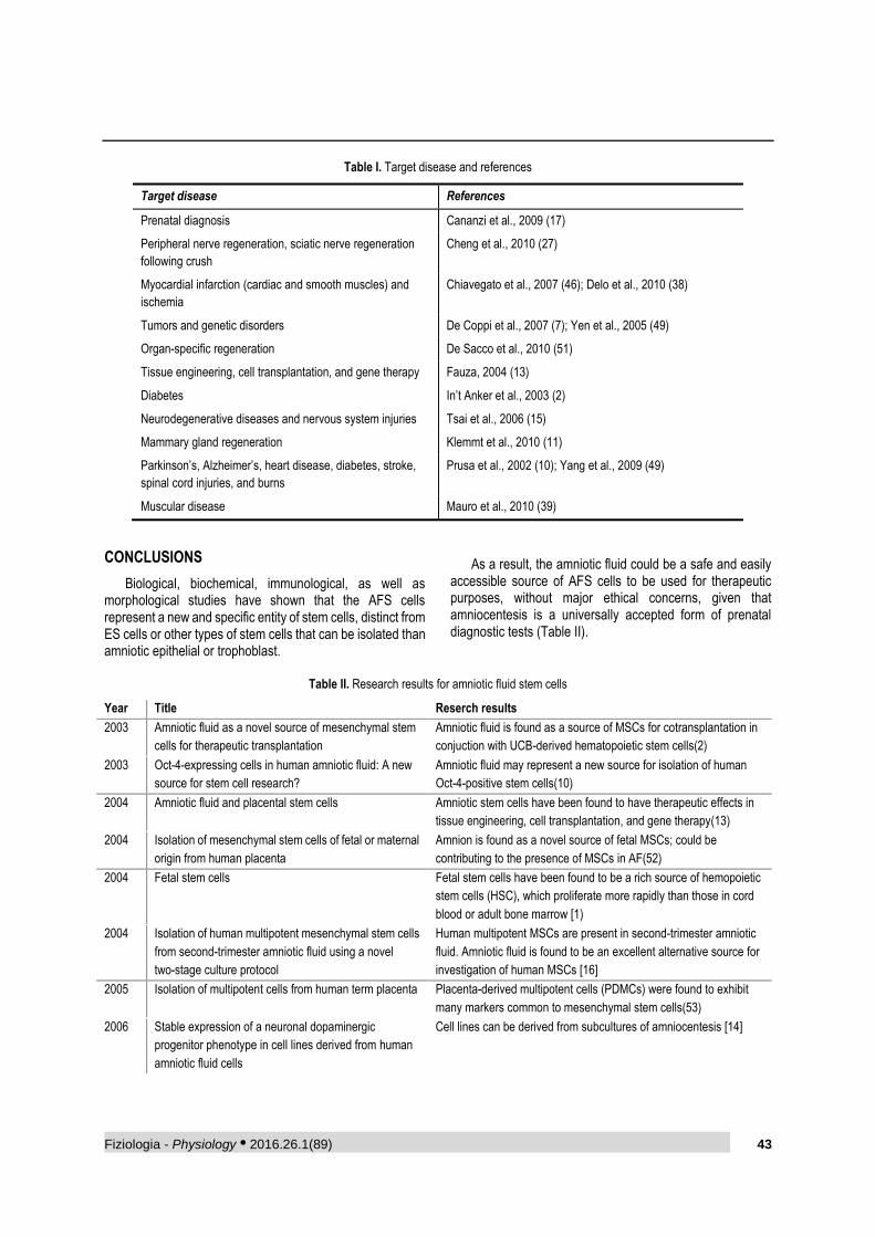
Fiziologia - Physiology • 2016.26.1(89) 43
Table I. Target disease and references
Target disease References
Prenatal diagnosis Cananzi et al., 2009 (17)
Peripheral nerve regeneration, sciatic nerve regeneration
following crush
Cheng et al., 2010 (27)
Myocardial infarction (cardiac and smooth muscles) and
ischemia
Chiavegato et al., 2007 (46); Delo et al., 2010 (38)
Tumors and genetic disorders De Coppi et al., 2007 (7); Yen et al., 2005 (49)
Organ-specific regeneration De Sacco et al., 2010 (51)
Tissue engineering, cell transplantation, and gene therapy Fauza, 2004 (13)
Diabetes In’t Anker et al., 2003 (2)
Neurodegenerative diseases and nervous system injuries Tsai et al., 2006 (15)
Mammary gland regeneration Klemmt et al., 2010 (11)
Parkinson’s, Alzheimer’s, heart disease, diabetes, stroke,
spinal cord injuries, and burns
Prusa et al., 2002 (10); Yang et al., 2009 (49)
Muscular disease Mauro et al., 2010 (39)
CONCLUSIONS
Biological, biochemical, immunological, as well as morphological studies have shown that the AFS cells represent a new and specific entity of stem cells, distinct from ES cells or other types of stem cells that can be isolated than amniotic epithelial or trophoblast.
As a result, the amniotic fluid could be a safe and easily accessible source of AFS cells to be used for therapeutic purposes, without major ethical concerns, given that amniocentesis is a universally accepted form of prenatal diagnostic tests (Table II).
Table II. Research results for amniotic fluid stem cells
Year Title Reserch results
2003 Amniotic fluid as a novel source of mesenchymal stem
cells for therapeutic transplantation
Amniotic fluid is found as a source of MSCs for cotransplantation in
conjuction with UCB-derived hematopoietic stem cells(2)
2003 Oct-4-expressing cells in human amniotic fluid: A new
source for stem cell research?
Amniotic fluid may represent a new source for isolation of human
Oct-4-positive stem cells(10)
2004 Amniotic fluid and placental stem cells Amniotic stem cells have been found to have therapeutic effects in
tissue engineering, cell transplantation, and gene therapy(13)
2004 Isolation of mesenchymal stem cells of fetal or maternal
origin from human placenta
Amnion is found as a novel source of fetal MSCs; could be
contributing to the presence of MSCs in AF(52)
2004 Fetal stem cells Fetal stem cells have been found to be a rich source of hemopoietic
stem cells (HSC), which proliferate more rapidly than those in cord
blood or adult bone marrow [1)
2004 Isolation of human multipotent mesenchymal stem cells
from second-trimester amniotic fluid using a novel
two-stage culture protocol
Human multipotent MSCs are present in second-trimester amniotic
fluid. Amniotic fluid is found to be an excellent alternative source for
investigation of human MSCs [16]
2005 Isolation of multipotent cells from human term placenta Placenta-derived multipotent cells (PDMCs) were found to exhibit
many markers common to mesenchymal stem cells(53)
2006 Stable expression of a neuronal dopaminergic
progenitor phenotype in cell lines derived from human
amniotic fluid cells
Cell lines can be derived from subcultures of amniocentesis [14]
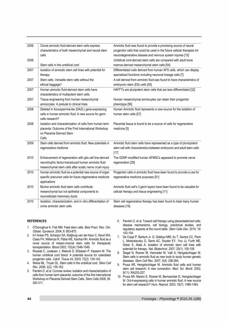
44 Fiziologia - Physiology • 2016.26.1(89)
2006 Clonal amniotic fluid-derived stem cells express
characteristics of both mesenchymal and neural stem
cells
Amniotic fluid was found to provide a promising source of neural
progenitor cells that could be used in the future cellular therapies for
neurodegenerative disease and nervous system injuries [15]
2006
Stem cells in the umbilical cord
Umbilical cord-derived stem cells are compared with adult bone
marrow-derived mesenchymal stem cells [54]
2007 Isolation of amniotic stem cell lines with potential for
therapy
Differentiated cells derived from human AFS cells, which can display
specialized functions including neuronal lineage cells [7]
2007 Stem cells. Versatile stem cells without the
ethical baggage?
A cell derived from amniotic fluid was found to have characteristics of
embryonic stem (ES) cells [55]
2007 Human amniotic fluid-derived stem cells have
characteristics of multipotent stem cells
HAFFTs are pluripotent stem cells that are less differentiated [32]
2007 Tissue engineering from human mesenchymal
amniocytes: A prelude to clinical trials
Human mesenchymal amniocytes can retain their progenitor
phenotype [56]
2008 Deleted in Azoospermia-ike (DAZL) gene-expressing
cells in human amniotic fluid: A new source for germ
cells research?
Human Amniotic fluid represents a new source for the isolation of
human stem cells [57]
2008 Isolation and characterization of cells from human term
placenta: Outcome of the First International Workshop
on Placenta Derived Stem
Cells
Placental tissue is found to be a source of cells for regenerative
medicine [5]
2009 Stem cells derived from amniotic fluid: New potentials in
regenerative medicine
Amniotic fluid stem cells have represented as a type of pluriopotent
stem cell with characteristics between embryonic and adult stem cells
[17]
2010 Enhancement of regeneration with glia cell line-derived
neurotrophic factor-transduced human amniotic fluid
mesenchymal stem cells after sciatic nerve crush injury
The GDNF-modified human AFMSCs appeared to promote nerve
regeneration [28]
2010 Human amniotic fluid as a potential new source of organ
specific precursor cells for future regenerative medicine
applications
Progenitor cells in amniotic fluid have been found to provide a use for
regenerative medicine purposes [51]
2010 Murine amniotic fluid stem cells contribute
mesenchymal but not epithelial components to
reconstituted mammary ducts
Amniotic fluid cell’s 3 germ layers have been found to be valuable for
cellular therapy and tissue engineering [11]
2010 Isolation, characterization, and in vitro differentiation of
ovine amniotic stem cells
Stem cell regenerative therapy has been found to treat many human
diseases [19]
REFERENCES
1. O’Donoghue K, Fisk NM. Fetal stem cells. Best Pract. Res. Clin. Obstet. Gynaecol. 2004; 6: 853-875.
2. In’t Anker PS, Scherjon SA, Kleijburg-van der Keur C, Noort WA, Claas FH, Willemze R, Fibbe WE, Kanhai HH. Amniotic fluid as a novel source of mesen-chymal stem cells for therapeutic transplantation. Blood 2003; 102(4):1548-1549.
3. Rosada C, Justesen J, Melsvik D, Ebbesen P, Kassem M. The human umbilical cord blood: A potential source for osteoblast progenitor cells. Calcif. Tissue Int. 2003; 72(2): 135-142.
4. Weiss ML, Troyer DL. Stem cells in the umbilical cord. Stem Cell Rev. 2006; 2(2): 155-162.
5. Parolini O, et al. Concise review: isolation and characterization of cells from human term placenta: outcome of the first international Workshop on Placenta Derived Stem Cells. Stem Cells 2008; 26: 300-311.
6. Parolini O, et al. Toward cell therapy using placentaderived cells: disease mechanisms, cell biology, preclinical studies, and regulatory aspects at the round table. Stem Cells Dev. 2010; 19: 143-154.
7. De Coppi P, Bartsch Jr. G, Siddiqui MM, Xu T, Santos CC, Perin L, Mostoslavsky G, Serre AC, Snyder EY, Yoo JJ, Furth ME, Soker S, Atala A. Isolation of amniotic stem cell lines with potential for therapy. Nat. Biotechnol. 2007; 25(1): 100-106.
8. Siegel N, Rosner M, Hanneder M, Valli A, Hengstschlager M. Stem cells in amniotic fluid as new tools to study human genetic diseases. Stem Cell Rev. 2007; 3(4): 256-264.
9. Prusa AR, Hengstschlager M. Amniotic fluid cells and human stem cell research: A new connection. Med. Sci. Monit. 2002; 8(11): RA253-257.
10. Prusa AR, Marton E, Rosner M, Bernaschek G, Hengstschlager M. Oct-4-expressing cells in human amniotic fluid: A new source for stem cell research? Hum. Reprod. 2003; 18(7): 1489-1493.
Fiziologia - Physiology • 2016.26.1(89) 45
11. Klemmt PA, Vafaizadeh V, Groner B. Murine amniotic fluid stem cells contribute mesenchymal but not epithelial components to reconstituted mammary ducts. Stem Cell Res. Ther. 2010; 1(4): 20.
12. De Coppi P, Bartsch G, Siddiqui MM, et al. Isolation of amniotic stem cell lines with potential for therapy,” Nat Biotechnol, 2007; 25(1): 100-106.
13. Fauza D. Amniotic fluid and placental stem cells. Best Pract. Res. Clin. Obstet. Gynaecol. 2004; 18(6): 877-891.
14. McLaughlin D, Tsirimonaki E, Vallianatos G, Sakellaridis N, Chatzistamatiou T, Stavropoulos-Gioka C, Tsezou A, Messinis I, Mangoura D. Stable expression of a neuronal dopaminergic progenitor phenotype in cell lines derived from human amniotic fluid cells. J. Neurosci. Res. 2006; 83(7): 1190-2006.
15. Tsai MS, Hwang SM, Tsai YL, Cheng FC, Lee JL, Chang YJ. Clonal amniotic fluidderived stem cells express characteristics of both mesenchymal and neural stem cells. Biol. Reprod. 2006; 74(3): 545-551.
16. Tsai MS, Lee JL, Chang YJ, Hwang SM. Isolation of human multipotent mesenchymal stem cells from second-trimester amniotic fluid using a novel two-stage culture protocol. Hum. Reprod. 2004; 19(6): 1450-56.
17. Cananzi M, Atala A, De Coppi P. Stem cells derived from amniotic fluid: New potentials in regenerative medicine. Reprod. Biomed. 2009; 18(1): 17-27.
18. Trounson A. A fluid means of stem cell generation. Nat. Biotechnol. 2007; 25(1): 62-63.
19. Mauro A, Turriani M, Ioannoni A, Russo V, Martelli A, Di Giacinto O, Nardinocchi D, Beradinelli P. Isolation, characterization, and in vitro differentiation of ovine amniotic stem cells. Vet. Res. Commun. 2010; 34(suppl. 1): S25-28.
20. Gundacker C, Dolznig H, Mikula M, Rosner M, Brandau O, Hengstschlager M. Amniotic fluid stem cell-based models to study the effects of genemutations and toxicants on male germ cell formation. Asian Journal of Andrology, 2012; 14: 247-250.
21. Rosner M, Dolznig H, Schipany K, Mikula M, Brandau O, Hengstschlager M. Human amniotic fluid stem cells as a model for functional studies of genes involved in human genetic diseases or oncogenesis. Oncotarget, 2011; 2: 705-712.
22. Kim K, Doi A, Wen B, et al. Epigenetic memory in induced pluripotent stem cells. Nature, 2010; 467(7313): 285-290.
23. Laurent LC, Ulitsky I, Slavin I, et al. Dynamic changes in the copy number of pluripotency and cell proliferation genes in human ESCs and iPSCs during reprogramming and time in culture. Cell Stem Cell, 2011; 8(1): 106-118.
24. Rosner M, Schipany K, Gundacker C, et al. Renal differentiation of amniotic fluid stem cells: perspectives for clinical application and for studies on specific human genetic diseases. Eur J Clin Invest. In press.
25. Hoehn H, Salk D. Morphological and biochemical heterogeneity of amniotic fluid cells in culture. Methods Cell Biol, 1982; 26: 12-34.
26. Gosden CM. Amniotic fluid cell types and culture. British Medical Bulletin, 1983; 39(4): 348-354.
27. Valli A, Rosner M, Fuchs C, et al. Embryoid body formation of human amniotic fluid stem cells depends on mTOR. Oncogene, 2010; 29(7): 966-977.
28. Cheng FC, Tai MH, Shu ML, Chen CJ, Yang DY, Su HL, Ho SP, Lai SZ, Pan HC. Enhancement of regeneration with glia cell line-derived neuro-trophic factor-transduced human amniotic fluid mesenchymal stem cells after sciatic nerve crush injury. J. Neurosurg. 2010; 112(4): 868-879.
29. Tsai MS, Lee JL, Chang YJ, Hwang SM. Isolation of human multipotent mesenchymal stem cells from second trimester amniotic fluid using a novel two-stage culture protocol. Human Reproduction, 2004; 19(6): 1450-56.
30. Karlmark KR, Freilinger A, Marton E, Rosner M, Lubec G, Hengstschlager M. Activation of ectopic Oct-4 and Rex-1 promoters in human amniotic fluid cells. Int J Mol Med., 2005; 16(6): 987-992.
31. Bossolasco P, Montemurro T, Cova L, et al. Molecular and phenotypic characterization of human amniotic fluid cells and their differentiation potential. Cell Res, 2006; 16(4): 329-336.
32. Kim J, Lee Y, Kim H, et al. Human amniotic fluid-derived stem cells have characteristics of multipotent stem cells. Cell Prolif, 2007; 40(1): 75-90.
33. Prusa AR, Marton E, Rosner M, et al. Neurogenic cells in human amniotic fluid. Am J Obstet Gynecol, 2004; 191(1): 309-314.
34. Rehni AK, Singh N, Jaggi AS, Singh M. Amniotic fluid derived stem cells ameliorate focal cerebral ischaemiareperfusion injury induced behavioural deficits in mice. Behav Brain Res, 2007; 183(1): 95-100.
35. Kolambkar YM, Peister A, Soker S, Atala A, Guldberg RE. Chondrogenic differentiation of amniotic fluidderived stem cells. J Mol Histolog, 2007; 38(5): 405-413.
36. Orciani M, Emanuelli M, Martino C, Pugnaloni A, Tranquilli AL, Di Primio R. Potential role of culture mediums for successful isolation and neuronal differentiation of amniotic fluid stem cells. Int J Immunopathol Pharmacol, 2008; 21(3): 595-602.
37. Rosner M, Mikula M, Preitschopf A, Feichtinger M, Schipany K, Hengstschlager M. Neurogenic differentiation of amniotic fluid stem cells. Amino Acids, 2011: 1-6.
38. Kaviani A, Perry TE, Dzakovic A, Jennings RW, Ziegler MM, Fauza DO. The amniotic fluid as a source of cells for fetal tissue engineering. J Pediatr Surg, 2001; 36(11): 1662-65.
39. Delo DM, Guan X, Wang Z, Groban L, Callahan M, Smith T, Sane DC, Payne RM, Atala A, Soker S. Calcification after myocardial infarction is independent of amniotic fluid stem cell injection. Cardiovasc. Pathol. 2011; 20(2): e69-e78.
40. Siegel N, Valli A, Fuchs C, Rosner M, Hengstschlager M. Induction ofmesenchymal/epithelial marker expression in human amniotic fluid stem cells. Reprod Biomed Online 2009; 19(6): 838-846.
41. Perin L, Giuliani S, Jin D, et al. Renal differentiation of amniotic fluid stem cells. Cell Prolif, 2007; 40(6): 936-948.
42. Siegel N, Rosner M, Unbekandt M, et al. Contribution of human amniotic fluid stem cells to renal tissue formation depends on mTOR. Hum Mol Genet, 2010; 19(17): 3320-31.
43. Perin L, Sedrakyan S, Giuliani S, et al. Protective effect of human amniotic fluid stem cells in an immunodeficient mouse model of acute tubular necrosis. PLoS ONE, 2010; 5(2): e9357.
44. Hauser PV, De Fazio R, Bruno S, et al. Stem cells derived from human amniotic fluid contribute to acute kidney injury recovery. Am J Pathol, 2010; 177(4): 2011-21.
45. Ghionzoli M, Cananzi M, Zani A, Rossi CA, Leon FF, Pierro A, Eaton S, De Coppi P. Amniotic fluid stem cell migration after intraperitoneal injection in pup rats: Implication for therapy. Pediatr. Surg. Int. 2010; 26(1):79- 84.
46. Antonucci I, Stuppia L, Kaneko Y, Yu S, Tajiri N, Bae EC, Chheda SH, Weinbren NL, Borlongan CV. Amniotic Fluid as a Rich Source of Mesenchymal Stromal Cells for Transplantation Therapy. Cell Transplant, 2011; 20: 789-95.
47. Chiavegato A, Bollini S, Pozzobon M, Callegari A, Gasparotto L, Taiani J, Piccoli M, Lenzini E, et al. Human amni-otic fluid-derived
46 Fiziologia - Physiology • 2016.26.1(89)
stem cells are rejected after transplantation in the myocardium of normal, ischemic, immunosuppressed or immunodeficient rat. J. Mol. Cell. Cardiol. 2007; 42(4):746-759.
48. Matikainen T, Laine J. Placenta - an alternative source of stem cells. Toxicol. Appl. Pharmacol. 2005; 207(2 Suppl.): 544-549.
49. Vieira NM, Brandalise V, Zucconi E, Secco M, Strauss BE, Zatz M. Isolation, characterization, and differentiation potential of canine adipose-derived stem cells. Cell Transplant. 2010; 19(3): 279-289.
50. Yang XX, Xue SR, Dong WL, Kong Y. Therapeutic effect of human amniotic epithelial cell transplantation into the lateral ventricle of hemiparkinson rats. Chin. Med. J. 2009; 122(20): 2449-2454.
51. De Sacco S, Sedrakyan S, Boldrin F, Giuliani S, Parnigotto P, Habibian R, Warburton D, De Filippo RE. Human amniotic fluid as a potential new source of organ specific precursor cells for future regenerative medicine applications. J. Urol. 2010; 183(3):1193-2000.
52. In’t Anker PS, Scherjon SA, Kleijburg-van der Keur C, de Groot-Swings GM, Claas FH, Fibbe WE, Kanhai H. Isolation of mesenchymal stem cells of fetal or maternal origin from human placenta. Stem Cells 2004; 22(7): 1338-1345.
53. Yen BL, Huang HI, Chien CC, Jui HY, Ko BS, Yao M, Shun CT, Yen ML, Lee MC, Chen YC. Isolation of multipotent cells from human term placenta. Stem Cells 2005; 23(1): 3-9.
54. Weiss ML, Troyer DL. Stem cells in the umbilical cord. Stem Cell Rev. 2006; 2(2):155-162.
55. Holden C. Stem cells. Versatile stem cells without the ethical baggage? Science 2007; 315(5809): 170.
56. Kunisaki SM, Armant M, Kao GS, Stevenson K, Kim H, Fauza DO. Tissue engineering from human mesenchymal amniocytes: A prelude to clinical trials. J. Pediatr. Surg. 2007; 42(6): 974-979.
57. Stefanidis K. Loutradis D, Koumbi L, Anastasiadou V, Dinopoulou V, Kiapekou E, Lavdas AA, Mesogitis S, Antsaklis A. Deleted in Azoospermia-Like (DAZL) gene-expressing cells in human amniotic fluid: A new source for germ cells research? Fertil. Steril. 2008; 90(3): 798-804.
CELULELE STEM DIN LICHIDUL AMNIOTIC –
UN INSTRUMENT ESENȚIAL PENTRU MEDICINA
REGENERATIVĂ
REZUMAT Încă de la raportarea existenţei celulelor stem amniotice, cunoștinţele despre aceste celule au crescut în mod evident. În ultimii ani a devenit din ce în ce mai limpede speranţa de a folosi celule stem şi celule precursoare umane pentru a înlocui țesuturile disfuncționale. Problema majoră în acest domeniu de cercetare este acela de a identifica noi surse pentru izolarea celulelor precursoare sau celule stem, care să nu ridice probleme etice. Ca o alternativă la alte surse de celule, lichidul amniotic este ușor de colectat prin puncție transabdominală ghidată ultrasonografic. Prin comparaţie cu celulele stem embrionare, celulele stem amniotice pot fi obținute fără a afecta embrionii umani, atenuând astfel controversele etice. Autorii au descris, de asemenea, potențialul proliferativ ridicat, precum și posibilitatea de cultivare a celulelor stem amniotice pentru perioade lungi de timp cu menţinerea stabilităţii genomice. Celulele AFS ar putea fi folosite pentru terapii pe bază de celule stem; în plus, sunt tot mai uşor acceptate ca un instrument optim pentru cercetarea de bază. Testele imunologice, biochimice, biologice şi investigațiile morfologice au arătat că celulele AFS sunt o entitate nouă și specifică, distinctă de celule stem embrionare sau alte tipuri de celule stem, cum ar fi cele care pot fi izolate din surse epiteliale sau țesutul trofoblastic. Cuvinte cheie: celule stem embrionare, celule stem amniotice, potențial proliferativ