EDITORIAL BOARD -...
44
2011.21.4 (72) Fiziologia - Physiology 1 ARDELEAN AUREL (Arad) BADIU GHEORGHE (Constanţa) BĂDĂRĂU ANCA (Bucureşti) BENEDEK GYÖRGY (Szeged) BENGA GHEORGHE (Cluj) BUNU CARMEN (Timişoara) COJOCARU MANOLE (Bucureşti) CUPARENCU BARBU (Oradea) CONSTANTIN NICOLAE (Bucureşti) HAULICĂ ION (Iaşi) IANCĂU MARIA (Craiova) MIHALAŞ GEORGETA (Timişoara) MUNTEAN DANINA (Timişoara) MUREŞAN ADRIANA (Cluj) NESTIANU VALERIU (Craiova) OPREA TUDOR (New Mexico) CHIEF EDITOR FRANCISC SCHNEIDER CO-CHIEF EDITORS IOANA SISKA CARMEN TATU ASSOCIATE EDITORS MIHAI NECHIFOR SORIN RIGA EXECUTIVE EDITORS FLORINA BOJIN GABRIELA TANASIE DACIANA NISTOR CALIN MUNTEAN EDITORIAL BOARD PĂUNESCU VIRGIL (Timişoara) PETROIU ANA (Timişoara) POPESCU LAURENŢIU (Bucureşti) RÁCZ OLIVER (Košice) RIGA DAN (Bucureşti) SABĂU MARIUS (Tg. Mureş) SIMIONESCU MAIA (Bucureşti) SIMON ZENO (Timişoara) SAULEA I. AUREL (Chişinău) SWYNGHEDAUW BERNARD (Paris) TANGUAY M. ROBERT (Canada) TATU FABIAN ROMULUS (Timişoara) VLAD AURELIAN (Timişoara) VOICU VICTOR (Bucureşti) ZĂGREAN LEON (Bucureşti) Publication data: Fiziologia (Physiology) is issued quarterly Subscription rates: Subscriptions run a full calendar year. Prices are give per volume, surface postage included. Personal subscription: Romania - 100 RON, Outside Romania - 35$ (must be in the name of, billed to, and paid by an individual. Order must be marked “personal subscription”) Institutional subscription: 50$ (regular rate) Single issues and back volumes: Information on availability and prices can be obtained through the Publisher. Change of address: Both old and new address should be stated and send to the subscription source. Bibliographic indices: We hope this journal will be regularly listed in bibliographic services, including “Current Contents” . Book Reviews: Books are accepted for review by special agreement. Advertising: Correspondence and rate requests should be addressed to the Publisher. 1. FOR SUBSCRIPTION ADDRESS HVB Bank TIMISOARA RO 21 BACX 0000000218508250 TIMISOARA – ROMANIA PENTRU REVISTA „FIZIOLOGIA – PHYSIOLOGY” 2. CORRESPONDENCE SHOULD BE ADDRESSED TO THE CHIEF EDITOR PROF. DR. FRANCISC SCHNEIDER PO BOX 135 300024 – TIMISOARA – ROMANIA e-mail: [email protected] Editura EUROSTAMPA Tel./fax: 0256-204816 ISSN 1223 – 2076 ACCREDITED BY CNCSIS - B CATEGORY - CODE 240 http://journals.indexcopernicus.com/karta.php?action=masterlist&id=4929 http://www.ebscohost.com/titleLists/a9h-journals.pdf CATEGORY - CODE 240 +
Transcript of EDITORIAL BOARD -...

2011.21.4 (72) Fiziologia - Physiology 1Fiziologia - Physiology 2010 supplement 1
Official Journal of the Romanian Society of Physiological Sciences
ARDELEAN AUREL (Arad)BADIU GHEORGHE (Constanţa)BĂDĂRĂU ANCA (Bucureşti)BENEDEK GYÖRGY (Szeged)BENGA GHEORGHE (Cluj)BUNU CARMEN (Timişoara)COJOCARU MANOLE (Bucureşti)CUPARENCU BARBU (Oradea)CONSTANTIN NICOLAE (Bucureşti)HAULICĂ ION (Iaşi)IANCĂU MARIA (Craiova)MIHALAŞ GEORGETA (Timişoara)MUNTEAN DANINA (Timişoara)MUREŞAN ADRIANA (Cluj)NESTIANU VALERIU (Craiova) OPREA TUDOR (New Mexico)
CHIEF EDITOR FRANCISC SCHNEIDERCO-CHIEF EDITORS IOANA SISKA CARMEN TATUASSOCIATE EDITORS MIHAI NECHIFOR SORIN RIGAEXECUTIVE EDITORS FLORINA BOJIN GABRIELA TANASIE DACIANA NISTOR CALIN MUNTEAN
E D I T O R I A L B O A R DPĂUNESCU VIRGIL (Timişoara)PETROIU ANA (Timişoara)POPESCU LAURENŢIU (Bucureşti)RÁCZ OLIVER (Košice)RIGA DAN (Bucureşti)SABĂU MARIUS (Tg. Mureş)SIMIONESCU MAIA (Bucureşti)SIMON ZENO (Timişoara)SAULEA I. AUREL (Chişinău)SWYNGHEDAUW BERNARD (Paris)TANGUAY M. ROBERT (Canada)TATU FABIAN ROMULUS (Timişoara)VLAD AURELIAN (Timişoara)VOICU VICTOR (Bucureşti)ZĂGREAN LEON (Bucureşti)
Publication data: Fiziologia (Physiology) is issued quarterly
Subscription rates: Subscriptions run a full calendar year. Prices
are give per volume, surface postage included.
Personal subscription: Romania - 100 RON, Outside
Romania - 35$ (must be in the name of, billed to, and paid by an
individual. Order must be marked “personal subscription”)
Institutional subscription: 50$ (regular rate)
Single issues and back volumes: Information on availability
and prices can be obtained through the Publisher.
Change of address: Both old and new address should be stated
and send to the subscription source.
Bibliographic indices: We hope this journal will be regularly listed
in bibliographic services, including “Current Contents”.
Book Reviews: Books are accepted for review by special
agreement.
Advertising: Correspondence and rate requests should be
addressed to the Publisher.
1. FOR SUBSCRIPTION ADDRESS
HVB Bank TIMISOARARO 21 BACX 0000000218508250
TIMISOARA – ROMANIAPENTRU REVISTA
„FIZIOLOGIA – PHYSIOLOGY”
2. CORRESPONDENCE SHOULD BE ADDRESSED TO THE CHIEF EDITOR
PROF. DR. FRANCISC SCHNEIDER PO BOX 135
300024 – TIMISOARA – ROMANIAe-mail: [email protected]
Editura EUROSTAMPATel./fax: 0256-204816
ISSN 1223 – 2076
ACCREDITED BY CNCSIS - B CATEGORY - CODE 240http://journals.indexcopernicus.com/karta.php?action=masterlist&id=4929
http://www.ebscohost.com/titleLists/a9h-journals.pdf
Fiziologia - Physiology 2010 supplement 1
Official Journal of the Romanian Society of Physiological Sciences
ARDELEAN AUREL (Arad)BADIU GHEORGHE (Constanţa)BĂDĂRĂU ANCA (Bucureşti)BENEDEK GYÖRGY (Szeged)BENGA GHEORGHE (Cluj)BUNU CARMEN (Timişoara)COJOCARU MANOLE (Bucureşti)CUPARENCU BARBU (Oradea)CONSTANTIN NICOLAE (Bucureşti)HAULICĂ ION (Iaşi)IANCĂU MARIA (Craiova)MIHALAŞ GEORGETA (Timişoara)MUNTEAN DANINA (Timişoara)MUREŞAN ADRIANA (Cluj)NESTIANU VALERIU (Craiova) OPREA TUDOR (New Mexico)
CHIEF EDITOR FRANCISC SCHNEIDERCO-CHIEF EDITORS IOANA SISKA CARMEN TATUASSOCIATE EDITORS MIHAI NECHIFOR SORIN RIGAEXECUTIVE EDITORS FLORINA BOJIN GABRIELA TANASIE DACIANA NISTOR CALIN MUNTEAN
E D I T O R I A L B O A R DPĂUNESCU VIRGIL (Timişoara)PETROIU ANA (Timişoara)POPESCU LAURENŢIU (Bucureşti)RÁCZ OLIVER (Košice)RIGA DAN (Bucureşti)SABĂU MARIUS (Tg. Mureş)SIMIONESCU MAIA (Bucureşti)SIMON ZENO (Timişoara)SAULEA I. AUREL (Chişinău)SWYNGHEDAUW BERNARD (Paris)TANGUAY M. ROBERT (Canada)TATU FABIAN ROMULUS (Timişoara)VLAD AURELIAN (Timişoara)VOICU VICTOR (Bucureşti)ZĂGREAN LEON (Bucureşti)
Publication data: Fiziologia (Physiology) is issued quarterly
Subscription rates: Subscriptions run a full calendar year. Prices
are give per volume, surface postage included.
Personal subscription: Romania - 100 RON, Outside
Romania - 35$ (must be in the name of, billed to, and paid by an
individual. Order must be marked “personal subscription”)
Institutional subscription: 50$ (regular rate)
Single issues and back volumes: Information on availability
and prices can be obtained through the Publisher.
Change of address: Both old and new address should be stated
and send to the subscription source.
Bibliographic indices: We hope this journal will be regularly listed
in bibliographic services, including “Current Contents”.
Book Reviews: Books are accepted for review by special
agreement.
Advertising: Correspondence and rate requests should be
addressed to the Publisher.
1. FOR SUBSCRIPTION ADDRESS
HVB Bank TIMISOARARO 21 BACX 0000000218508250
TIMISOARA – ROMANIAPENTRU REVISTA
„FIZIOLOGIA – PHYSIOLOGY”
2. CORRESPONDENCE SHOULD BE ADDRESSED TO THE CHIEF EDITOR
PROF. DR. FRANCISC SCHNEIDER PO BOX 135
300024 – TIMISOARA – ROMANIAe-mail: [email protected]
Editura EUROSTAMPATel./fax: 0256-204816
ISSN 1223 – 2076
ACCREDITED BY CNCSIS - B CATEGORY - CODE 240http://journals.indexcopernicus.com/karta.php?action=masterlist&id=4929
http://www.ebscohost.com/titleLists/a9h-journals.pdf
+

Fiziologia - Physiology 2011.21.4 (72)2 Fiziologia - Physiology 2010 supplement2
Official Journal of the Romanian Society of Physiological Sciences
Submission: Only original papers in English are considered and should be sent to:
Prof. dr. Francisc SchneiderChief Editor of “Fiziologia”PO Box 135300024, TIMISOARA, ROMANIATel./Fax: 40-256/490507
Manuscripts should be submitted in triplicate sets of illustrations (of which one is an original), typewritten doublespaced on one side of the paper, with a wide margin.
Conditions: All manuscripts are subject to editorial review. Manuscripts are received with the explicit understanding that they are not under simultaneous consideration by any other publication. Submission of an article for publication implies the transfer of the copyright from the author to the publisher upon acceptance. Accepted papers become the permanent property of “Fiziologia” (Physiology) and may not be reproduced by any means, in whole or in part, without the written consent of the publisher. It is the author’s responsibility to obtain permission to reproduce illustrations, tables, etc. from other publications.
Arrangement:Title page: The first of each paper should indicate the title
(main title underlined), the authors’ names, and the institute where the work was conducted. A short title for use as running head is also required.
Keywords: for indexing purposes, a list of 3-10 keywords in English and Romanian is essential.
Abstract: Each paper needs abstract and title in Romanian and English language, fonts size 9, Arial Narrow.
Bady text: fonts size 10, Arial Narrow.Small type: Paragraphs which can or must be set in smaller
type (case histories, test methods, etc.) should be indicated with a „p” (petit) in the margin on the left-hand side.
Footnotes: Avoid footnotes. When essential, they are numbered consecutively and typed at the foot of the appropriate page, fonts size 8, Arial Narrow.
Tables and illustrations: Tables (numbered in Roman numerals) and illustrations (numbered in Arabic numerals) should be prepared on separate sheets, fonts size 9, Arial Narrow. Tables require a heading, and figures a legend, also prepared on a separate sheet. For the reproduction of illustrations, only good drawings and original photographs can be accepted; negatives or photocopies cannot be used. When possible, group several illustrations on one block for reproduction (max. size 140x188 mm) or provide crop marks. On the back of each illustration indicate its number, the author’s name, and article title. Colour
illustration are reproduced at the author’s expense.References: In the text identify references by Arabic
figures, (in brackets), fonts size 9, Arial Narrow. Material submitted for publication but not yet accepted should be noted as “unpublished data” and not be included in the reference list. The list of references should include only those publications which are cited in the text. The references should be numbered and arranged alphabetically by the authors’ names. The surnames of the authors followed by initials should be given. There should be no punctuation signs other than a comma to separate the authors. When there are more than 3 authors, the names of the 3 only are used, followed by “et al”. abbreviate journal names according to the Index Medicus system. (also see International Committee of Medical Journal Editors: Uniform Requirements for manuscripts submitted to biomedical journals. Ann Intern Med 1982; 96: 766 – 771).
Examples:(a) Papers published in periodicals: Kauffman HF, van der
Heide S, Beaumont F, et al: Class-apecific antibody determination against Aspergillus fumigatus by mean of the enzyme-linked immunosorbent assay. III. Comparative study: IgG, IgA, IgM, ELISA titers, precipitating antibodies and IGE biding after fractionation of the antigen. Int Arch Allergy Appl Immunol 1986; 80: 300 – 306.
(b) Monographs; Matthews DE, Farewell VT: Using and Understanding Medical Statistics. Basel, Karger, 1985.
(c) Edited books: Hardy WD Jr, Essex M: FeLV-inducted feline acquired immune deficiency syndrome: A model for human AIDS; in Klein E(ed): Acquired Immunodeficiency Syndrome. Prog Allergy, Busel, Karger, 1986, vol 37, 353 – 376.
Full address: The exact postal address complete with postal code of the senior author must be given; if correspondence is handled by someone else, indicate this accordingly. Add the E-mail address if possible.
Page charges: There is no page charge for papers of 4 or fewer printed pages (including tables, illustrations and references).
Galley proofs: unless indicated otherwise, galley proofs are sent to the first-named author and should be returned with the least possible delay. Alternations made in galley proofs, other than the corrections of printer’s errors, are charged to the author. No page proofs are supplied.
Reprints: Order forms and a price list are sent with the galley proofs. Orders submitted after the issue is printed are subject to considerably higher prices. Allow five weeks from date of publication for delivery of reprints.
Instructions to Authors

2011.21.4 (72) Fiziologia - Physiology 3
CONTENTS
1. Controversies Related to Cell Cultures Obtained from Various Tissular Samples
Florina Bojin, Oana Gavriliuc, Valentin Ordodi, Mirabela Cristea, Simona Anghel, Daniela Crisnic, Daciana Nistor, Carmen Tatu, Gabriela Tanasie, Carmen Panaitescu, Virgil Paunescu ........................4
2. Comparative Analysis of Bone Marrow-Derived Mesenchymal Stem Cells and Immortalized Mesenchymal Cell Line
Marusciac Laura, Panaitescu Carmen, Paunescu Virgil ............................................................................................................................................................................................................................. 10
3. Prevalence of Obesity in Patients with Various Changes of Glycemic Balance and Thyroid Diseases
Adriana Gherbon, Lavinia Noveanu, Georgeta Mihalas ............................................................................................................................................................................................................................ 15
4. Cardiovascular Manifestations in Autoimmune Rheumatic Diseases
Manole Cojocaru, Inimioara Mihaela Cojocaru, Violeta Sapira .................................................................................................................................................................................................................. 20
5. The Assessment of ERG Differences between Red and White Stimuli
Alexandru D, Catalin B, Georgescu M, Georgescu D, Iancau Maria ............................................................................................................................................................................................................ 24
6. Strategy to Predict Potential for a “Regular Donor Career” in First Time Blood Donors
Alina Mirella Dobrota, Ileana Ion, Lavinia Voineagu ................................................................................................................................................................................................................................. 29
7. The Use of Keratinocyte and Fibroblast Cultures in Treatment of Burns and Chronic Wounds
Simona Vermesan, Tiberiu Bratu, Virgil Paunescu ..................................................................................................................................................................................................................................... 36
8. Markers of Oxidative Stress in Chronic Renal Failure
Lavinia Voineagu, Cecilia Adumitresi, Ileana Ion, Alina Dobrota, Victoria Badea, Liliana Tuta ................................................................................................................................................................. 40
CUPRINS
1. Controverse legate de culturile celulare obtinute din diferite surse tisulare
Florina Bojin, Oana Gavriliuc, Valentin Ordodi, Mirabela Cristea, Simona Anghel, Daniela Crisnic, Daciana Nistor, Carmen Tatu, Gabriela Tanasie, Carmen Panaitescu, Virgil Paunescu ........................4
2. Analiza comparativa a celulelor stem mezenchimale obtinute din maduva osoasa hematogena si a liniei de celule stem mezenchimale imortalizate
Marusciac Laura, Panaitescu Carmen, Paunescu Virgil ............................................................................................................................................................................................................................. 10
3. Prevalenta obezitatii la pacientii cu variatii ale echilibrului glicemic si afectiuni tiroidiene
Adriana Gherbon, Lavinia Noveanu, Georgeta Mihalas ............................................................................................................................................................................................................................ 15
4. Manifestări cardiovasculare in bolile reumatice autoimune
Manole Cojocaru, Inimioara Mihaela Cojocaru, Violeta Sapira .................................................................................................................................................................................................................. 20
5. Evaluarea diferentelor ERG prin stimulare cu lumina rosie, monocromatica si cu lumina alba
Alexandru D, Catalin B, Georgescu M, Georgescu D, Iancau Maria ............................................................................................................................................................................................................ 24
6. Strategie pentru estimarea potentialului donatorilor de sange initiali pentru o cariera de “donator cu donari regulate”
Alina Mirella Dobrota, Ileana Ion, Lavinia Voineagu ................................................................................................................................................................................................................................. 29
7. Folosirea culturilor autologe de keratinocite si fibroblaste in tratamentul arsurilor si plagilor cronice
Simona Vermesan, Tiberiu Bratu, Virgil Paunescu ..................................................................................................................................................................................................................................... 36
8. Markeri ai stresului oxidativ in insuficienta renala cronica
Lavinia Voineagu, Cecilia Adumitresi, Ileana Ion, Alina Dobrota, Victoria Badea, Liliana Tuta ................................................................................................................................................................. 40
Fiziologia - Physiology 2010 supplement2
Official Journal of the Romanian Society of Physiological Sciences
Submission: Only original papers in English are considered and should be sent to:
Prof. dr. Francisc SchneiderChief Editor of “Fiziologia”PO Box 135300024, TIMISOARA, ROMANIATel./Fax: 40-256/490507
Manuscripts should be submitted in triplicate sets of illustrations (of which one is an original), typewritten doublespaced on one side of the paper, with a wide margin.
Conditions: All manuscripts are subject to editorial review. Manuscripts are received with the explicit understanding that they are not under simultaneous consideration by any other publication. Submission of an article for publication implies the transfer of the copyright from the author to the publisher upon acceptance. Accepted papers become the permanent property of “Fiziologia” (Physiology) and may not be reproduced by any means, in whole or in part, without the written consent of the publisher. It is the author’s responsibility to obtain permission to reproduce illustrations, tables, etc. from other publications.
Arrangement:Title page: The first of each paper should indicate the title
(main title underlined), the authors’ names, and the institute where the work was conducted. A short title for use as running head is also required.
Keywords: for indexing purposes, a list of 3-10 keywords in English and Romanian is essential.
Abstract: Each paper needs abstract and title in Romanian and English language, fonts size 9, Arial Narrow.
Bady text: fonts size 10, Arial Narrow.Small type: Paragraphs which can or must be set in smaller
type (case histories, test methods, etc.) should be indicated with a „p” (petit) in the margin on the left-hand side.
Footnotes: Avoid footnotes. When essential, they are numbered consecutively and typed at the foot of the appropriate page, fonts size 8, Arial Narrow.
Tables and illustrations: Tables (numbered in Roman numerals) and illustrations (numbered in Arabic numerals) should be prepared on separate sheets, fonts size 9, Arial Narrow. Tables require a heading, and figures a legend, also prepared on a separate sheet. For the reproduction of illustrations, only good drawings and original photographs can be accepted; negatives or photocopies cannot be used. When possible, group several illustrations on one block for reproduction (max. size 140x188 mm) or provide crop marks. On the back of each illustration indicate its number, the author’s name, and article title. Colour
illustration are reproduced at the author’s expense.References: In the text identify references by Arabic
figures, (in brackets), fonts size 9, Arial Narrow. Material submitted for publication but not yet accepted should be noted as “unpublished data” and not be included in the reference list. The list of references should include only those publications which are cited in the text. The references should be numbered and arranged alphabetically by the authors’ names. The surnames of the authors followed by initials should be given. There should be no punctuation signs other than a comma to separate the authors. When there are more than 3 authors, the names of the 3 only are used, followed by “et al”. abbreviate journal names according to the Index Medicus system. (also see International Committee of Medical Journal Editors: Uniform Requirements for manuscripts submitted to biomedical journals. Ann Intern Med 1982; 96: 766 – 771).
Examples:(a) Papers published in periodicals: Kauffman HF, van der
Heide S, Beaumont F, et al: Class-apecific antibody determination against Aspergillus fumigatus by mean of the enzyme-linked immunosorbent assay. III. Comparative study: IgG, IgA, IgM, ELISA titers, precipitating antibodies and IGE biding after fractionation of the antigen. Int Arch Allergy Appl Immunol 1986; 80: 300 – 306.
(b) Monographs; Matthews DE, Farewell VT: Using and Understanding Medical Statistics. Basel, Karger, 1985.
(c) Edited books: Hardy WD Jr, Essex M: FeLV-inducted feline acquired immune deficiency syndrome: A model for human AIDS; in Klein E(ed): Acquired Immunodeficiency Syndrome. Prog Allergy, Busel, Karger, 1986, vol 37, 353 – 376.
Full address: The exact postal address complete with postal code of the senior author must be given; if correspondence is handled by someone else, indicate this accordingly. Add the E-mail address if possible.
Page charges: There is no page charge for papers of 4 or fewer printed pages (including tables, illustrations and references).
Galley proofs: unless indicated otherwise, galley proofs are sent to the first-named author and should be returned with the least possible delay. Alternations made in galley proofs, other than the corrections of printer’s errors, are charged to the author. No page proofs are supplied.
Reprints: Order forms and a price list are sent with the galley proofs. Orders submitted after the issue is printed are subject to considerably higher prices. Allow five weeks from date of publication for delivery of reprints.
Instructions to Authors

Fiziologia - Physiology 2011.21.4 (72)4
CONTROVERSIES RELATED TO CELL CULTURES OBTAINED FROM VARIOUS TISSULAR SAMPLES
FLORINA BOJIN, OANA GAVRILIUC, VALENTIN ORDODI, MIRABELA CRISTEA, SIMONA ANGHEL, DANIELA CRISNIC, DACIANA NISTOR, CARMEN TATU, GABRIELA TANASIE, CARMEN PANAITESCU, VIRGIL PAUNESCUDepartment of Functional Sciences, “Victor Babes” University of Medicine and Pharmacy Timisoara
Received August 10th 2011. Accepted September 5th 2011. Address for correspondence: Florina Bojin, MD, PhD, Physiology Department, “Victor Babes” University of Medicine and Pharmacy Timisoara, Spl. Tudor Vladimirescu no. 14A, phone/fax: +40256490507, e-mail: [email protected]
ABSTRACTMesenchymal stem cells (MSCs) are nonhematopoietic stromal cells that are capable of differentiating into, and contribute to the regeneration of, mesenchymal tissues such as bone, cartilage, muscle, ligament, tendon, and adipose. MSCs are identified by the expression of many molecules including CD105 (SH2) and CD73 (SH3/4) and are negative for the hematopoietic markers CD34, CD45, and CD14. We isolated two cellular populations of MSCs, from bone marrow (BM), and umbilical cord (UC), and we assessed them comparatively for presence of phenotypical markers, trilineage potential and morphological characteristics. Although, both BM-MSCs and UC-MSCs presented similar phenotypical pattern and morphological characteristics (in optic microscopy), UC-MSCs failed to differentiate into adipocytes, osteoblasts and chondrocytes, thus suggesting their poor developed or modified functionality. A more detailed analysis on morphologic appearance (electron microscopy) demonstrated presence of intercellular junctions - desmosomes - which could account for their behavior. Whether the UC-isolated cells acquired this phenotype, or the isolation method failed to separate only mesenchymal cells, is still to be determined. However, the “stemness” characteristics should relay more on function than on presence of phenotypical markers, since stem cell types are considered for clinical applications. Key words: BM-MSCs, UC-MSCs, differentiation, function
INTRODUCTIONStroma is in most of the cases seen as a connecting “device”
for the specific structures of an organ. Usually, people perceive interstitial cells as being mainly fibroblasts and great confusion still exists amongst cell biologists and other specialists interested in regenerative medicine regarding the in vivo identity of human bone marrow (BM) mesenchymal stem cells (MSCs). Contrary to views of many scientists, methods for the robust identification and purification of BM-MSCs are now well established. Human BM-MSCs represent a phenotypically homogeneous cell population that share an identical phenotype with marrow adventitial reticular cells, which are stromal cells similar in nature to pericytes. When an extensive panel of markers is used to characterize BM-MSCs, it appears that the diverse MSC markers described in different laboratories are expressed on the same cell population. Rare cell phenotypical analysis and in vitro colony forming unit-fibroblast (CFU-F) assays produce no compelling evidence that BM-MSCs circulate in healthy man. Furthermore, although investigators speak of a number of specific MSC markers, a true marker of MSC ‘stemness’ and multipotentiality has not yet been defined since culture-expanded MSCs may lose some of these markers, but remain multipotential. This knowledge provides a platform for understanding MSCs in vivo leading to novel approaches for therapy development, including in situ tissue engineering.
The concept of a mesenchymal stem cell (MSC) arose from the work of Friedenstein and colleagues four decades ago (1). They noted that upon plastic adherence of bone marrow (BM)
cells, a rare cell population developed into colony forming units that were fibroblastic (CFU-F) (2). Following in vitro culture ex-pansion, clonal cultures derived from individual CFU-Fs could be introduced into diffusion chambers in experimental models where the formation of bone, cartilage and stromal elements was observed (3,4).
The interest in MSCs increased greatly almost a decade ago with the reporting of novel markers for culture-expanded MSCs including CD73 and CD105 and the development of robust in vitro assays of MSC tripotentiality (5). Some investigators sug-gested that these findings were erroneously celebrated by the scientific community and media as the happy outcome of an extraordinary hunt for MSCs (6). Indeed, the studies in question described the same culture-expanded CFU-F population that went back as far as Friedenstein’s work, and the identity of the unknown ancestral cell remained enigmatic. The only firm clue to the in vivo identity of BM MSCs came from the work of Sim-mon’s group (7) who showed that an antibody Stro-1 could be used to enrich CFU-Fs approximately 100-fold; however, their purification was still not feasible.
The ongoing confusion in the MSC field has been contributed to by the assumption that any marker expressed on culture-ex-panded MSCs was also likely to be present in vivo. Consequently, independent laboratories have begun to use different markers of expanded MSCs to search for MSCs in vivo (8, 9). This has resulted in the perception that these in vivo MSCs were a hetero-geneous cell population, and could be distinct from Stro-1þ stromal

2011.21.4 (72) Fiziologia - Physiology 5
cells and progenitors. Indeed, the confusion to the in vivo identity of the BM-MSC has lead to difficulty with terminology whereby the MSC acronym continues to signify both MSCs and marrow stromal stem cells (5, 6, 10). In addition to the identification of MSCs based on their morphologic or phenotypic characteristics, a further way to identify supposed MSC populations is by their capacity to be induced to differentiate into bone, fat, and cartilage in vitro.
Based on our studies of in vitro MSCs and related litera-ture, the purpose of this article is to reconcile these apparent contradictions and to discuss their implications for further use in clinical applications.
MATERIALS AND METHODS
Cell isolation and cultureUnprocessed bone marrow (10 ml) obtained from 10 hu-
man adult subjects free of hematological disorders was used for isolation of mesenchymal stem cells (MSCs). Bone marrow was placed in culture plates, and the fibroblastic-like, plastic adherent fraction, was isolated following multiple passages and used in our experiments. The BM-MSCs were further cultured and expanded in alpha-minimum essential medium (MEM; Gibco BRL, Invitrogen, Carlsbad, CA, USA), supplemented with 10% fetal calf serum (FCS; PromoCell, Heidelberg, Germany) and 2% Penicillin/Streptomycin mixture (Pen/Strep, 10,000 IU/ml; PromoCell), by incubation at 37oC in 5% CO2 atmosphere. Medium replacement was performed every third days and when reaching 80-90% confluence, the cells were passed using 0.25% Trypsin-EDTA solution (Sigma Aldrich Company, Ayrshire, UK) followed by centrifugation (10 minutes, 300g) and replated in T75 culture flasks at a density of 10,000 cells/cm2.
Human umbilical cords (n = 10) were collected from full-term births with informed consent of the mother after either Caesarean section or normal vaginal delivery and stored at 4 oC up to 12h in sterile physiological saline prior to processing. Following disinfec-tion in 75% ethanol for 30 s, the umbilical cord was rinsed several times with PBS-Buffer (Sigma Aldrich Company). The cord blood was drained and clots flushed from the vessels. The umbilical cord was dissected into cubes of approximately 1cm3 and the vessels were stripped manually from these cord segments. This umbilical cord tissue was then diced into pieces of about 0.2 cm and treated with an enzyme cocktail for 3 h at 37 oC. The enzyme cocktail consisted of 4mg/ml BSA, 4mg/ml Collagenase (Sigma Aldrich Company), 1mg/ml Hyaluronidase, and 0.1mg/ml Trypsin-Inhibitor (all substances were purchased from Sigma Aldrich Company). The dissociated mesenchymal cell solution was diluted with PBS (1:10), pelleted twice by low speed centrifugation (300 x g for 10 min) and suspended in fresh media. The UC-MSCs were counted under the microscope with the aid of a hemocytometer and were subsequently used for cell cultures.
Osteogenic, chondrogenic and adipogenic differentia-tion experiments
The trilineage potential of BM-MSC and UC-MSC to differ-entiate into adipogenic, osteogenic and chondrogenic lineages
was assessed at different passage levels, starting with passage 2 for each cellular type. Cells were seeded in 4-well Lab-Tek glass chamber slides (Nunc, Rochester, NY, USA) at a cellular density of 10,000 cells/cm2 in standard growth medium until they reached confluence, being then stimulated to differentiate under appropriate medium conditions. Nonhematopoietic stem cell medium for generation of osteoblasts, chondrocytes and adipocytes (Miltenyi Biotec, Bergisch Gladbach, Germany) was used, supplemented with 1% Penicillin/Streptomycin.
Flow-cytometryBM-MSCs and UC-MSCs in culture reaching 80-90%
confluence were detached using 0.25% Trypsin-EDTA (Sigma Aldrich Company), washed two times with PBS, resuspended in 100 μl PBS at a concentration of 105 cells/ml and incubated in the dark at room temperature for 30 minutes with mouse anti-human fluorochrome-conjugated antibody at a dilution specified in manufacturer’s protocol. Cells were then washed twice with 1 ml Cell Wash Solution (BD Biosciences, San Jose, CA, USA) each and resuspended in 500 μl of the same solution for further analysis on a four color capable FACSCalibur (Becton-Dickinson) flow-cytometer. Conjugated antibodies utilized included PE-con-jugated CD14 (BD Pharmingen™), CD117 (BD Pharmingen™), α-SMA (BD Biosciences), CD29, CXCR4, Nestin, VEGF-R1 (Flt-1), VEGF-R2 (Kdr), E-Cadherin, TGF-β RII, TGF-β RIII (R&D Systems) as well as FITC-conjugated CD34, CD44, CD45, CD73, CD90, CD106, HLA-DR (BD Pharmingen™), Cytokeratin (R&D Systems) and APC-conjugated CD31 (BD Pharmingen™). Acquisition and data analyses were performed using CellQuest Pro software (BD).
Immunohistochemical analysisImmunohistochemistry was performed for BM-MSCs and
UC-MSCs. Cells prepared for these analyses were grown in 4-well glass chamber slides, and 3-5 days from plating medium was removed, cells were washed, fixed with 4% paraformal-dehyde and permeabilized with 0.1% Triton X-100 and then investigated for expression of the proteins of interest, using for labeling the following antibodies: monoclonal mouse anti-swine Vimentin (clone V9), monoclonal anti-human endoglin, CD105 (clone SN6h), monoclonal mouse anti-human cytokeratin (clone MNF116). All primary antibodies were provided by DakoCytoma-tion (Glostrup, Denmark) and tested for human specificity and cross-reactivity. Staining protocol continued with secondary biotinylated antibody binding, substrate addition, and hematoxylin counterstaining of the nuclei (LSAB2 System-HRP, Dako) follow-ing the manufacturer procedures.
BM-MSCs and UC-MSCs differentiation experiments to-wards adipocytes, chondrocytes and osteoblasts was assessed using anti-mFABP4, anti-hAggrecan, and anti-hOsteocalcin, primary antibodies, respectively antibodies from the Human Mesenchymal Stem Cell Functional Identification Kit (R&D Sys-tems), while the visualization system was LSAB2 System-HRP (Dako). Microscopy analysis was performed on a Nikon Eclipse E800 microscope.
Fiziologia - Physiology 2011.21.4 (72)6
Electron microscopyScanning electron microscopy (SEM) was performed
for identification of morphological changes between BM and UC-derived MSCs. Cells were cultured at cellular density of 10,000 cells/cm2 in 24-well format cell culture inserts (BD). After 24 hours cells were pre-fixed for 1 hour with 2.5% buffered glutaraldehyde (in PBS), rinsed three times in PBS, and the 0.4 μm pore-sized membranes were detached from the culture inserts. For better image quality, cells fixed on the membranes were sputter-coated with platinum-palladium and examined with a FEI Quanta 3D FEG electron microscope (FEI Company, Eindhoven, The Netherlands) generating digital electron micrographs.
Transmission electron microscopy (TEM) was used to compare MSCs’ ultrastructural characteristics. For TEM analysis, cells were spined down and immediately fixed with 4% buffered glutaraldehyde. They were then postfixed with 1% OsO4 in 0.1M cacodylate buffer, included in agar, ethanol dehydrated and then embedded in Epon 812 at 60ºC for 48 hours. The ultrathin sec-tions were cut using a diamond knife and double stained with uranyl acetate and lead citrate. Ultrathin sections were examined using a Morgagni 286 TEM (FEI Company, Eindhoven, Neder-lands) at 60 kV. Digital electron micrographs were recorded with a MegaView III CCD using iTEM-SIS software (Olympus, Soft Imaging System GmbH, Germany).
RESULTS
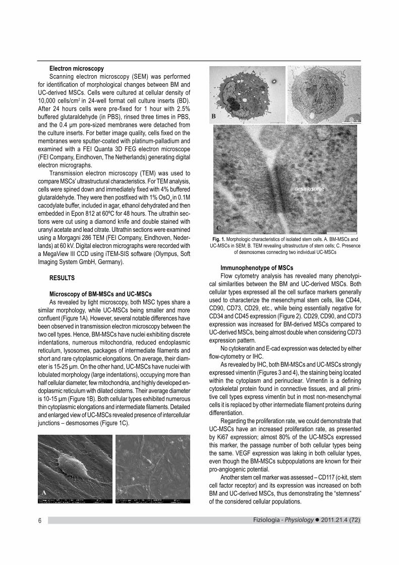
Microscopy of BM-MSCs and UC-MSCsAs revealed by light microscopy, both MSC types share a
similar morphology, while UC-MSCs being smaller and more confluent (Figure 1A). However, several notable differences have been observed in transmission electron microscopy between the two cell types. Hence, BM-MSCs have nuclei exhibiting discrete indentations, numerous mitochondria, reduced endoplasmic reticulum, lysosomes, packages of intermediate filaments and short and rare cytoplasmic elongations. On average, their diam-eter is 15-25 µm. On the other hand, UC-MSCs have nuclei with lobulated morphology (large indentations), occupying more than half cellular diameter, few mitochondria, and highly developed en-doplasmic reticulum with dilated cisterns. Their average diameter is 10-15 µm (Figure 1B). Both cellular types exhibited numerous thin cytoplasmic elongations and intermediate filaments. Detailed and enlarged view of UC-MSCs revealed presence of intercellular junctions – desmosomes (Figure 1C).
Fig. 1. Morphologic characteristics of isolated stem cells. A. BM-MSCs and UC-MSCs in SEM; B. TEM revealing ultrastructure of stem cells; C. Presence
of desmosomes connecting two individual UC-MSCs
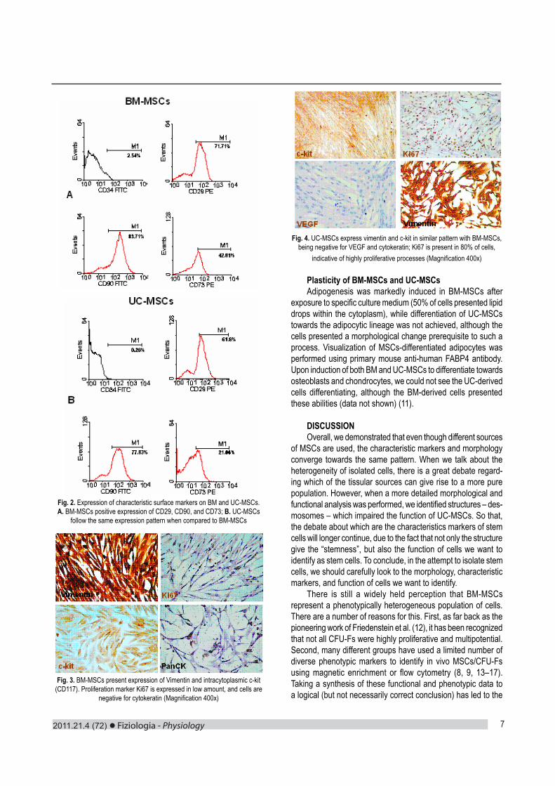
Immunophenotype of MSCsFlow cytometry analysis has revealed many phenotypi-
cal similarities between the BM and UC-derived MSCs. Both cellular types expressed all the cell surface markers generally used to characterize the mesenchymal stem cells, like CD44, CD90, CD73, CD29, etc., while being essentially negative for CD34 and CD45 expression (Figure 2). CD29, CD90, and CD73 expression was increased for BM-derived MSCs compared to UC-derived MSCs, being almost double when considering CD73 expression pattern.
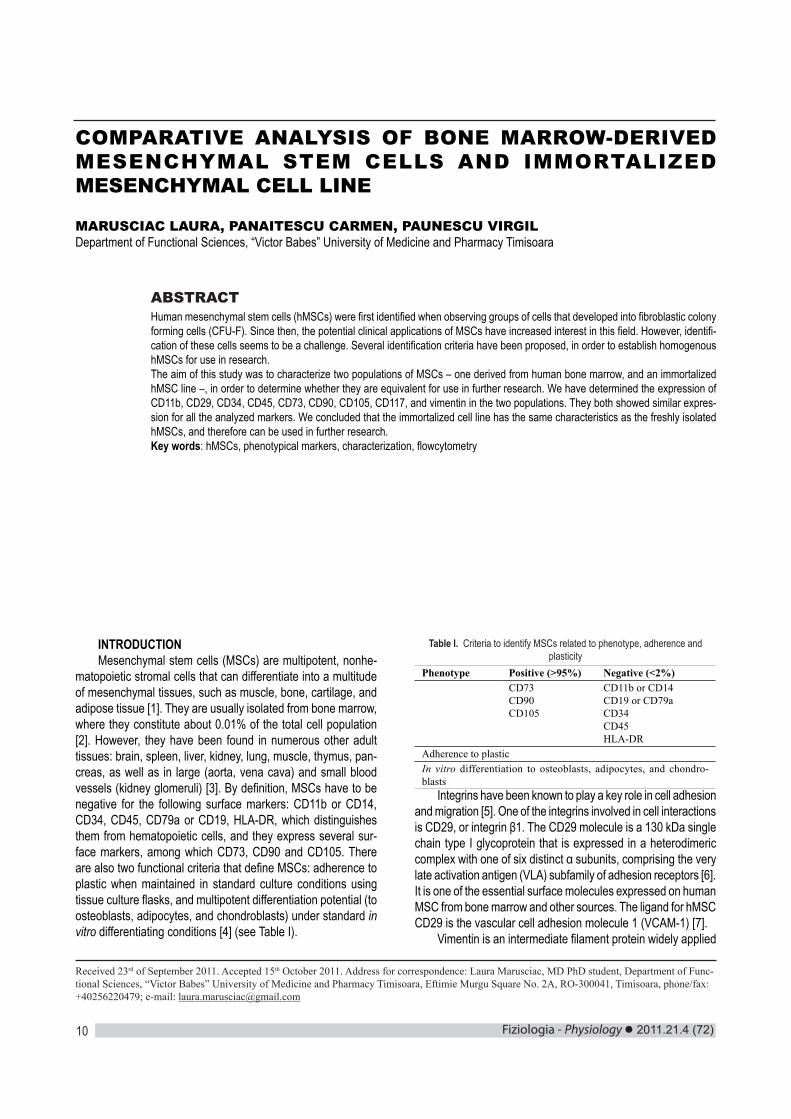
No cytokeratin and E-cad expression was detected by either flow-cytometry or IHC.
As revealed by IHC, both BM-MSCs and UC-MSCs strongly expressed vimentin (Figures 3 and 4), the staining being located within the cytoplasm and perinuclear. Vimentin is a defining cytoskeletal protein found in connective tissues, and all primi-tive cell types express vimentin but in most non-mesenchymal cells it is replaced by other intermediate filament proteins during differentiation.
Regarding the proliferation rate, we could demonstrate that UC-MSCs have an increased proliferation rate, as presented by Ki67 expression; almost 80% of the UC-MSCs expressed this marker, the passage number of both cellular types being the same. VEGF expression was laking in both cellular types, even though the BM-MSCs subpopulations are known for their pro-angiogenic potential.
Another stem cell marker was assessed – CD117 (c-kit, stem cell factor receptor) and its expression was increased on both BM and UC-derived MSCs, thus demonstrating the “stemness” of the considered cellular populations.
A
B
C
2011.21.4 (72) Fiziologia - Physiology 7
Fig. 2. Expression of characteristic surface markers on BM and UC-MSCs. A. BM-MSCs positive expression of CD29, CD90, and CD73; B. UC-MSCs
follow the same expression pattern when compared to BM-MSCs
Fig. 3. BM-MSCs present expression of Vimentin and intracytoplasmic c-kit (CD117). Proliferation marker Ki67 is expressed in low amount, and cells are
negative for cytokeratin (Magnification 400x)
Fig. 4. UC-MSCs express vimentin and c-kit in similar pattern with BM-MSCs, being negative for VEGF and cytokeratin; Ki67 is present in 80% of cells,
indicative of highly proliferative processes (Magnification 400x)
Plasticity of BM-MSCs and UC-MSCs Adipogenesis was markedly induced in BM-MSCs after
exposure to specific culture medium (50% of cells presented lipid drops within the cytoplasm), while differentiation of UC-MSCs towards the adipocytic lineage was not achieved, although the cells presented a morphological change prerequisite to such a process. Visualization of MSCs-differentiated adipocytes was performed using primary mouse anti-human FABP4 antibody. Upon induction of both BM and UC-MSCs to differentiate towards osteoblasts and chondrocytes, we could not see the UC-derived cells differentiating, although the BM-derived cells presented these abilities (data not shown) (11).
DISCUSSIONOverall, we demonstrated that even though different sources
of MSCs are used, the characteristic markers and morphology converge towards the same pattern. When we talk about the heterogeneity of isolated cells, there is a great debate regard-ing which of the tissular sources can give rise to a more pure population. However, when a more detailed morphological and functional analysis was performed, we identified structures – des-mosomes – which impaired the function of UC-MSCs. So that, the debate about which are the characteristics markers of stem cells will longer continue, due to the fact that not only the structure give the “stemness”, but also the function of cells we want to identify as stem cells. To conclude, in the attempt to isolate stem cells, we should carefully look to the morphology, characteristic markers, and function of cells we want to identify.
There is still a widely held perception that BM-MSCs represent a phenotypically heterogeneous population of cells. There are a number of reasons for this. First, as far back as the pioneering work of Friedenstein et al. (12), it has been recognized that not all CFU-Fs were highly proliferative and multipotential. Second, many different groups have used a limited number of diverse phenotypic markers to identify in vivo MSCs/CFU-Fs using magnetic enrichment or flow cytometry (8, 9, 13–17). Taking a synthesis of these functional and phenotypic data to a logical (but not necessarily correct conclusion) has led to the

Fiziologia - Physiology 2011.21.4 (72)8
impression that MSCs were both functionally and phenotypically heterogeneous.
To clarify this, we used multiparameter flow cytometry and cross-tested different MSC markers and purification methods, including plastic adherence for their selectivity and specificity for in vivo BM-MSCs (14, 18). We found that all these methods identified a phenotypically identical rare cell population that was distinct from BM hematopoietic cells by their very low CD45 expression and a larger cell size.
Nevertheless contrary to some currently propagated views (19, 20), a phenotypically distinct, in vivo BM- MSC population has now been identified. Importantly, a striking consensus re-garding the morphology of fresh MSCs is emerging, regardless of the method of isolation used. They appear as large cells that have prominent nucleoli and bleb-like projections, which extend further as MSCs adhere-this is different from spindle-shaped morphology of typical cultured MSCs (14, 15, 21). Based on functional assays, the presence of MSCs in extra skeletal loca-tions including synovium, fat and even placental tissue and umbilical cord has been firmly established. Identifying the MSC population from the much larger stromal fraction will be a more formidable challenge compared with MSC identification in the marrow. There has been a common opinion that CD73, CD105, CD90 and CD44 are highly specific for MSCs, and hence can discriminate multipotential cells from the more mundane tissue resident fibroblasts. More recently, however, several studies showed that these markers were ubiquitously expressed on stromal cells from many locations as well as on skin fibroblasts (22–24), and at best they only inform an investigator that the phenotyped cells are non-hematopoietic and stromal in origin.
Despite their functional heterogeneity, MSC populations obtained from most tissues commonly express a number of surface receptors including CD29, CD44, CD49a-f, CD51, CD73, CD105, CD106, CD166, and Stro1 and lack expression of defini-tive hematopoietic lineage markers including CD11b, CD14, and CD45. Recent studies have shown cells that express the afore-mentioned surface markers and are capable of differentiating into connective tissue cell types can be enriched from peripheral and umbilical cord blood by selection for CD133 and from bone mar-row by selection for stage-specific embryonic antigen (SSEA)-1, SSEA-4, or the nerve growth factor receptor CD271.
In the BM, where the overwhelming majority of cells are hematopoietic, these markers may indeed be useful, but in con-nective tissues, where most of the cells are fibroblastic, their utility for the isolation of resident MSCs will be limited and a search for new, more specific markers, if they indeed exist, is needed. For the isolation of MSCs from post-partum tissues, such as placenta, an embryonic stem cell marker SSEA-4 was found to be useful and, more recently, it was successfully applied for the isolation of MSCs from adult BM. Another important issue to bear in mind is the stability of putative MSC markers in culture. Despite the loss of certain markers following passaging (7, 14) and the gain of others (18), MSC cultures remain multipotential, indicating that these markers are unlikely to be reflective of the MSC’s true ‘stem cell’ nature or its multipotentiality. More likely, many
markers present on MSCs in vivo may be induced by the BM microenvironment or be reflective of some other MSC function in vivo that is lost upon plastic adherence and exposure to culture media. At this stage, it would appear that the heterogeneity in the MSC proliferative and differentiation capacities, first noted by Friedenstein et al. (1) cannot be explained on the basis of known surface markers alone.
However, it is important to realize that no single isolation method is regarded as a standard in the field. Therefore, the varied approaches used to culture-expand and select for MSCs make it difficult to directly compare experimental results. More-over, some isolation schemes introduce epigenetic and genetic changes in cells that may dramatically affect their plasticity and therapeutic utility. Finally, human MSCs exhibit some variation in their pattern of expressed genes among different donor prepara-tions using the same isolation protocols, and larger variations as sparse cultures become confluent and are expanded by serial passage and approach senescence (23). These subtleties have been overlooked in several publications in which high density and confluent human MSC cultures were assumed to consist of homogeneous cell populations.
ACKNOWLEDGMENTSThis work was supported by CNCSIS-UEFISCSU, project
number PNII-IDEI 1748/2008 and by the Sectorial Operational Programme for Human Resources Development, financed from the European Social Fund, FSE POSDRU/89/1.5/S/60746.
REFERENCES1. Friedenstein AJ, Chailakhjan RK, Lalykina KS. The development of fibroblast colonies in monolayer cultures of guinea-pig bone marrow and spleen cells. Cell Tissue Kinet 1970; 3: 393-403.2. Friedenstein AJ, Latzinik NV, Gorskaya YF, Luria EA, Moskvina IL. Bone-marrow stromal colony formation requires stimulation by hematopoietic-cells. Bone Mineral 1992; 18: 199-213.3. Castro-Malaspina H, Gay RE, Resnick G et al. Characterisation of human bone marrow fibroblast colony-forming cells (CFU-F) and their progeny. Blood 1980; 56: 289-301.4. Civin CI, Trischmann T, Kadan NS et al. Highly purified CD34-positive cells reconstitute hematopoiesis. J Clin Oncol 1996; 14: 2224-33.5. Pittenger MF, Mackay AM, Beck SC et al. Multilineage potential of adult human mesenchymal stem cells. Science 1999; 284: 143-7.6. Bianco P, Robey PG. Marrow stromal stem cells. J Clin Invest 2000; 105: 1663-8.7. Simmons PJ, Torokstorb B. Identification of stromal cell precursors in human bone-marrow by a novel monoclonal-antibody, Stro-1. Blood 1991; 78: 55-62.8. Majumdar MK, Thiede MA, Haynesworth SE, Bruder SP, Gerson SL. Human marrow-derived mesenchymal stem cells (MSCs) express hematopoietic cytokines and support long-term hematopoiesis when differentiated toward stromal and osteogenic lineages. J Hematother Stem Cell Res 2000; 9: 841-8.9. Boiret N, Rapatel C, Veyrat-Masson R et al. Characterization of nonexpanded mesenchymal progenitor cells from normal adult human bone marrow. Exp Hematol 2005; 33: 219-25.10. Prockop DJ. Marrow stromal cells as steam cells for nonhematopoi-Prockop DJ. Marrow stromal cells as steam cells for nonhematopoi-etic tissues. Science 1997; 276: 71-4.11. Paunescu V, Bojin F, Tatu CA, Gavriliuc OI, et al. Tumour-associated

2011.21.4 (72) Fiziologia - Physiology 9
fibroblasts and mesenchymal stem cells: more similarities than differ-ences. J Cell Mol Med., 2011; 15(3):635-646.12. Friedenstein AJ, Chailakhyan RK, Gerasimov UV. Bone-marrow osteogenic stem cells - In vitro cultivation and transplantation in diffusion chambers. Cell Tissue Kinet 1987; 20: 263-72.13. Quirici N, Soligo D, Bossolasco P, Servida F, Lumini C, Deliliers GL. Isolation of bone marrow mesenchymal stem cells by anti-nerve growth factor receptor antibodies. Exp Hematol 2002; 30: 783-91.14. Jones EA, Kinsey SE, English A et al. Isolation and characterization of bone marrow multipotential mesenchymal progenitor cells. Arthritis Rheum 2002; 46: 3349-60.15. Gronthos S, Zannettino ACW, Hay SJ et al. Molecular and cellular characterisation of highly purified stromal stem cells derived from human bone marrow. J Cell Sci 2003; 116: 1827-35.16. Shi S, Gronthos S. Perivascular niche of postnatal mesenchymal stem cells in human bone marrow and dental pulp. J Bone Mineral Res 2003; 18: 696-704.17. Deschaseaux F, Gindraux F, Saadi R, Obert L, Chalmers D, Herve P. Direct selection of human bone marrow mesenchymal stem cells using an anti-CD49a antibody reveals their CD45(med,low) phenotype. Br J Haematol 2003; 122: 506-17.18. Jones EA, English A, Kinsey SE et al. Optimization of a flow
cytometry-based protocol for detection and phenotypic characterization of multipotent mesenchymal stromal cells from human bone marrow. Cytometry Part B: Clin Cytometry 2006; 70B: 391-9.19. Tuan R. Stemming cartilage degeneration: Adult mesenchymal stem cells as a cell source for articular cartilage tissue engineering. Arthritis Rheum 2006; 54: 3075-8.20. Kolf C, Cho E, Tuan R. Mesenchymal stromal cells. Biology of adult mesenchymal stem cells: regulation of niche, self–renewal and differentiation. Arthritis Res 2007; 9: 204-304.21. Buhring H-J, Battula VL, Treml S, Schewe B, Kanz L, Vogel W. Novel markers for the prospective isolation of human MSC. Ann NY Acad Sci 2007; 1106: 262–71.22. Wagner W, Wein F, Seckinger A et al. Comparative characteristics of mesenchymal stem cells from human bone marrow, adipose tissue, and umbilical cord blood. Exp Hematol 2005; 33: 1402-16.23. Ishii M, Koike C, Igarashi A et al. Molecular markers distinguish bone marrow mesenchymal stem cells from fibroblasts. Biochem Biophys Res Commun 2005; 332: 297-303.24. Jones EA, English A, Henshaw K et al. Enumeration and phenotypic characterization of synovial fluid multipotential mesenchymal progenitor cells in inflammatory and degenerative arthritis. Arthritis Rheum 2004; 50: 817-27.
CONTROVERSE LEGATE DE CULTURILE CELULARE OBTINUTE DIN DIFERITE SURSE TISULARE
REZUMATCelulele stem mezenchimale (MSC) sunt celule stromale non-hematopoietice, care sunt capabile sa se diferentieze si sa contribuie la regenerarea tesuturilor mezenchimale, cum ar fi tesutul osos, cartilaj, muschi, ligamente, tendoane si tesut adipos. MSC sunt identificate prin expresia moleculelor de suprafata, cum ar fi CD105 (SH2) si CD73 (SH3/4), fiind negative pentru markerii celulelor hematopoietice CD34, CD45 si CD14. In prezentul studiu am izolat doua populatii celulare, din maduva osoasa hematogena (BM) si cordonul ombilical (UC) si am investigat comparativ prezenta markerilor fenotipici, potentialul de diferentiere spre trei linii celulare (trilineage) si caracteristicile morfologice. Cu toate ca atat BM-MSC, cat si UC-MSC au prezentat un profil fenotipic similar si caracteristici morfologice comune (in microscopie optica), nu am reusit diferentierea UC-MSC spre adipocite, condrocite si osteoblaste, ceea ce sugereaza functionalitatea scazuta sau modificata a acestora. Analiza detaliata a aspectului morfologic (microscopie electronica) a demonstrat prezenta jonctiunilor intercelulare de tipul desmozomilor, care ar putea explica partial comportamentul in vitro al acestor celule. Trebuie investigat daca celulele stromale izolate din cordonul ombilical au dobandit acest fenotip in conditii de cultivare in vitro, sau daca metoda utilizata pentru izolarea acestora nu a fost cea potrivita. Totusi, caracteristicile “stemness” ar trebui sa fie bazate mai mult pe functia celulara si mai putin pe caracteristicile fenotipice celulare, deoarece aceste celule sunt candidate potentiale pentru terapiile regenerative si aplicatii clinice. Cuvinte cheie: BM-MSCs, UC-MSCs, diferentiere, functie
Fiziologia - Physiology 2011.21.4 (72)10
INTRODUCTIONMesenchymal stem cells (MSCs) are multipotent, nonhe-
matopoietic stromal cells that can differentiate into a multitude of mesenchymal tissues, such as muscle, bone, cartilage, and adipose tissue [1]. They are usually isolated from bone marrow, where they constitute about 0.01% of the total cell population [2]. However, they have been found in numerous other adult tissues: brain, spleen, liver, kidney, lung, muscle, thymus, pan-creas, as well as in large (aorta, vena cava) and small blood vessels (kidney glomeruli) [3]. By definition, MSCs have to be negative for the following surface markers: CD11b or CD14, CD34, CD45, CD79a or CD19, HLA-DR, which distinguishes them from hematopoietic cells, and they express several sur-face markers, among which CD73, CD90 and CD105. There are also two functional criteria that define MSCs: adherence to plastic when maintained in standard culture conditions using tissue culture flasks, and multipotent differentiation potential (to osteoblasts, adipocytes, and chondroblasts) under standard in vitro differentiating conditions [4] (see Table I).
Table I. Criteria to identify MSCs related to phenotype, adherence and plasticity
Phenotype Positive (>95%) Negative (<2%)CD73CD90CD105
CD11b or CD14CD19 or CD79aCD34CD45HLA-DR
Adherence to plasticIn vitro differentiation to osteoblasts, adipocytes, and chondro-blasts
Integrins have been known to play a key role in cell adhesion and migration [5]. One of the integrins involved in cell interactions is CD29, or integrin β1. The CD29 molecule is a 130 kDa single chain type I glycoprotein that is expressed in a heterodimeric complex with one of six distinct α subunits, comprising the very late activation antigen (VLA) subfamily of adhesion receptors [6]. It is one of the essential surface molecules expressed on human MSC from bone marrow and other sources. The ligand for hMSC CD29 is the vascular cell adhesion molecule 1 (VCAM-1) [7].
Vimentin is an intermediate filament protein widely applied
COMPARATIVE ANALYSIS OF BONE MARROW-DERIVED MESENCHYMAL STEM CELLS AND IMMORTALIZED MESENCHYMAL CELL LINE
MARUSCIAC LAURA, PANAITESCU CARMEN, PAUNESCU VIRGILDepartment of Functional Sciences, “Victor Babes” University of Medicine and Pharmacy Timisoara
ABSTRACTHuman mesenchymal stem cells (hMSCs) were first identified when observing groups of cells that developed into fibroblastic colony forming cells (CFU-F). Since then, the potential clinical applications of MSCs have increased interest in this field. However, identifi-cation of these cells seems to be a challenge. Several identification criteria have been proposed, in order to establish homogenous hMSCs for use in research. The aim of this study was to characterize two populations of MSCs – one derived from human bone marrow, and an immortalized hMSC line –, in order to determine whether they are equivalent for use in further research. We have determined the expression of CD11b, CD29, CD34, CD45, CD73, CD90, CD105, CD117, and vimentin in the two populations. They both showed similar expres-sion for all the analyzed markers. We concluded that the immortalized cell line has the same characteristics as the freshly isolated hMSCs, and therefore can be used in further research.Key words: hMSCs, phenotypical markers, characterization, flowcytometry
Received 23rd of September 2011. Accepted 15th October 2011. Address for correspondence: Laura Marusciac, MD PhD student, Department of Func-tional Sciences, “Victor Babes” University of Medicine and Pharmacy Timisoara, Eftimie Murgu Square No. 2A, RO-300041, Timisoara, phone/fax: +40256220479; e-mail: [email protected]

2011.21.4 (72) Fiziologia - Physiology 11
as a mesenchymal indicator [8]. It is functionally involved in maintaining the structure of mesenchymal cells [9]. In addition to serving as a marker in the epithelial to mesenchymal transi-tion, it plays a versatile role in cancer cell motility [10]. In normal tissue injuries, vimentin-deficient mice suffer from delayed wound healing due to the failure of mesenchymal contraction at the wound site [11] and impairment of fibroblast migration [12]. Vimentin seems to be related to the activation of mesenchymal cells, but little is known about the relationship between vimentin expression and normal cell activation [13].
CD117, also known as the mast/stem cell growth factor recep-tor (SCFR), proto-oncogene c-Kit or tyrosine-protein kinase Kit, is a protein that in humans is encoded by the KIT gene. It functions as a cytokine receptor, and signalling through CD117 has been shown to play a role in cell survival, proliferation, and differentiation [14]. CD117 has been used to identify and characterize different types as stem and progenitor cells, including hematopoietic stem cells [15] and mesenchymal stem cells [16, 17].
The aim of this study was to characterize human mesen-chymal stem cells in regard to surface markers, and to provide a comparison between the characteristics of isolated hMSCs and immortalized hMSCs.
MATERIAL AND METHODS
1. Isolation and culture of human MSCs
Human MSCs were obtained from the iliac crest bone marrow of healthy male donors, with ages between 18 and 40 years. The donors had been previously evaluated for the presence of hepatitis B surface antigens (HbSAg), hepatitis C antibodies (HCV Ab), human immunodeficiency virus antibodies (HIV Ab), and cytomegalovirus antibodies. All samples of bone marrow were collected after informed consent was obtained in accordance with the guidelines on the use of human subjects, and approval by the ethics committee. 10-20 ml of bone marrow was collected from each donor in heparin-coated tubes.
The bone marrow was filtered through 100 μm sieves (BD Falcon, San Jose, CA, USA), diluted with Phosphate Buffer Solu-tion (PBS, Invitrogen, Carlsbad, CA, USA) in a 1:1 ratio, and then collected into sterile 50 ml Falcon tubes (BD Falcon). Then Biocoll density gradient (Biochrome AG, Germany), with a density of 1,077 g/ml was added carefully at the bottom of the tube, underneath the bone marrow, in a 1:2 ratio. The tubes were then centrifuged for 30 minutes, at 1800 rpms, at room temperature.
The mononuclear cell layer (“buffy coat”) was then carefully collected in new sterile 50 ml Falcon tubes, and diluted with Dulbecco’s Modified Eagle Medium (DMEM, Invitrogen) in a 1:1 ratio. The tubes were then centrifuged at 2000 rpms, at room temperature. After centrifugation, the supernatant was discarded and the cells were resuspended in 1 ml DMEM and counted. The mononuclear cells were then cultured in T75 culture flasks (BD Falcon), at a density of 1x10⁵ cells/cm2, at 37 °C, in a humidified atmosphere that contained 5% CO2. The culture medium that was used contained DMEM supplemented with 20% fetal calf serum
(FCS, Invitrogen), 1% penicillin/streptomycin mixture (Pen/Strep, 10,000 IU/ml; PromoCell, Heidelberg, Germany). The culture medium was changed to remove the remaining nonadherent cells 24 hours after the initial plating. Thereafter, the culture medium was replaced twice per week.
The cells were processed no longer than 4 hours after bone marrow harvesting. In parallel, human MSC line was obtained from Vitro BioPharma (Native Human MSC, CO, USA).
2. Immunophenotypical characterization of hMSCsFor flowcytometry, human MSCs were labeled with conju-
gated antibodies against several human proteins to analyze the cell surface expression of typical MSC antigens, as well as the absence of antigen expression for other CD molecules, considered nega-tive for mesenchymal stem cells. The antibodies were conjugated with Allophycocyanin (APC), Fluorescein isothiocyanate (FITC), or Phycoerythrin (PE) as follows: CD29-PE, CD73-PE, CD90-APC, CD105-FITC, CD117-APC, CD11b-APC, CD34-PE, CD45-APC. All antibodies were purchased from BD Pharmingen.
Human MSCs from passages 2-5 were used when they reached a confluence of 70-80%. They were trypsinized, counted and then resuspended in 1 ml staining buffer, containing 0.5% bovine albumin serum (BSA), 2 mM EDTA, pH=7.2, and 0.05% azide. For each monoclonal antibody, 1x10⁵ cells were put into Eppendorf tubes, staining buffer was added up to 500 µl, and then 2 µl of monoclonal antibody were added to each tube. The tubes were incubated in the dark, at 4 °C, for 20 minutes. The cells were then washed twice with 1000 µl of staining buffer, and centrifuged at 300g, for 5 minutes, at 4 °C. The cells were then resuspended in 200 µl of staining buffer and, for each antibody, 1x10⁴ labeled cells were analyzed using a flow cytometer. Data acquisition was performed using a FACSCanto II flow cytometer (Becton Dickinson, Franklin Lakes, NJ, USA), and the data was analyzed by FlowJo software, version 7.6 (Flowjo, Ashland, Oregon, USA).
For immunofluorescence assays, human MSCs were labeled with antibodies against CD117 (c-kit) and vimentin. For CD117, the primary antibody consisted of a polyclonal rabbit anti-human antibody (clone A4502, Dako, Glostrup, Denmark) and for vimentin of a monoclonal mouse anti-swine antibody (clone V9, Dako). The secondary antibody consisted of an anti-rabbit antibody for CD117, and an anti-mouse antibody for vimentin. Both secondary antibodies were conjugated with Alexa Fluor 488. The antibodies were diluted with PBS in a ratio of 1:300 before use.
Human MSCs from passage 2 were cultured in cover slip slides until they reached a confluence of 80-90%. After discarding the growth medium, the cells were washed with PBS, and then fixed with 4% formaldehyde, for 8 minutes, at 4 °C. The cells were then washed with PBS for 5 minutes, and the primary antibody was added. The slides were then incubated for 24 hours, at 4 °C, in the dark. The cells were then washed twice with PBS, the fluorescent secondary antibody was added, and the slides were incubated for 1 hour, at room temperature, in the dark. The nuclei were then counterstained for 1 minute, using 4,6’-diamidino-2-phenylindole (DAPI, 1mg/ml; Sigma-Aldrich Company, Ayrshire, UK), diluted with PBS, in a ration of 1:5000. The cells were then

Fiziologia - Physiology 2011.21.4 (72)12
washed twice with PBS and left to dry. The slides were mounted using cover slip (ESCO microscope cover glass, Erie Scientific Company, Portsmouth, N.H., USA) and fluorescence mounting medium was then added (ProLong® Gold anti-fade reagent, Invitrogen Molecular Probes™). The slides were analyzed using a fluorescence microscope (Nikon Eclipse E800).
For immunohistochemistry assays, human MSCs were labeled with antibodies against vimentin (Dako) and CD29 (R&D Systems, Minneapolis, MN, USA). Human MSCs from passage 2 were cul-tured in Nunc plates (Thermo Fisher Scientific Inc., Hennigsdorf, Germany) until they reached a confluence of 80-90%. They were then trypsinized and 500 μl cellular suspension were cytospun and cytospin slides were obtained by 6 minutes centrifugation at 600 rpm in Shandon Cytospin 4 (Thermo Fisher Scientific). Slides were air-dried for 10 minutes and then used for immunocytochemistry procedure. The cells were then fixed with 4% formaldehyde, for 8 minutes, at 4 °C. The cells were then washed with PBS for 5 minutes, and the primary antibody was added. The cells were then incubated on an orbital shaker, at 200 rpm, for 30 minutes, at room temperature. The cells were then washed again with PBS, and the secondary antibody was added.
For staining for vimentin, the Dako EnVision+ System-HRP kit for use with mouse antibodies (Dako) was used. The cells were incubated with 1 drop of Labelled Polymer-HRP Anti-Mouse secondary antibody, on the orbital shaker, at 200 rpm, for 30 minutes, at room temperature. The substrate was prepared, using 500 µl of DAB+ substrate buffer and 1 drop of DAB+ Chromogen. The slides were then washed again with PBS, and 50 µl of sub-strate were added, followed by incubation on the orbital shaker, at 200 rpm, for 10-20 minutes, at room temperature, depending on the intensity of the staining.
For staining for CD29, the Cell & Tissue Staining Kit, HRP-AEC system (R&D Systems) was used. The cells were incubated with 1 drop of Biotinylated Secondary Antibody, on the orbital shaker, at 200 rpm, for 30 minutes, at room temperature. The slides were washed with PBS, and then the cells were incubated with 1 drop of HSS-HRP on the orbital shaker, at 200 rpm, for 30 minutes, at room temperature. The substrated was prepared, using 500 µl of Chromogen buffer and 1 drop of AEC Chromogen. After washing with PBS, the cells were incubated with 50 µl of substrate, the orbital shaker, at 200 rpm, for 10-20 minutes, at room temperature, depending on the intensity of the staining.
For both vimentin and CD29 staining, the slides were then washed with running tap water, and stained for 5 minutes with 50 µl of hematoxylin solution (Hematoxylin, Mayer’s Lillie’s Modification, Dako, Glostrup, Denmark) diluted with tap water in a ration of 1:5. After another washing with running tap water, one drop of mounting media was added to the slides, and the cover slips were put into position.
RESULTS AND DISCUSSION
Isolation and cultivationThe success rate for isolating bone marrow MSCs was 100%
(8 out of 8 donors). The average donor age was 26.375±7.76 years old. The average quantity of bone marrow collected from
each donor was 16 ± 3 ml. The average number of mononuclear cells isolated from the donors was 85x106±22.8 x10⁶.
Optical microscopyBoth types of human MSCs – isolated from the bone
marrow of donors, and derived from cell lines – had a typical morphology, exhibiting a fibroblast-like, and spindle shape, characterized by a small cell body, with few long, thin cell processes. The cell body contained a large, round nucleus. The cells adhered to the flask. When reaching confluence, they exhibited a “whirl” arrangement in culture. Immortalized MSCs from cell lines had a more elongated cell body, with longer cell processes (Figure 1).
Fig. 1. A. Isolated MSCs, passage 2, 40% confluence; B. MSCs cell line, pas-sage 7, 40% confluence. Magnification 100X
Immunophenotypical characterization of isolated MSCs
Bone marrow-derived MSCs presented a configuration of positive characteristic markers, including CD73, CD90, and CD105. Negative CD molecules were CD11b, CD34, and CD45 (Figure 2). CD90 positive population is divided in two subpopu-lation, suggesting that within the heterogenous MSCs some of the cells are more mature than others, CD90 being a marker of cellular immaturity.

2011.21.4 (72) Fiziologia - Physiology 13
Fig. 2. Flowcytometric characterization of bone marrow-isolated MSCs. MSCs
cellular line expressed similar proportion of characteristic markers.
MSCs and cell line-obtained MSCs immune stainingBoth bone-marrow derived human MSCs and the MSC line
expressed of vimentin (Figure 3). The expression was not in abundance, due to the fact that the cells were not permeabilized beforehand.
CD117 was positive for both bone marrow-derived and MSCs cell line, but in a small amount of cells (Figure 4). For flowcytometric procedure, cells were not permeabilized, so that detection level could not exceed the cell surface. Given the bipolar structure of this marker (stem cell factor receptor, c-kit), we expected to find an increased expression of CD117 within the cellular cytoplasm, using other phenotypical analyses.
Immunohistochemistry showed an abundance of CD29 marker on the cell surface, in both isolated hMSCs and hMSC line (Figure 5, A and B). This result is confirmed by the flowcytometric results, where the majority of the cell population is positive for CD29 (Figure 5, C and D).
Integrin receptors have major importance in cell signaling, one of their functions being the modulation of the activity and expression of intracellular proteins and signaling factor, includ-ing kinases and scaffold proteins [18]. The assembly of protein complexes determines the activation of downstream signaling pathways, some of which overlap with pathways mediated by growth factors; therefore, integrin signaling is required for adhe-sion-dependent survival, growth, and migration of cells [19].
There are 18 different α and 8 different β subunits, which can combine in various ways. The largest subgroup is formed by the β1 subunit, whose members bind to different extracellular matrix molecules, such as collagen, laminin, fibronectin, but also interact with cellular receptors like VCAM-1 [20].
Integrin β1 has an important role in cell invasion, especially in a 3D matrigel. Inhibition of integrin β1can restore the ability of some tumor cells to form acinar structures, which represents an indicator of reduced tumorigenicity, as well as a return to more
normal cell phenotype [18]. It has also been shown to have a critical role in pancreatic cancer progression and metastasis, by inhibiting pancreatic cancer cell adhesion, proliferation and migration [21].
These observations highlight the importance of investigating the role of adhesion molecules in the appropriate context.
Fig. 3. Comparative expression of cytoskeleton protein Vimentin in isolated MSCs (A) and hMSC line (B). Note that majority of cells are positive for this marker. The cell nuclei are stained with DAPI and appear blue-white under
fluorescent light. Magnification 400x.
Fig. 4. CD117 expression on both types of MSCs - isolated hMSCs (A) and hMSC line (B) - shows abundance of this marker within the cellular cytoplasm . Magnification 400x. Flowcytometry shows that only a small percentage of the cells are CD117+ (4.56%, respectively 12.73%) (C
and D).
Fig. 3. Comparative expression of cytoskeleton protein Vimentin in isolated MSCs (A) and hMSC line (B). Note that majority of cells are positive for this marker. The cell nuclei are stained with DAPI and appear blue-white under fluorescent light. Magnification 400x.
Fig. 4. CD117 expression on both types of MSCs - isolated hMSCs (A) and hMSC line (B) - shows abundance of this marker within the cellular cytoplasm . Magnification 400x. Flowcytometry shows that only a small percentage of the cells are CD117+ (4.56%, respectively 12.73%) (C and D).
A B
A B
A B
C D

Fiziologia - Physiology 2011.21.4 (72)14
Fig. 5. CD29 expression on MSCs shows abundance of this marker in both isolated hMSC (A) and MSC line (B). Please note the whirl-like arrangement of the cells. Magnification 100x. This result is confirmed by the flowcytometric
results (C and D).
CONCLUSIONOur results confirm previous studies, which show expression
of vimentin, CD117, and CD29 on human mesenchymal stem cells. The immortalized hMSC line stains true for all these markers, and seems to be a good candidate for use in further experiments.
ACKNOWLEDGMENTSThis work was supported by CNCSIS-UEFISCSU, project
number PNII-IDEI 1748/2008 and by the Sectorial Operational Programme for Human Resources Development, financed from the European Social Fund, FSE POSDRU/89/1.5/S/60746.
REFERENCES1. Pittenger MF, Mackay AM, Beck SC, et al. Multilineage Potential of Adult Human Mesenchymal. Science. 1999; 284(5411): 143-7.2. Chamberlain G, Fox J, Ashton B, Middleton J. Concise review: mesenchymal stem cells: their phenotype, differentiation capacity, immunological features, and potential for homing. Stem Cells. 2007; 25(11): 2739-49.3. da Silva Meirelles L, Chagastelles PC, Nardi NB. Mesenchymal stem cells reside in virtually all post-natal organs and tissues. J Cell Sci. 2006; 119(Pt 11): 2204-13.4. Dominici M, Le Blanc K, Mueller I, et al. Minimal criteria for defining multipotent mesenchymal stromal cells. The International Society for Cellular Therapy position statement. Cytotherapy. 2006; 8(4): 315-7.5. Imhof BA, Aurrand-Lions M. Adhesion mechanisms regulating the migration of monocytes. Nat Rev Immunol. 2004; 4(6): 432-44.6. Shiokawa S, Yoshimura Y, Nagamatsu S, et al. Expression of beta
1 integrins in human endometrial stromal and decidual cells. J Clin Endocrinol Metab. 1996; ;81(4): 1533-40.7. Margadant C, Monsuur HN, Norman JC, et al. Mechanisms of integrin activation and trafficking. Curr Opin Cell Biol. 2011; 23(5): 607-14. 8. P M, S H, R M, et al. Adult mesenchymal stem cells and cell surface characterization - a systematic review of the literature. Open Orthop J. 2011; 5(Suppl 2): 253-60.9. Mendez MG, Kojima S, Goldman RD. Vimentin induces changes in cell shape, motility, and adhesion during the epithelial to mesenchymal transition. FASEB J. 2010; 24(6): 1838-51.10. Li B, Zheng YW, Sano Y, et al. Evidence for mesenchymal-epithelial transition associated with mouse hepatic stem cell differentiation. PLoS One. 2011; 11;6(2): e17092.11. Eckes B, Colucci-Guyon E, Smola H, et al. Impaired wound healing in embryonic and adult mice lacking vimentin. J Cell Sci. 2000; 113 (Pt 13): 2455-62.12. Eckes B, Dogic D, Colucci-Guyon E, et al. Impaired mechanical stability, migration and contractile capacity in vimentin-deficient fibro-blasts. J Cell Sci. 1998; 111 (Pt 13): 1897-907.13. Shirahata A, Sakata M, Sakuraba K, et al. Vimentin methylation as a marker for advanced colorectal carcinoma. Anticancer Res. 2009; 29(1): 279-81.14. Ashman LK. The biology of stem cell factor and its receptor C-kit. Int J Biochem Cell Biol. 1999; 31(10): 1037-51.15. Lin KK, Goodell MA. Detection of hematopoietic stem cells by flow cytometry. Methods Cell Biol. 2011;103: 21-30.16. Deng W, St Hilaire RC, Chattergoon NN, et al. Inhibition of vascular smooth muscle cell proliferation in vitro by genetically engineered mar-row stromal cells secreting calcitonin gene-related peptide. Life Sci. 2006; 13; 78(16): 1830-8.17. Moorefield EC, McKee EE, Solchaga L, et al. Cloned, CD117 se-lected human amniotic fluid stem cells are capable of modulating the immune response. PLoS One. 2011; 6(10): e26535.18. 18. Schooley AM, Andrews NM, Zhao H, et al. β1 integrin is required for anchorage-independent growth and invasion of tumor cells in a con-text dependent manner. Cancer Lett. 2012 Mar 28; 316(2): 157-67.19. 19. Scales TM, Parsons M. Spatial and temporal regulation of integrin signalling during cell migration. Curr Opin Cell Biol. 2011 Oct; 23(5): 562-8.20. 20. Brakebusch C, Fässler R. beta 1 integrin function in vivo: adhesion, migration and more. Cancer Metastasis Rev. 2005 Sep; 24(3): 403-11.21. 21. Grzesiak JJ, Tran Cao HS, Burton DW, et al. Knockdown of the β(1) integrin subunit reduces primary tumor growth and inhibits pancre-atic cancer metastasis. Int J Cancer. 2011 Dec 15; 129(12): 2905-15.
Fig. 5. CD29 expression on MSCs shows abundance of this marker in both isolated hMSC (A) and MSC line (B). Please note the whirl-like arrangement of the cells. Magnification 100x. This result is confirmed by the flowcytometric results (C and D). CONCLUSION Our results confirm previous studies, which show expression of vimentin, CD117, and CD29 on human mesenchymal stem cells. The immortalized hMSC line stains true for all these markers, and seems to be a good candidate for use in further experiments. REFERENCES 1. Pittenger MF, Mackay AM, Beck SC, et al. Multilineage Potential of Adult Human Mesenchymal. Science.
1999; 284(5411): 143-7. 2. Chamberlain G, Fox J, Ashton B, Middleton J. Concise review: mesenchymal stem cells: their phenotype,
differentiation capacity, immunological features, and potential for homing. Stem Cells. 2007; 25(11): 2739-49. 3. da Silva Meirelles L, Chagastelles PC, Nardi NB. Mesenchymal stem cells reside in virtually all post-natal
organs and tissues. J Cell Sci. 2006; 119(Pt 11): 2204-13. 4. Dominici M, Le Blanc K, Mueller I, et al. Minimal criteria for defining multipotent mesenchymal stromal cells.
The International Society for Cellular Therapy position statement. Cytotherapy. 2006; 8(4): 315-7. 5. Imhof BA, Aurrand-Lions M. Adhesion mechanisms regulating the migration of monocytes. Nat Rev Immunol.
2004; 4(6): 432-44. 6. Shiokawa S, Yoshimura Y, Nagamatsu S, et al. Expression of beta 1 integrins in human endometrial stromal
and decidual cells. J Clin Endocrinol Metab. 1996; ;81(4): 1533-40. 7. Margadant C, Monsuur HN, Norman JC, et al. Mechanisms of integrin activation and trafficking. Curr Opin
Cell Biol. 2011; 23(5): 607-14. 8. P M, S H, R M, et al. Adult mesenchymal stem cells and cell surface characterization - a systematic review of
the literature. Open Orthop J. 2011; 5(Suppl 2): 253-60. 9. Mendez MG, Kojima S, Goldman RD. Vimentin induces changes in cell shape, motility, and adhesion during
the epithelial to mesenchymal transition. FASEB J. 2010; 24(6): 1838-51. 10. Li B, Zheng YW, Sano Y, et al. Evidence for mesenchymal-epithelial transition associated with mouse hepatic
stem cell differentiation. PLoS One. 2011; 11;6(2): e17092. 11. Eckes B, Colucci-Guyon E, Smola H, et al. Impaired wound healing in embryonic and adult mice lacking
vimentin. J Cell Sci. 2000; 113 (Pt 13): 2455-62. 12. Eckes B, Dogic D, Colucci-Guyon E, et al. Impaired mechanical stability, migration and contractile capacity in
vimentin-deficient fibroblasts. J Cell Sci. 1998; 111 (Pt 13): 1897-907. 13. Shirahata A, Sakata M, Sakuraba K, et al. Vimentin methylation as a marker for advanced colorectal
carcinoma. Anticancer Res. 2009; 29(1): 279-81. 14. Ashman LK. The biology of stem cell factor and its receptor C-kit. Int J Biochem Cell Biol. 1999; 31(10):
1037-51.
C D
ANALIZA COMPARATIVA A CELULELOR STEM MEZENCHIMALE OBTINUTE DIN MADUVA OSOASA HEMATOGENA SI A LINIEI DE CELULE STEM MEZENCHIMALE IMORTALIZATE
REZUMATCelulele stem mezenchimale umane au fost identificate pentru prima oară în momentul în care s-au observat populaţii celulare care au suferit o transformare în unităţi formatoare de colonii fibroblastice (CFU-F). Din acel moment, potenţialele aplicaţii clinice ale MSC-urilor au menţinut un interes crescut pentru acest domeniu. Cu toate acestea, identificarea MSC-urilor pare să reprezinte o provocare. Au fost propuse mai multe criterii de identificare, pentru a stabili uzul unor populaţii omogene de MSC-uri în studii.Scopul acestui studio a fost de a characteriza două populaţii de MSC-uri – una izolată din măduvă hematopoietică, iar cealaltă fiind constituită dintr-o linie imortalizată de MSC-uri – pentru a determina dacă acestea sunt echivalente în ceea ce priveşte uzul lor în cercetări ulterioare. Am determinat expresia CD11b, CD29, CD34, CD45, CD73, CD90, CD105, CD117, şi vimentină în cele două populaţii. Acestea au prezentat expresie similară pentru markerii analizaţi. Concluzia studiului este că linia celulară imortalizată prezintă aceleaşi caracteristici ca MSC-urile proaspăt izolate, şi astfel pot fi folosite în cercetări ulterioare.Cuvinte cheie: hMSC, markeri fenotipici, caracterizare, flowcitometrie
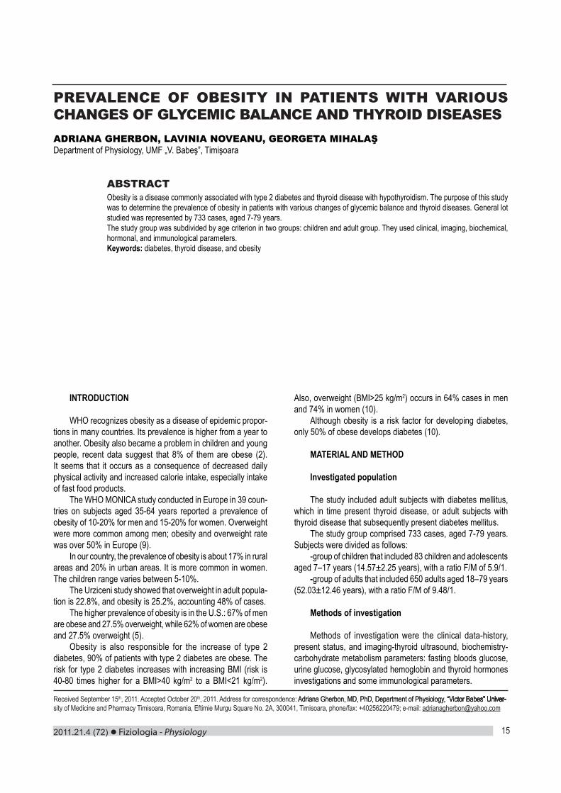
2011.21.4 (72) Fiziologia - Physiology 15
INTRODUCTION
WHO recognizes obesity as a disease of epidemic propor-tions in many countries. Its prevalence is higher from a year to another. Obesity also became a problem in children and young people, recent data suggest that 8% of them are obese (2). It seems that it occurs as a consequence of decreased daily physical activity and increased calorie intake, especially intake of fast food products.
The WHO MONICA study conducted in Europe in 39 coun-tries on subjects aged 35-64 years reported a prevalence of obesity of 10-20% for men and 15-20% for women. Overweight were more common among men; obesity and overweight rate was over 50% in Europe (9).
In our country, the prevalence of obesity is about 17% in rural areas and 20% in urban areas. It is more common in women. The children range varies between 5-10%.
The Urziceni study showed that overweight in adult popula-tion is 22.8%, and obesity is 25.2%, accounting 48% of cases.
The higher prevalence of obesity is in the U.S.: 67% of men are obese and 27.5% overweight, while 62% of women are obese and 27.5% overweight (5).
Obesity is also responsible for the increase of type 2 diabetes, 90% of patients with type 2 diabetes are obese. The risk for type 2 diabetes increases with increasing BMI (risk is 40-80 times higher for a BMI>40 kg/m2 to a BMI<21 kg/m2).
Also, overweight (BMI>25 kg/m2) occurs in 64% cases in men and 74% in women (10).
Although obesity is a risk factor for developing diabetes, only 50% of obese develops diabetes (10).
MATERIAL AND METHOD
Investigated population
The study included adult subjects with diabetes mellitus, which in time present thyroid disease, or adult subjects with thyroid disease that subsequently present diabetes mellitus.
The study group comprised 733 cases, aged 7-79 years. Subjects were divided as follows:
-group of children that included 83 children and adolescents aged 7–17 years (14.57±2.25 years), with a ratio F/M of 5.9/1.
-group of adults that included 650 adults aged 18–79 years (52.03±12.46 years), with a ratio F/M of 9.48/1.
Methods of investigation
Methods of investigation were the clinical data-history, present status, and imaging-thyroid ultrasound, biochemistry-carbohydrate metabolism parameters: fasting bloods glucose, urine glucose, glycosylated hemoglobin and thyroid hormones investigations and some immunological parameters.
Received September 15th, 2011. Accepted October 20th, 2011. Address for correspondence: Adriana Gherbon, MD, PhD, Department of Physiology, “Victor Babes” Univer- Adriana Gherbon, MD, PhD, Department of Physiology, “Victor Babes” Univer-“Victor Babes” Univer-s” Univer-” Univer-sity of Medicine and Pharmacy Timisoara, Romania, Eftimie Murgu Square No. 2A, 300041, Timisoara, phone/fax: +40256220479; e-mail: [email protected]
PREVALENCE OF OBESITY IN PATIENTS WITH VARIOUS CHANGES OF GLYCEMIC BALANCE AND THYROID DISEASES ADRIANA GHERBON, LAVINIA NOVEANU, GEORGETA MIHALAŞDepartment of Physiology, UMF „V. Babeş”, Timişoara
ABSTRACTObesity is a disease commonly associated with type 2 diabetes and thyroid disease with hypothyroidism. The purpose of this study was to determine the prevalence of obesity in patients with various changes of glycemic balance and thyroid diseases. General lot studied was represented by 733 cases, aged 7-79 years.The study group was subdivided by age criterion in two groups: children and adult group. They used clinical, imaging, biochemical, hormonal, and immunological parameters.Keywords: diabetes, thyroid disease, and obesity

Fiziologia - Physiology 2011.21.4 (72)16
Glucose determination was performed by enzymatic techniques with glucose oxidase. Were considered normal fasting blood glucose between 70-110 mg%, diabetes mellitus-fasting blood glucose values above 126 mg%, impaired glucose tolerance-fasting blood glucose values between 110-126 mg% and the oral glucose tolerance test (OGTT) at 2 h between 140-200 mg% and fasting impaired glucose tolerance-fasting blood glucose values between 110-126 mg% and OGTT at 2 h under 140 mg%.
Determination of glycosylated hemoglobin (HbA1c) was achieved through the DiaStat program for glycosylated hemo-globin HbA1c that measures the ratio of glycated hemoglobin to total HbA.
Determination of serum levels of TSH, free fraction of serum level of triiodothyronine (FT3), free fraction of thyroxin (FT4) were ARCHITECT quantitative method, which is an im-munologic determination by chemiluminescence’s with small Chemilumnescent Micro particle Immunoassay (CMIA). The following values were considered normal: TSH=0.465 to 4.68 mIU/ml, FT3=3.69-10.4 pmol/l, FT4=10 to 28.2 pmol/l.
Immunological parameters were represented by some mark-ers of thyroid autoimmunity-antiperoxydase (antiTPO) and anti-thyroglobulin (antiTG) antibodies (AB). To determine the serum titers of antiTPO AB AxSYM antiTPO kit was used, the method is enzyme immunoassay with micro particles, Meia (Micro particle Enzyme Immunoassay). It was considered normal: antiTPO AB <35 IU/ml. To determine the serum titers of antiTG AB AxSYM antiTG kit was used, the method is enzyme immunoassay with micro particles, Meia (Micro particle Enzyme Immunoassay). It was considered normal: antiTG AB <55 IU/ml.
Thyroid ultrasound performed in all cases is a non-invasive method of exploration that allows measurement of thyroid vol-ume, thyroid study report with cervical anatomical structures and thyroid parenchyma changes.
The appearance of normal thyroid parenchyma is character-ized by a high intensity echogenic, homogeneous, easily distin-guishable from the neck muscles which look hypoecogenic.
Inflammatory and autoimmune processes are hypoeco-genic. The degree of thyroid hypoecogenity was assessed as: discreet +, moderate + + and marked + + +.
In autoimmune thyroid disease is found hipoecogenity of thyroid parenchyma.
Graves’ disease appears: thyroid volume generally incre-ased and hipoecogenity with different intensities with variable homogeneity.
Chronic autoimmune thyroiditis appears: hipoecogenity generally uneven and normal or increased thyroid volume.
The appreciation of overweight and obesity in children and adolescents was based on BMI, whose value was correlated with age and sex (Table I). According to BMI, the weight of a child older than 2 years is estimated as follows: underweight at a BMI under percentile 5, overweight at a BMI equal to or above the 85th percentile, but under the 95th percentile and obese at a BMI above the 95th percentile.
Table I. Definition of child overweight and obesity based on BMI (kg/m2) (6, 3)Age
(years)The value of BMI which estimated overweight (85th
percentile)
The value of BMI which estimated obe-sity (95th percentile)
Boys Girls Boys Girls2.0 18.41 18.02 20.09 19.812.5 18.13 17.76 19.80 19.553.0 17.89 17.56 19.57 19.363.5 17.69 17.40 19.39 19.234.0 17.55 17.28 19.29 19.154.5 17.47 17.19 19.26 19.125.0 17.42 17.15 19.30 19.175.5 17.45 17.20 19.47 19.346.0 17.55 17.34 19.78 19.656.5 17.71 17.53 20.23 20.087.0 17.92 17.53 20.63 20.517.5 18.16 18.03 21.09 21.018.0 18.44 18.35 21.60 21.578.5 18.76 18.69 22.17 22.189.0 19.10 19.07 22.77 22.819.5 19.46 19.45 23.39 23.4610.0 19.84 19.86 24.00 24.1110.5 20.20 20.29 24.57 24.7711.0 20.55 20.74 25.10 25.4211.5 20.89 21.20 25.58 26.0512.0 21.22 21.68 26.02 26.6712.5 21.56 22.14 26.43 27.2413.0 21.91 22.58 26.84 27.7613.5 22.27 22.98 27.25 28.2014.0 22.62 23.34 27.63 28.5714.5 22.96 23.66 27.98 28.8715.0 23.29 23.94 28.30 29.1115.5 23.60 24.17 28.60 29.2916.0 23.90 24.37 28.88 29.4316.5 24.19 24.54 29.14 29.5617.0 24.46 24.70 29.41 29.6917.5 24.73 24.85 29.70 29.84≥ 18 25 25 30 30
In the case of an adult, depending on height and weight it was determinate body mass index classified by WHO and International Obesity Task Force as follows (Table II).
Table II. Weight status classification after WHO and Obesity Task Force (7)
Classification BMI (kg/m2)Underweight < 18.5Normal 18.5 -24.9Overweight 25 – 29.9Obese ≥ 30- type I 30 – 34.9- type II 35 – 39.9- type III ≥ 40
RESULTS AND DISCUSSIONThe children’s group included 83 subjects, aged 7–17
years (Table III). All studied children had type 1 diabetes.
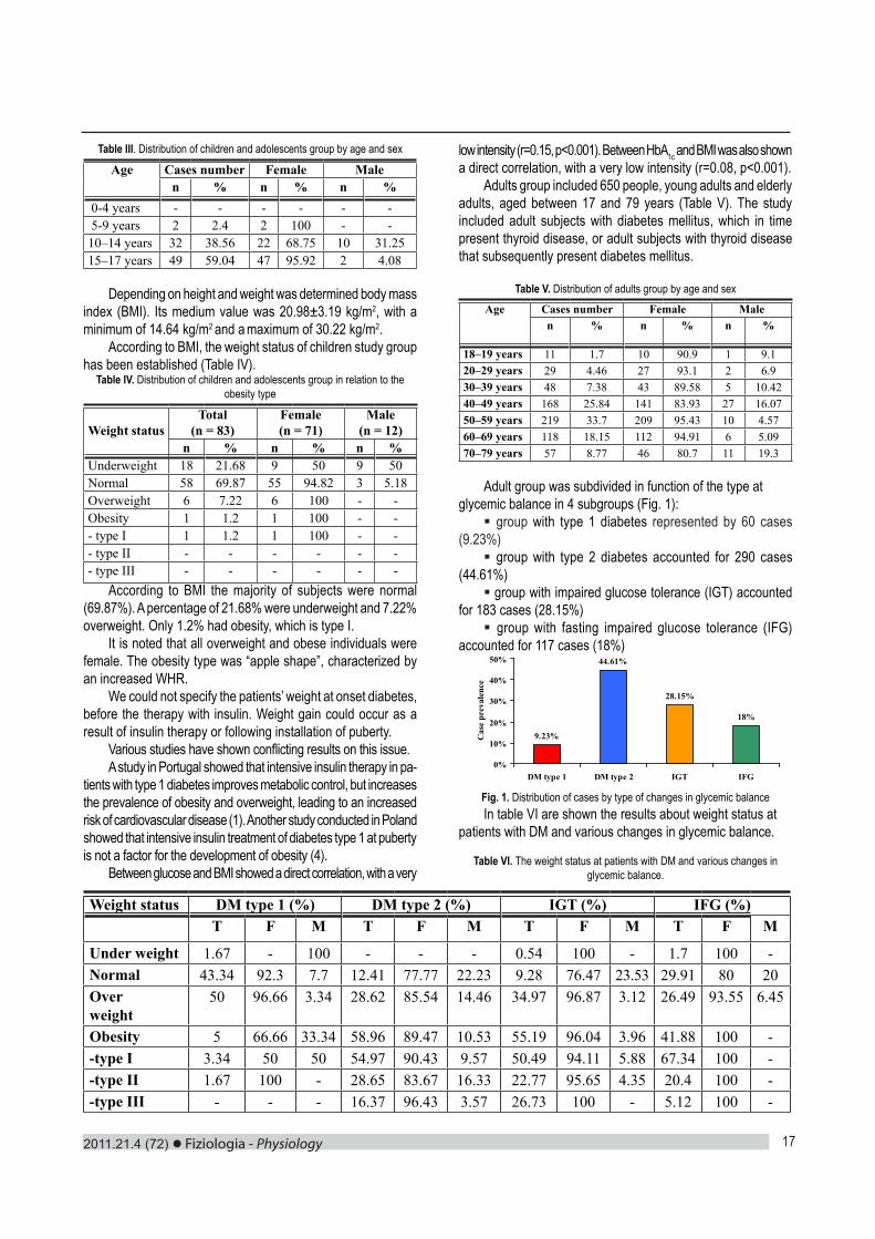
2011.21.4 (72) Fiziologia - Physiology 17
Table III. Distribution of children and adolescents group by age and sex
Age Cases number Female Malen % n % n %
0-4 years - - - - - - 5-9 years 2 2.4 2 100 - -10–14 years 32 38.56 22 68.75 10 31.2515–17 years 49 59.04 47 95.92 2 4.08
Depending on height and weight was determined body mass index (BMI). Its medium value was 20.98±3.19 kg/m2, with a minimum of 14.64 kg/m2 and a maximum of 30.22 kg/m2.
According to BMI, the weight status of children study group has been established (Table IV).
Table IV. Distribution of children and adolescents group in relation to the obesity type
Weight statusTotal
(n = 83)Female(n = 71)
Male(n = 12)
n % n % n %Underweight 18 21.68 9 50 9 50Normal 58 69.87 55 94.82 3 5.18Overweight 6 7.22 6 100 - -Obesity 1 1.2 1 100 - -- type I 1 1.2 1 100 - -- type II - - - - - -- type III - - - - - -
According to BMI the majority of subjects were normal (69.87%). A percentage of 21.68% were underweight and 7.22% overweight. Only 1.2% had obesity, which is type I.
It is noted that all overweight and obese individuals were female. The obesity type was “apple shape”, characterized by an increased WHR.
We could not specify the patients’ weight at onset diabetes, before the therapy with insulin. Weight gain could occur as a result of insulin therapy or following installation of puberty.
Various studies have shown conflicting results on this issue.A study in Portugal showed that intensive insulin therapy in pa-
tients with type 1 diabetes improves metabolic control, but increases the prevalence of obesity and overweight, leading to an increased risk of cardiovascular disease (1). Another study conducted in Poland showed that intensive insulin treatment of diabetes type 1 at puberty is not a factor for the development of obesity (4).
Between glucose and BMI showed a direct correlation, with a very
low intensity (r=0.15, p<0.001). Between HbA1c and BMI was also shown a direct correlation, with a very low intensity (r=0.08, p<0.001).
Adults group included 650 people, young adults and elderly adults, aged between 17 and 79 years (Table V). The study included adult subjects with diabetes mellitus, which in time present thyroid disease, or adult subjects with thyroid disease that subsequently present diabetes mellitus.
Table V. Distribution of adults group by age and sexAge Cases number Female Male
n % n % n %
18–19 years 11 1.7 10 90.9 1 9.120–29 years 29 4.46 27 93.1 2 6.930–39 years 48 7.38 43 89.58 5 10.4240–49 years 168 25.84 141 83.93 27 16.0750–59 years 219 33.7 209 95.43 10 4.5760–69 years 118 18.15 112 94.91 6 5.0970–79 years 57 8.77 46 80.7 11 19.3
Adult group was subdivided in function of the type at glycemic balance in 4 subgroups (Fig. 1): group with type 1 diabetes represented by 60 cases
(9.23%) group with type 2 diabetes accounted for 290 cases
(44.61%) group with impaired glucose tolerance (IGT) accounted
for 183 cases (28.15%) group with fasting impaired glucose tolerance (IFG)
accounted for 117 cases (18%)
9.23%
44.61%
28.15%
18%
0%
10%
20%
30%
40%
50%
DM type 1 DM type 2 IGT IFG
Cas
e pr
eval
ence
Fig. 1. Distribution of cases by type of changes in glycemic balanceIn table VI are shown the results about weight status at
patients with DM and various changes in glycemic balance.
Table VI. The weight status at patients with DM and various changes in glycemic balance.
Weight status DM type 1 (%) DM type 2 (%) IGT (%) IFG (%)T F M T F M T F M T F M
Under weight 1.67 - 100 - - - 0.54 100 - 1.7 100 -Normal 43.34 92.3 7.7 12.41 77.77 22.23 9.28 76.47 23.53 29.91 80 20Overweight
50 96.66 3.34 28.62 85.54 14.46 34.97 96.87 3.12 26.49 93.55 6.45
Obesity 5 66.66 33.34 58.96 89.47 10.53 55.19 96.04 3.96 41.88 100 --type I 3.34 50 50 54.97 90.43 9.57 50.49 94.11 5.88 67.34 100 --type II 1.67 100 - 28.65 83.67 16.33 22.77 95.65 4.35 20.4 100 --type III - - - 16.37 96.43 3.57 26.73 100 - 5.12 100 -

Fiziologia - Physiology 2011.21.4 (72)18
In the case of the studied group with type 1 diabetes, the medium value of BMI was 25.1±3.71 kg/m2, with a minimum of 18.22 kg/m2 and a maximum of 35.08 kg/m2.
According to BMI, 50% of patients are overweight. The percentage of normal weight was 43.34% and the percentage of underweight was 1.67% cases. Obesity was present in 5% cases. It is noted that obesity and overweight was more encounter in 56.36% women and 40% men (p=0.48, X2=0.5). The obesity type was “apple shape”, characterized by a WHR>0.85. The prevalence of obesity in type 1 diabetes is very rare. It is not known the weight at onset of diabetes.
It is possible that patients with type 1 diabetes become obese due to insulin therapy. Increased fat mass increases insulin resistance. This occurs due to decreased number of insulin receptors in adipose tissue and other tissues. A number of studies show that adipose tissue is higher in the elderly. If a person has the same body weight at 65 years to 25 years in this period it is doubled fat mass (8).
In the case of the studied group with type 2 diabetes, the medium value of BMI was 31.94±6,53 kg/m2, with a minimum of 20 kg/m2 and a maximum of 59.11 kg/m2. According to BMI was establish the weight status of the studied subjects with DM type 2 (Tab.VI).
According to BMI, 58.96% of patients are obese. Normal weight subjects were 12.41%. It was not found the presence of underweight subjects. Overweight were present in 28.62% of cases.
Regarding to obesity type, 54.97% had obesity type I, 28.65% obesity type II and 16.37% obesity type III.
In the type 2 diabetes group, obesity and overweight encounter in 88.88% women and 78.94% men (p=0.08, X2=3). Obesity type which predominated was “apple shape” character-ized by a WHR>0.85 (1.18% “pear shape” and 98.82% “apple shape”, p<0.001, X2 = 484.28).
In the case of the studied group with IGT, the medium value of BMI was 31.89±6.72 kg/m2, with a minimum of 16 kg/m2 and a maximum of 55.63 kg/m2. The results about weight status in patients with IGT are shown in Tab.VI.
According to BMI, 55.19% of patients are obese. Normal weight subjects were 9.28% of cases, and underweight subjects were 0.54% cases. Overweight were present in 34.97% of cases. Regarding the obesity type, 50.49% had obesity type I, 22.77% obesity type II and 26.73% obesity type III.
In the IGT group, obesity and overweight encounter in 91.9% women and 60% men (p = 0.0009, X2=10.85). Obesity type which predominated was “apple shape” characterized by a WHR>0.85 (3.03% “pear shape” and 76.97% “apple shape”, p<0.001, X2 = 291.21).
In the case of the studied group with IFG, the medium value of BMI was 28.8±5.96 kg/m2, with a minimum of 17.64 kg/m2 and a maximum of 50.6 kg/m2. According to BMI was establish the weight status of subjects with IFG (Tab.VI).
According to BMI, 41.88% of patients were obese. Normal weight subjects were 29.91% of cases, and underweight were 1.7% cases. Overweight were present in a proportion of 26.49%
of cases. Regarding the obesity type, 67.34% had obesity type I, 20.4% obesity type II and 5.12% obesity type III.
In the IFG group, obesity and overweight encounter in 72.22% women and 22.22% men (p=0.0019, X2=9.61%). Obesity type was “apple shape” characterized by a WHR>0.85.
The role of obesity as a risk factor for type 2 diabetes has been shown by many prospective studies that included Cau-casian populations in Norway, Sweden, Israel and the U.S., Mexicans in Texas, U.S., and Pima Indians (5).
In another two studies conducted in Israel and at the Pima Indians, it was found that the duration and degree of obesity are risk factors for type 2 diabetes. In the Pima Indian group, the risk of type 2 diabetes is two-fold in individuals with a history of obesity for 10 years and over this period compared to those with a history of less than 5 years (5).
Central distribution of intra-abdominal fat is a major risk fac-tor for type 2 diabetes independent of obesity degree, hypothesis demonstrated by prospective studies conducted on populations from Sweden, Japan and Pima Indians.
In the Pima Indians group, 5-year longitudinal studies have shown the association of abnormal glucose tolerance transition with weight gain, decreased insulin secretion and action. No changes were observed in hepatic glucose production. Empha-sizing excess weight and defect of synthesis/action of insulin and increased hepatic glucose production is characteristic of abnormal glucose tolerance progression to diabetes (5).
Between BMI and blood glucose in all types of changes in glycemic balance of the studied group was a direct correla-tion, very weak (r=0.048, p<0.001 for the group with type 1 diabetes, r=0.19, p<0.001 for the group with type 2 diabetes, r=0.13, p<0.001 for the IGT group and r=0.09, p<0.001 for the IFG group). Also, between BMI and HbA1c was found a direct correlation, very weak (r=0.36, p<0.001 for the group with type 1 diabetes and r=0.18, p<0.001 for the group with type 2 diabetes).
Significant differences between patients with different changes in glycemic control about obesity prevalence are show in table VII.
Table VII. Significant differences between patients with different changes in glycemic control about obesity prevalenceDM
type IDM type
2IGT IFG DM
childrenDM type I - p<0.001
X2=35.77p<0.001
X2=37.03p=0.07
X2=3.07p<0.001
X2=37.48DM type 2 p<0.001
X2=35.77- p=0.39
X2=0.74p<0.001X2=20.09
p<0.001X2=192.42
IGT p<0.001X2=37.03
p=0.39X2=0.74
- p<0.001X2=22.63
p<0.001X2=166.92
IFG p=0.07X2=3.07
p<0.001X2=20.09
p<0.001X2=22.63
- p<0.001X2=70.98
DM children
p<0.001X2=37.48
p<0.001X2=192.42
p<0.001X2=166.92
p<0.001X2=70.98
-
Depending on glycemic balance, obesity and overweight predominate in people with type 2 diabetes and IGT to those with type 1 diabetes. There were no significant differences

2011.21.4 (72) Fiziologia - Physiology 19
between DM type 1 and IFG. Also, obesity and overweight predominate in people with type 2 diabetes to those with IFG, but was no significant differences between DM type 2 and IGT. Also, between IGT and IFG, obesity and overweight predominate in people with IGT.
In function of age, there were significant differences between DM type 1 at children and different changes in glycemic control at adult (obesity and overweight prevailed at adults).
CONCLUSIONS
In patients with various changes in glycemic balance and thyroid disorders, obesity is more common in women in case of IGT and IFG. In case of DM type 1 and DM type 2, there was no significance difference between men and women.
Depending on glycemic balance, it predominates in people with type 2 diabetes and IGT to those with type 1 diabetes, in people with type 2 diabetes to those with IFG, and to those with IGT vs. IFG.
In function of age, there was a net prevalence of obesity and overweight at adult with different changes in glycemic control vs. children with DM type 1.
Depending on obesity type, the predominant type was “apple shape”, its predominance shown an increased risk of cardiovascular morbidity-mortality, especially for atherosclerotic cardiovascular disease.
Association of thyroid disease, which in time can evolve with hypothyroidism, is an additional risk factor for atherosclerotic cardiovascular disease.
That’s why it is indicated the screening of thyroid diseases in patients with various changes in glycemic balance in order to diagnose these conditions early and to institute appropriate treatment.
REFERENCES1. Arcanjo CL, Piccirillo LJ, Machado Ido V, Andrade CR Jr, Clemente EL, Gomes Mde B. Lipid profile and anthropometrical evaluation in type 1 diabetes. Arq Bras Endocrinol Metabol, 2005; 49 (6): 951-8. 2. Bundred P, Kitchiner D, Buchan I. Prevalence of overweight and obese children between 1989 and 1998: population based series of cross sectional studies. BMJ, 2001; 322: 326-8. 3. Cole TJ. Establishing a standard definition for child overweight and obesity worldwide: international survey. Br Med J, 2000; 320: 1240-43. 4. Noczynska A, Filipowski H, Wasikowa R. Is overweight in patients with type 1 diabetes in the puberty a problem ? Endokrynol Diabetol Chor Przemiany Materii Wieku Rozw, 2003; 9 (2): 89-92.5. Pencea C, Ionescu-Târgovişte C. Obezitatea. În: Tratat de diabet Paulescu, Editura Academiei Române, 2004: 701-725. 6. Popa I, Brega D, Drăgan M, Alexa A, Raica M. Obezitatea copilului şi ţesutul adipos. Editura Mirton, 2001, pag. 24-42 7. Şerban V, Babeş AP. Clinică Medicală. Teorie şi practică. Editura de Vest, 1999; 1: 11-149. 8. Şerban V, Vlad A, Sima A. Diabetul zaharat al vârstnicului, Editura Brumar, 2003 9. Uwaifo GI, Arioglu E. Obesity. In Endocrinology (elec-Uwaifo GI, Arioglu E. Obesity. In Endocrinology (elec-tronic book), 2002.9. Wilding JPH. Obesity and nutritional factors in the pathogenesis of type 2 diabetes mellitus. In: Textbook of diabetes. 3rd ed., Blackwell Science Ltd, 2003: 21.1-21.16.
PREVALENTA OBEZITATII LA PACIENTII CU VARIATII ALE ECHILIBRULUI GLICEMIC SI AFECTIUNI TIROIDIENE
REZUMATObezitatea este o afecţiune frecvent asociată diabetului zaharat tip 2, precum şi unor afecţiuni tiroidiene însoţite de hipotiroidie. Scopul acestui studiu a fost de a determina prevalenţa obezităţii la pacienţii cu diferite modificări ale echilibrului glicemic şi afecţiuni tiroidiene. Lotul general studiat a fost reprezentat de 733 cazuri, cu vârste cuprinse între 7-79 ani. Lotul studiat a fost subîmpărţit după criteriul vârstei în 2 loturi: lotul de copii şi lotul de adulţi. S-au folosit parametrii clinici, imagistici, biochimici, hormonali, imunologici.Cuvinte cheie: diabet zaharat, afecţiuni tiroidiene, obezitate

Fiziologia - Physiology 2011.21.4 (72)20
INTRODUCTION
In the last several decades, the increased prevalence of cardiovascular manifestation in patients with autoimmune rheumatic diseases (ARD) was observed (1). Heart disease in ARD develops through several pathophysiological mechanisms including myocardial inflammation and/or fibrosis, infiltration by granulomatous tissue, vasculitis, thrombus formation, of acceler-ated coronary atherosclerosis. Beyond this, pulmonary arterial hypertension leads to significant cardiac dysfunction and rapid progression of heart failure. Whilst it is likely that chronic systemic inflammation promotes accelerated atherosclerosis in these patients (2,3). Cardiac manifestations range from unusual to prominent and from mild to dramatic. In particular, coronary heart disease seems to be associated with inflammatory rheumatic conditions (4,5). Autoantibodies can damage all heart structures (Figure 1) (3). Autoantibodies may interact with target tissue or induce downstream mechanisms leading to cardiac lesions (6). Rhythm and conduction disorders are not only a frequent clinical manifestation of cardiovascular involvement in ARD but also a possible cause of sudden death. The conduction disorders are more frequent in ARD than the cardiac arrhythmias. Cardio-vascular autonomic dysfunction was revealed in the majority of autoimmune patients (7). Another difficulty is to differentiate cardiovascular effects of drugs from ARD involvement. Basic
research in imaging and rheumatology might provide a better insight into ARD pathogenesis and might guide development of targeted disease-specific therapies (8,9).
Fig. 1. Involvement of the heart structures in autoimmune rheumatic diseases (3)
Rheumatoid arthritisCardiovascular features in rheumatoid arthritis (RA) are
common, including pericarditis, cardiomyopathy/myocarditis, car-diac amyloidosis, coronary vasculitis, arrythmia, valve diseases and, most importantly, congestive heart failure and ischaemic
Received October 5th, 2011. Accepted November 20th, 2011. Address for correspondence: Manole Cojocaru, MD, PhD, ”Titu Maiorescu” University, Faculty of Medi-”Titu Maiorescu” University, Faculty of Medi-cine, Department of Physiology, Center for Rheumatic Diseases, Bucharest, Thomas Masaryk No. 5 Street, Sector 2, RO-020983, Bucharest, Romania, phone/fax: +4021.324.30.13, e-mail: [email protected]
CARDIOVASCULAR MANIFESTATIONS IN AUTOIMMUNE RHEUMATIC DISEASES M. COJOCARU1, INIMIOARA MIHAELA COJOCARU2, VIOLETA ŞAPIRA3
1”Titu Maiorescu” University, Faculty of Medicine, Department of Physiology, Center for Rheumatic Diseases, Bucharest2“Carol Davila” University of Medicine and Pharmacy, Department of Neurology, Colentina Clinical Hospital, Bucharest3“Ovidius” University, Faculty of Dental Medicine and Faculty of General Medicine, Department of Pathophysiology, Constanţa
ABSTRACTCardiovascular manifestations in autoimmune rheumatic diseases (ARD) have been infrequently recognized and mainly neglected in the medical community. Cardiac involvement may represent the initial manifestation of an ARD. Cardiac manifestations range from unusual to prominent and from mild to dramatic. Although ARD affects all cardiac structures, pericarditis is the most common manifestation. Early detection of cardiac abnormalities may have important therapeutic or prognostic implications. Aggressive anti-inflammatory therapy might additionally reduce clinical manifestations of cardiovascular disease. We review the cardiovascular manifestations of more common ARD in the light of the reported literature.Keywords: cardiovascular manifestations, autoimmune rheumatic diseases
2011.21.4 (72) Fiziologia - Physiology 21
was related to the presence of congestive heart failure, conduc-tion abnormalities, ventricular arrhythmias, and cardiovascular death (30). The large artery stiffening parallels that described above in RA and SLE. Stiffening of the conduit arteries develops as a consequence of inflammation and/or microvascular disease (31,32). Pulmonary arterial hypertension is a major issue in ARD, especially SSc significantly affecting quality of life and prognosis of the patients. The most frequent cardiac rhythm disturbances in SSc are premature ventricular contractions, often appearing as monomorphic, single ventricular contractions, or rarely as bigeminy, trigeminy or pairs (33). Conduction disturbances in SSc are due to fibrosis of sinoatrial node, presenting as abnormal ECG, bundle and fascicular blocks (34).
Dermatomyositis and polymyositis.Cardiac involvement is now well recognized as a clinically
important manifestation in patients with polymyositis (PM) or der-matomyositis (DM), although its actual frequency is still uncertain. Despite the fact that clinical manifestations are relatively rare, asymptomatic cardiovascular features are frequently reported in patients with PM and DM and are predominated by conduc-tion abnormalities and arrhythmias (35). The most frequently reported clinically overt manifestations are congestive heart failure, conduction abnormalities, that may lead to complete heart block, and coronary artery disease. Cardiac arrhythmias (including conduction disturbances and abnormal systolic time intervals) can occur but are often asymptomatic. These patients can manifest with AV blocks and ventricular or supraventricular tachyarrhythmias. Subclinical manifestations, characterized by conduction abnormalities and arrhythmias are frequently observed. In DM and PM, heart involvement may have fatal consequences, despite clinically overt involvement of the heart is seldom evident (36).
Psoriatic ArthritisPremature atherosclerosis has been recognized as an
important co-morbidity in patients with psoriatic arthritis (PA). Patients with PA had higher prevalences of both cardiovascular disease risk factors, and ischemic heart disease, peripheral vas-cular disease, and congestive heart failure. The risk of incident myocardial infarction was increased (37).
Ankylosing spondylitisPatients with ankylosing spondylitis (AS) may develop
cardiovascular manifestations ranging from asymptomatic forms to life threatening conditions. Heart disease is a well recognized complication of AS (38). The spectrum of disease is wide and includes mitral valve disease, cardiomyopathy, and pericarditis. The most common cardiac lesions in patients with AS are aortic regurgitation and conduction abnormalities. Conduction disease may develop as a consequence of postinflammatory scarring of myocardial tissue (39). First-degree atrioventricular block is most common; however, higher-grade atrioventricular block and right and left bundle-branch block have also been reported. Heart block appears to occur more frequently in HLA-B27-positive
heart disease (10-13). Chronic inflammation is primarily respon-sible for accelerated atherosclerosis in RA (14-16). Clinically significant valvular disease attributable to RA appears to be un-common (17). Fibrinous pericarditis may be detected at autopsy in RA patients but is generally not of clinical relevance. Arterial stiffness is increased in RA. It is related to disease duration (18). Rheumatoid arthritis is associated with an increased risk of congestive heart failure. The prevalence of left ventricular systolic dysfunction was to be higher in patients with RA (19). Coronary arteritis and fibrinous pericarditis may be detected at autopsy in RA patients. Important for clinical practice is the positive effect of methotrexate therapy on the reduction of cardiovascular mortality and the contraindication for TNF-α antagonists in patients with severe heart failure (20). In patients with RA, a major cause of sudden cardiac death is atherosclerotic coronary artery disease, leading to acute coronary syndrome and ventricular arrhythmias (21). In RA, infiltration of the atrioventricular node can cause right bundle branch block. AV block is rare in RA, and is usually complete. In RA patients, conduction abnormalities may regress when the underlying disease is controlled (22,23).
Systemic lupus erythematosusThe premature development of atherosclerotic coronary
artery disease has been documented in autopsy. Arte-rial stiffness is increased in SLE and is related to disease duration and to circulating levels of C-reactive protein and interleukin-6. It is associated with active disease, including myositis and serositis. SLE-related factors associated with clinical manifestations of coronary artery disease include older age at diagnosis, longer duration of SLE, higher damage score, longer duration of steroid therapy, and higher levels of oxidized low-density lipoprotein cholesterol and homocysteine. Valvular disease associated with Libman-Sacks lesions, se-rositis resulting in pericardial disease, and venous and arterial thromboses associated with the presence of antiphospholipid antibodies are well-established cardiovascular manifestations of SLE (24). Myocarditis is rarely diagnosed clinically and val-vular nodules have been described in the majority of patients at autopsy. The premature development of atherosclerotic coronary artery disease has been documented in autopsy (25). Pericardial disease, as a manifestation of serositis, is a diagnostic feature of SLE. Pericardial effusions occur most commonly in the setting of active disease (flares) (26). In SLE, sinus tachycardia, atrial fibrillation and atrial ectopic beats are the major cardiac arrhythmias (27).
Systemic sclerosisThe most prominent cardiovascular abnormalities associ-
ated with systemic sclerosis (SSc) are microvascular perfusion abnormalities of the ventricular myocardium resulting in ischemia, fibrosis, systolic dysfunction, and conduction disease (28). Myo-cardial disease in SSc may be related to associated pulmonary or renal involvement or to hypertension (29). The severity of these lesions was unrelated concomitant pulmonary and systemic hypertension, Raynaud’s phenomenon, and renal disease but

Fiziologia - Physiology 2011.21.4 (72)22
individuals, even in the absence of clinical manifestations of AS. Diastolic filling abnormalities have been described in AS. Aortitis of the ascending aorta may lead to distortion of the aortic ring, causing aortic regurgitation (40). Fibrosis of the conduction system may result in various degrees of atrioventricular block, including complete heart block. Aortic aneurysmal dilation is a recognized rare association with AS, and bicuspid aortic valve or coronary artery disease is only occasionally present. Decreased aortic elasticity with impaired endothelial function could be responsible for the development of the aortic complication in AS patients (41).
CONCLUSION
Cardiovascular manifestations are known to occur in pa-tients with ARD often in subclinical form. Cardiovascular morbid-ity and mortality have been found to be increased in association with many of the ARD. A thorough history for cardiac symptoms is very important to prevent any future major cardiac event. Rheumatologists should carefully consider, both in late and in early phases of the disease, the occurrence of cardiovascular manifestations in patients with ARD.
REFERENCES1. Roman MJ, Salmon JE. Cardiovascular manifestations of rheuma-Cardiovascular manifestations of rheuma-tologic diseases. Circulation, 2007; 116: 2346-55.2. Goodson NJ, Solomon DH. The cardiovascular manifestations of rheumatic diseases. Current Opinion in Rheumatology, 2006; 18: 135-140.3. Matucci-Cerinic M, Seferović PM. Heart involvement in autoimmune rheumatic diseases: the “phantom of the opera”. Rheumatology, 2006; 45: iv1-3.4. Seferović PM, Ristić AD, Maksimović R, Simeunović DS, Ristić GG, Radovanović G, Seferović D, Maisch B, Matucci-Cerinic M. Cardiac arrhythmias and conduction disturbances in autoimmune rheumatic diseases. Rheumatology (Oxford), 2006; 45(Suppl. 4): iv39-42.5. Cutolo M, Sulli A, Secchi ME, Paolino S, Pizzorni C. Nailfold capil-laroscopy is useful for the diagnosis and follow up of autoimmune rheumatic diseases. A future tool for the analysis of microvascular heart involvement? Rheumatology (Oxford), 2006; 45(Suppl.4): iv44-47.6. Scherer Y, Shoenfeld Y. Mechanisms of disease: atherosclerosis in autoimmune diseases. Nat Clin Pract Rheumatol, 2006; 2: 99-106.7. Stojanovich L. Autonomic dysfunction in autoimmune rheumatic disease. Autoimmun Rev, 2009; 8(7): 569-72. 8. Hansson G. Inflammation, atherosclerosis, and coronary artery disease. N Engl J Med, 2005; 352: 1685-95. 9. Maksimović M, Seferović PM, Ristić AD, Vujisić-Tesić B, Simeunović DS, Radovanović G, Matucci-Cerinic M, Maisch B. Cardiac imaging in autoimmune rheumatic diseases. Rheumatology (Oxford), 2006; 45(Suppl. 4): iv26-31. 10. Stojanovich L. Milovanovich B, de Luka SR, Popovich-Kuzmanovich D, Bisenich V, Djukanovich B, Randjelovich T, Krotin M. Cardiovascular autonomic dysfuntion in systemic lupus, rheumatoid arthritis, primary Sjögren syndrome and other autoimmune diseases. Lupus, 2007; 16: 181-85. 11. Roman MJ, Moeller E, Davis A, Paget SA, Crow MK, Lockshin MD, Sammaritano L, Devereux RB, Schwartz JE, Levine DM, Salmon JE. Preclinical carotid atherosclerosis in patients with rheumatoid
arthritis: prevalence and associated factors. Ann Intern Med, 2006; 144: 249-56.12. Voskuyl AE. The heart and cardiovascular manifestations in rheu-matoid arthritis. Rheumatology (Oxford), 2006; 45 Suppl. 4: iv4-7.13. Liang KP, Liang KV, Matteson EL, McClelland RL, Christianson TJ, Turesson C. Incidence of noncardiac vascular disease in rheumatoid arthritis and relationship to extra-articular disease manifestations. Arthritis Rheum, 2006; 54: 642-48.14. Sarzi-Puttini P, Atzeni F, Gerli R, Bartolini E, Doria A, Barskova T, Matucci-Cerinic M, Sitia S, Tomasoni L, Turiel M. Cardiac involvement in systemic rheumatic diseases: An update. Autoimmun Rev, 2010; 9(12): 849-52.15. Ciftci, O, Yilmaz S, Topcu M, Caliskan M, Gullu H, Erdogan D, Pamuk BO, Yildirir A, Muderrisoglu H. Impaired coronary microvascular function and increased intima-media thickness in rheumatoid arthritis. Atherosclerosis, 2008; 198: 332-7.16. Bhatia GS, Sosin MD, Patel JV, Grindulis KA, Khattak FH, Hughes EA, Lip GYH, Davis RC. Left ventricular systolic dysfunction in rheu-matoid disease: an unrecognized burden? J Am Coll Cardiol, 2006; 4: 1169-74.17. Turiel M, Sitia S, Atzeni F, Tomasoni L, Gianturco L, Giuffrida M, De Gennaro Colonna V, Sarzi-Puttini P. The heart in rheumatoid arthritis. Autoimmun Rev, 2010; 9(6): 414-18.18. Nicola PJ, Maradit-Kremers H, Roger VL, Jacobsen SJ, Crowson CS, Ballman KV, Gabriel SE. The risk of congestive heart failure in rheumatoid arthritis: a population-based study over 46 years. Arthritis Rheum, 2005; 52: 412-20. 19. Han C, Robinson DW Jr, Hackett MV, Paramore LC, Fraeman KH, Bala MV. Cardiovascular disease and risk factors in patients with rheumatoid arthritis, psoriatic arthritis, and ankylosing spondilytis. J Rheumatol, 2006; 33(11): 2167-7220. Pham T, Gossec L, Constantin A, Pavy S, Bruckert E, Cantagrel A, Combe B, Flipo RM, Goupille P, Le Loêt X, Mariette X, Puéchal X, Schaeverbeke T, Sibilia J, Tebib J, Wendling D, Dougados M. Car-diovascular risk and rheumatoid arthritis: clinical practice guidelines based on published evidence and expert opinion. Joint Bone Spine, 2006; 73(4): 379-87.21. Voskuyl AE. The heart and cardiovascular manifestations in rheu-matoid arthritis. Rheumatology (Oxford), 2006; 45(Suppl.4): iv4-7.22. Chung CP, Oeser A, Raggi P, Gebretsadik T, Shintani AK, Sooka T, Pincus T, Avalos I, Stein CM. Increased coronary-artery atherosclerosis in rheumatoid arthritis: relationship to disease duration and cardiovas-cular risk factors. Arthritis Rheum, 2005; 52: 3045-53.23. Chogle AR, Chakravarty A. Cardiovascular events in systemic lupus erythematosus and rheumatoid arthritis: Emerging concepts, early diag-nosis and management. J Assoc Physicians India, 2007; 55: 32-40.24. Ben-Menachem E. Systemic lupus erythematosus: a review for anesthesiologists. Anesthesia & Analgesia, 2010; 111: 665-76.25. Pons-Estel GJ, Gonzalez LA, Zhang J, Burgos PI, Reveille JD, Vila LM, Alarcon GC. Predictors of cardiovascular damage in patients with systemic lupus erythematosus: data from LUMINA (LXVIII0, a multiethnic US cohort. Rheumatology, 2009; 48: 817-22.26. Ahmad Y, Shelmerdine J, Bodill H. Lunt M, Patrick MG, Teh LS, Menstein RM, Walker M, Bruce IN. Subclinical atherosclerosis in sys-temic lupus erythematosus (SLE): the relative contribution of classic risk factors and the lupus phenotype. Rheumatology, 2007; 46: 983-8.27. Tincani A, Biasini Rebaioli C, Taglietti M, Shoenfeld Y. Heart involve-ment in systemic lupus erythematosus, antiphospholipid syndrome and neonatal lupus. Rheumatology (Oxford), 2006; 45(Suppl.4): iv8-13.28. Vacca A, Siotto P, Cauli A, Montisci R, Garau P, Ibba V, Mameli A, Passiu G, Iliceto S, Mathieu A. Absence of epicardial coronary stenosis

2011.21.4 (72) Fiziologia - Physiology 23
in patients with systemic sclerosis and severe impairment of coronary flow reserve. Ann Rheum Dis, 2006; 65: 274-5. 29. Moyssakis I, Gialafos E, Vassiliou V, Taktikou E, Katsiari C, Pa-padopoulos DP, Sfikakis PP. Aortic stiffness in systemic sclerosis in increased independently of the extend of skin involvement. Rheumatol-ogy, 2005; 44: 251-4. 30. Ioannidis JPA, Vlachoyiannopoulos PG, Haidich A-B, Medsger TA Jr, Lucas M, Michet CJ, Kuwana M, Yasuoka H, van den Hoogen F, te Boome L, van Laar JM, Verbeet NL, Matucci-Cerinic M, Georgountzos A, Moutsopoulos HM. Mortality in systemic sclerosis: an international meta-analysis of individual patient data. Am J Med. 2005; 118: 2-10.31. Kahan A, Allanoire Y. Primary myocardial involvement in systemic sclerosis. Rheumatology (Oxford), 2006; 45 Suppl. 4: iv14-17. 32. Akram MR, Handler CE, Williams M, Carulli MT, Andron M, Black CM, Denton CP, Coghlan JG. Angiographically proven coronary artery disease in scleroderma. Rheumatology, 2006; 45: 1395-8. 33. Hsaio SH, Lee CY, Chang SM, Lin SK, Liu CP. Right heart function in scleroderma: insights from myocardial Doppler tissue imaging. J Am Soc Echocardiogr, 2006; 19: 507-14. 34. Meune C, Allanore Y, Devaux JY, Dessault O, Duboc D, Weber S, Kahan A. High prevalence of right ventricular systolic dysfunction in early systemic sclerosis. J Rheumatol, 2004; 31:1941-5.35. Lundberg IE. Cardiac involvement in autoimmune myositis and
mixed connective tissue disease. Lupus, 2005; 14(9): 708-12. 36. Lundberg I. The heart in dermatomyositis and polymyositis. Rheu-matology (Oxford), 2006; 45(Suppl. 4): iv22-25.37. Madland TM, Apalset EM, Johannessen AE, Rossebo B, Brun JG. Prevalence, disease manifestations, and treatment of psoriatic arthritis in Western Norway. J Rheumatol, 2005; 32: 1918-22.38. Caliskan M, Erdogan D, Gullu H, Yilmaz S, Gursoy Y, Yildirir A, Yucel E, Muderrisoglu H. Impaired coronary microvascular and left ventricular diastolic functions in patients with ankylosing spondylitis. Atherosclerosis, 2008; 196: 306-12.39. Divecha H, Sattar N, Rumley A, Cherry L, Lowe GD, Stur-rock R. Cardiovascular risk parameters in men with ankylosing spondylitis in comparison with non-inflammatory control subjects: relevance of systemic inflammation. Clin Sci (Lond), 2005; 109: 171-6.40. Brunner F, Kunz A, Weber U, Kissling R. Ankylosing spondylitis and heart abnormalities: do cardiac conduction disorders, valve regurgita-tion and diastolic dysfunction occur more often in male patients with diagnosed ankylosing spondylitis for over 15 years than in the normal population? Clin Rheumatol, 2006; 25(1): 24-29.41. Yuan S-M. Cardiovascular involvement of ankylosing spondylitis: Report of three cases. Vascular, 2009; 17: 342-54.
MANIFESTĂRI CARDIOVASCULARE ÎN BOLILE REUMATICE AUTOIMUNE
REZUMATManifestările cardiovasculare în bolile reumatice autoimmune nu sunt frecvente şi de obicei sunt subdiagnosticate. Afectarea cardiacă poate să reprezinte manifestarea iniţială a unei boli reumatice autoimune. Manifestările cardiace variază de la neobişnuite la evidente şi de la uşoare la dramatice. Deşi bolile reumatice autoimmune afectează toate structurile cardiace, pericardita este cea mai frecventă manifestare. Evidenţierea precoce a tulburărilor cardiace este importantă pentru terapie sau prognostic. Terapia antiinflamatorie agresivă reduce manifestările clinice ale bolii cardiovasculare. Autorii prezintă manifestările cardiovasculare mai frecvente ale bolilor reumatismale autoimmune în lumina datelor din literatură.Cuvinte cheie: manifestări cardiovasculare, boli reumatice autoimmune
Fiziologia - Physiology 2011.21.4 (72)24
INTRODUCTION
Although the cellular structure of the retina is known in detail for over 100 years, the interactions between different cell types and the roles of their subtypes are still far from being completely characterized. The information they generate and the way this information is processed at the retina level are topics more actual than ever, because of the improvement of research methods, due to the development of information technology and that of bio-signal recording techniques.
Image generation has been highly studied and recent dis-coveries regarding the image processing that begins in the retina or the cellular pathways identified at this level are arguments that this area is in constant development.
Still, basic aspects of light perception are not well docu-mented in the literature, such as the general behavior of the retina in response to selective color stimuli, in normal conditions.
As a result of this observation, we conducted an elec-troretinography study, meant to highlight the differences between the ERG parameters obtained by stimulating the retina with monochromatic (red) and white light. The study consisted of two tests:
- in the first test, we recorded Flash ERG in photopic con-ditions, in response to a series of 8 increasing light intensities, stimulation being done with white light, using a strobe lamp;
- in the second one, we recorded Flash ERG in photopic conditions, in response to a series of 8 increasing light intensi-ties, stimulation being done with red light (660nm), using LED goggles.
In both tests, the eyes were light adapted and pupils were not dilated, to recreate normal daytime conditions. Minimum intensity was -1 log cd·s/m2 (0.1 cd·s/m2), which allowed us to
obtain both ”a” and ”b” ERG waves for red light stimuli.
MATERIALS AND METHOD
We performed these studies according to the ethical and moral principles of the Helsinki Declaration of Human Rights. The most important factors taken into account were the well being and safety of subjects. Before testing, each subject was given a detailed presentation of the purpose and the conduct of the experiment, clearly specifying what data will be recorded and how it will be used. All subjects have agreed to voluntary participation.
At the time of the experiments no subject was suffering from eye disorders, acute or chronic. Volunteers have made a commitment to come to experiments rested, without consuming coffee, alcoholic beverages or any substances affecting attention and reaction time, for at least 12 hours before the experiment, for the results to be scientific relevant.
In both tests, subjects were prepared for the recordings as follows:
1. to reduce the discomfort of the recording electrode, the subject was given a drop of lidocaine clorhydrate (XILINE) 4% solution in the lower conjunctival sac
2. to prevent irritation and infection of the conjunctiva, the subject was given a drop of tobramycin + dexamethasone (TOBRADEX) solution in the lower fornix
3. we attached the required electrodes for recording the ERG:
- the recording electrode, HK-loop type, previously wetted with physiological serum to not irritate the conjunctiva, was introduced into the lower conjunctival sac of the right eye and fixed on the lower eyelid with medical adhesive tape
Received October 20th, 2011. Accepted November 3rd, 2011. Address for correspondence: Teaching assistant Dragoş Alexandru, MD, PhD, University of Medicine and Pharmacy of Craiova, Faculty of Medicine, Department of Medical Informatics, 2 Petru Rareş Street, 200349 , Craiova, Romania, e-mail: [email protected]
THE ASSESSMENT OF ERG DIFFERENCES BETWEEN RED AND WHITE STIMULI ALEXANDRU D.1, CĂTĂLIN B.2, GEORGESCU M.2, GEORGESCU D.1, IANCĂU MARIA2
1Department of Medical Informatics, University of Medicine and Pharmacy of Craiova, Faculty of Medicine2Department of Physiology, University of Medicine and Pharmacy of Craiova, Faculty of Medicine
ABSTRACTIn the study featured in this article we investigated the differences between the series of amplitudes and latencies of ERG waves recorded to flash stimuli, produced by different light, red (monochromatic) or white, using increasing intensities, from -1 to 2 log with steps of 0.5 log, plus the 1.7 log intensity (50 cd·s/m2). We obtained significantly different amplitudes for the “a” waves only for flash intensities over 1.5 log cd·s/m2 and for the “b” waves only for the maximum amplitude of the series, measured at 0.5 log cd·s/m2. Analyzing the latencies, we found highly significant statistical differences for all the intensities used.Keywords: photopic ERG, red light, white light, amplitude, latency

2011.21.4 (72) Fiziologia - Physiology 25
- the reference electrode, self adhesive, was set to the ipsilateral external cantus
- the grounding electrode, self-adhesive, was placed on the forehead
4. we proceeded with light adaptation, for 10 minutes5. we applied the stimuli, each repeated 10 times, thus doing
the recording of ERG responses. The end result was achieved through the coherent averaging of the 10 samples.
On the Flash ERG recordings many waves are described; we were interested in the first negative wave, denoted “a” and in the first positive wave, denoted “b”. Parameters used to describe them were the amplitude and latency.
For this, we marked their extreme points that are the minimum of the “a” wave and the maximum of the “b” wave. We measured the amplitude of “a” as difference between the average voltage measured before applying the stimulus and the peak of the wave, and the amplitude of “b” as difference between the „b” and the “a” peaks („trough to peak” amplitude). Latencies of the “a” and “b” waves were reported as the differ-ence between the time from the stimulus onset and the time when the waves peak.
For stimulation and data recording we used the Neuropack M1 MEB-9100 system (Nihon Kohden Corp., Tokyo, Japan). Red light stimuli were obtained with special LED goggles - LS-102J, part of the system (Nihon-Kohden Corp, Tokyo, Japan), emitting a narrow band of frequencies, with peak wavelength of 660nm, and white light stimuli were produced with a TSD122b strobe lamp (Biopac Systems Inc., Goleta, CA, USA).
Table I. Characteristics of the used stimuli
Luminance(log cd·s/m2)
Luminance (cd·s/m2)
Inter Stimuli Interval
(s)-1 0.1 10
-0.5 0.3 100 1 10
0.5 3 101 10 15
1.5 30 151.7 50 152 100 15
The recording electrodes were HK-loop type („Hawlina-Konec loop”)(1). This electrode consists of a thin silver wire, shaped to form a loop that is bent and inserted into the lower conjunctival sac. The wire is electrically insulated with Teflon, except for three windows at the top of the loop, as opposed to the electrode connector. Electrical contact with conjunctiva is made by the not insulated, exposed portion of the wire. Its advantages are stability similar to surface electrodes and sensitivity compa-rable to corneal contact electrodes, foil or wire type (DTL).
A BFig.1. HK loop electrodes (A) and their fixation in the conjunctival fornix, for
recordings (B)
The reference electrode was self-adhesive, Ag-AgCl type, with foam, model PG10S (FIAB SpA, Florence, Italy), while the grounding one was also Ag-AgCl type, model VivoMed Solid Gel (Servoprav GmbH, Wesel, Germany).
Data processing and computing of classical statistical indica-tors (arithmetic mean, standard deviation, coefficient of variation), as well as tests for comparing averages (Student) were per-formed using Microsoft Excel 2003 (Microsoft Corp., Redmond, WA, USA). To compute data normality tests (Shapiro-Wilks and Anderson-Darling) and nonparametric tests for comparing aver-ages (Mann-Whitney-Wilcoxon), we used the XLSTAT add-on (Addinsoft SARL, Paris, France).
In the first test we performed ERG recordings on 31 vol-unteers (9 men and 22 women), using white light stimulation, under photopic conditions, in response to a series of flashes with increasing intensities, between -1 and 2 log, from 0.5 to 0.5 log, plus the 1.7 log intensity (50 cd·s/m2).
For the second test of the study, 20 ERG recordings were made (for 6 men and 14 women, 11 persons from the first experi-ment missing), in response to a series of red light flash stimuli, using the same intensities as above.
Women, 22,
70.97%
Men, 9,
29.03%
Fig.2.Gender distribution in the first test
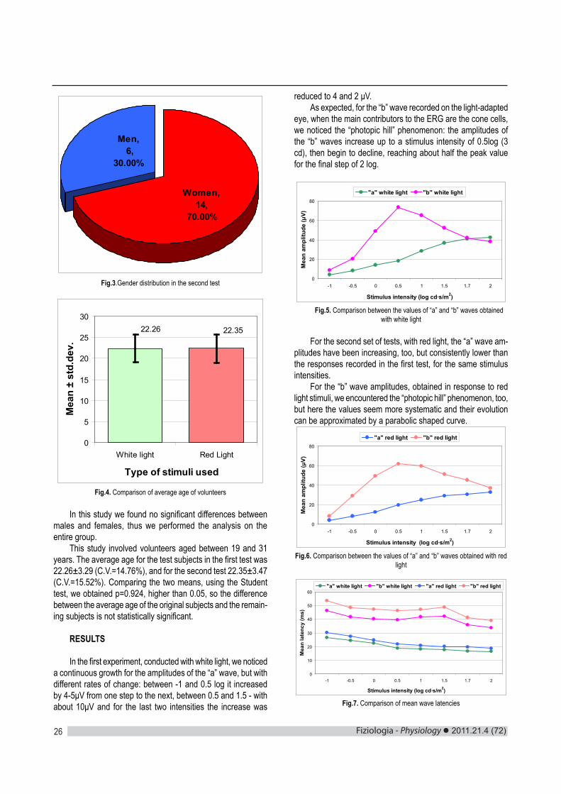
Fiziologia - Physiology 2011.21.4 (72)26
Men, 6,
30.00%
Women, 14,
70.00%
Fig.3.Gender distribution in the second test
22.26 22.35
0
5
10
15
20
25
30
White light Red Light
Type of stimuli used
Mea
n ±
std.
dev.
Fig.4. Comparison of average age of volunteers
In this study we found no significant differences between males and females, thus we performed the analysis on the entire group.
This study involved volunteers aged between 19 and 31 years. The average age for the test subjects in the first test was 22.26±3.29 (C.V.=14.76%), and for the second test 22.35±3.47 (C.V.=15.52%). Comparing the two means, using the Student test, we obtained p=0.924, higher than 0.05, so the difference between the average age of the original subjects and the remain-ing subjects is not statistically significant.
RESULTS
In the first experiment, conducted with white light, we noticed a continuous growth for the amplitudes of the “a” wave, but with different rates of change: between -1 and 0.5 log it increased by 4-5μV from one step to the next, between 0.5 and 1.5 - with about 10μV and for the last two intensities the increase was
reduced to 4 and 2 μV.As expected, for the “b” wave recorded on the light-adapted
eye, when the main contributors to the ERG are the cone cells, we noticed the “photopic hill” phenomenon: the amplitudes of the “b” waves increase up to a stimulus intensity of 0.5log (3 cd), then begin to decline, reaching about half the peak value for the final step of 2 log.
0
20
40
60
80
-1 -0.5 0 0.5 1 1.5 1.7 2
Stimulus intensity (log cd·s/m2)M
ean
ampl
itude
(μV)
"a" white light "b" white light
Fig.5. Comparison between the values of “a” and “b” waves obtained with white light
For the second set of tests, with red light, the “a” wave am-plitudes have been increasing, too, but consistently lower than the responses recorded in the first test, for the same stimulus intensities.
For the “b” wave amplitudes, obtained in response to red light stimuli, we encountered the “photopic hill” phenomenon, too, but here the values seem more systematic and their evolution can be approximated by a parabolic shaped curve.
0
20
40
60
80
-1 -0.5 0 0.5 1 1.5 1.7 2
Stimulus intensity (log cd·s/m2)
Mea
n am
plitu
de (μ
V)
"a" red light "b" red light
Fig.6. Comparison between the values of “a” and “b” waves obtained with red light
0
10
20
30
40
50
60
-1 -0.5 0 0.5 1 1.5 1.7 2
Stimulus intensity (log cd·s/m2)
Mea
n la
tenc
y (m
s)
"a" white light "b" white light "a" red light "b" red light
Fig.7. Comparison of mean wave latencies

2011.21.4 (72) Fiziologia - Physiology 27
Analyzing the evolution of the latencies of the “a” and “b” waves, we found highly significant statistical differences for all the intensities used (Mann-Whitney p<0.001), but we also noticed a trend of parallel variation in the recorded values.
Calculating the differences between the latencies of the “a” waves recorded in the two tests, respectively between the laten-cies of the “b” waves, we observed only little variation, close to 3 ms for the “a” waves and to 6 ms for the “b” waves. Therefore, it is possible that the significant differences we found between latencies to be due to the different conditions and equipment used, and not to reflect an objective difference between white and red light stimulation.
Table II. Values and ratios of the measured amplitudes
Table III. Differences between the pairs of recorded latencies
DISCUSSIONS
It should be noted that the study was performed on light adapted eyes, but without dilating the pupils, as is usually done, because we were interested in recording normal retinal activity, comparable to everyday situations. The range of used intensities also contributes to this end, the lower value being close to the limit for perceiving a color scene, while the upper one is equiva-lent to the light produced by a standard 60-75W bulb.
The multiple interactions between retinal cells, that occur mainly for the photopic sight, cause a highly variable behavior of the series of amplitudes recorded in this study. For example, for stimuli implying high intensities, the amplitudes of the “a” waves equalize or even surpass the amplitudes of the “b” waves, and the amplitudes of the “b” waves are not significantly higher for white light tan for red light, except for the maximum values of the series, obtained for 0.5 log cd·s/m2, for the other intensities the amplitudes’ ratio being close to 1. The only thing proven, corresponding to what we expected to happen, is that “a” waves produced with white light, at high intensities, are significantly larger “a” waves obtained with red light (Table II). Amplitude values measured in these experiments have not been edifying, showing statistically significant differences only for few intensities and mainly for “a” waves, although the results were almost always higher for white light than those recorded for red light.
Recorded latencies have shown that, for white light stimuli,
Intensity -1 -0.5 0 0.5 1 1.5 1.7 2„a” white 4.13 8.25 13.88 18.35 28.58 36.75 40.83 42.47„a” red 4.08 8.17 12.38 19.55 24.75 28.88 30.53 33.00
a white /a red 1.01 1.01 1.12 0.94 1.15 1.27 1.34 1.29p Student 0.923 0.906 0.138 0.429 0.088 0.002 0.000 0.000„b” white 8.74 20.42 49.00 73.49 65.33 52.18 42.26 38.38„b” red 8.23 28.88 49.51 61.85 59.75 51.27 45.38 37.13
b white /b red 1.06 0.71 0.99 1.19 1.09 1.02 0.93 1.03p Student 0.459 0.000 0.881 0.031 0.227 0.804 0.418 0.719
Int. -1 -0.5 0 0.5 1 1.5 1.7 2 Mean Std.dev. C.V.(%)Dif.a 3.69 2.98 2.37 2.96 2.80 2.46 3.06 2.36 2.83 0.45 15.83%Dif.b 7.28 6.88 6.98 6.69 5.52 6.57 5.40 5.30 6.33 0.79 12.56%
the time to peak, both for “a” and “b” waves, was significantly lower than the implicit time for red light (Table III). Because in some studies cited (6,7) we have not encountered this situation and because the differences were relatively constant (2.83±0.45 ms for the “a” waves – C.V. equal to 15.83%, and 6.33±0.79 ms for the “b” waves – C.V. equal to 12.56%), we suspect an interference due to the different equipment used for stimulation, a strobe lamp for white light stimuli and LED goggles for red light stimuli.
In regard to a comparative analysis of the absolute values of wave amplitudes recorded, with data from other studies (2,3,4,5), the method does not seem viable because of large variations due to different ERG stimulation and recording systems, and,
last but not least, the characteristics of the population that the subjects are part of. For these reasons we turned to the use of the ratios between the amplitudes of “b” and “a” waves, from the same experiment. Most articles that refer to series of ERG responses, describe only general behavior trends or the functions used to estimate the series of values recorded, exemplifying only the results for the maximum amplitude, so a detailed comparison is not possible.
In two studies that examine various aspects of ERG ob-tained with white and monochromatic light stimuli (6,7), we found results consistent with those from our study. Stimulus intensity, for which results are reported, is the one corresponding to the maximum amplitude of the “b” waves series.
Table IV. Comparison of the „b”/„a” amplitude ratios for the maximal responses
bmax/a Intensity White light Red light
Present study 0.5 log 4 3.16Rufiange 2005 0.5 log 3.49 3.1Sustar 2009 0.4 log 3.75 3.37Sustar 2009 0.4 log 3.41 2.77
Unfortunately, a thorough numerical comparison of the behavior of the latencies with data from literature is not possible, most authors preferring to ignore the details concerning the mea-

Fiziologia - Physiology 2011.21.4 (72)28
EVALUAREA DIFERENŢELOR ERG PRIN STIMULARE CU LUMINĂ ROŞIE, MONOCROMATICĂ ŞI CU LUMINĂ ALBĂ
REZUMAT
În cercetările descrise în articolul de faţă am investigat ce diferenţe există între seriile de amplitudini şi latenţe ale undelor ERG înregistrate pentru stimuli de tip flash, produşi cu lumină diferită, roşie (monocromatică) sau albă, folosind intensităţi crescătoare, între -1 şi 2 log, din 0.5 în 0.5 log, plus intensitatea de 1.7 log (50 cd·s/m2). Am obţinut amplitudini diferite semnificativ pentru undele „a” doar pentru stimulii de peste 1.5 log cd·s/m2, iar pentru undele „b” doar în cazul amplitudinii maxime a seriei, măsurată la 0.5 log cd·s/m2. Analizând evoluţiile latenţelor, am identificat diferenţe înalt semnificative statistic pentru toate intensităţile folosite.Cuvinte cheie: ERG fotopică, lumină roşie, lumină albă, amplitudini, latenţe
sured times or showing them only as charts, from which we can only see the general trend of evolution. That kind of information is also useful, because the data from our research are congruent with ideas found in similar studies (5,6,7).
CONCLUSIONS
1. By measuring the amplitudes of the ERG waves obtained with white and red light stimuli we obtained significantly different results for the amplitudes of the “a” waves only for stimuli of more than 1.5 log cd·s/m2 and for the “b” waves only for the maximum amplitude of the series, measured at 0.5 log cd·s/m2 - in all these cases, the amplitude of the waves recorded with white light was higher than for red light.
2. Stimulation with white light systematically produced significantly lower latencies than the red light, for “a” waves, as well as for “b” waves (p<0.001). Still, further investigations should be made to rule out any influences due to the equipment used for producing the stimuli.
REFERENCES1. Hawlina M, Konec B. New noncorneal HK-loop electrode for clinical electroretinography. Doc Ophthalmol., 1992; 81(2): 253-259. 2. Robson JG, Saszik SM, Ahmed J, Frishman LJ. Rod and cone contributions to the a-wave of the electroretinogram of the macaque. J Physiol., 2003; 547.2:509-530. 3. Euler T, Masland RH. Light-evoked responses of bipolar cells in a mammalian retina. J. Neurophysiol., 2000; 83:1817-29. 4. Hamilton R, Bees MA, Chaplin CA, McCulloch DL. The luminance–response function of the human photopic electroretinogram: A math-ematical model. Vision Research, 2007; 47:2968-72.5. Al Abdlseaed A, McTaggart Y, Ramage T, Hamilton R, McCulloch DL. Light- and dark-adapted electroretinograms (ERGs) and ocular pigmentation: comparison of brown- and blue-eyed cohorts. Doc Oph-thalmol., 2010; 121:135-46. 6. Rufiange M, Dumont M, Lachapelle P. Modulation of the human photopic ERG luminance-response function with the use of chromatic stimuli. Vision Research, 2005; 45, 2321-30.7. Sustar M, Cvenkel B, Brecelj J. The effect of broadband and monochromatic stimuli on the photopic negative response of the elec-troretinogram in normal subjects and in open-angle glaucoma patients. Doc Ophthalmol., 2009; 118: 167-77.

2011.21.4 (72) Fiziologia - Physiology 29
INTRODUCTIONEnsuring an appropriate blood supply for a community is the
main goal for any blood establishment. This may be achieved through 3 actions: sustained promotion of voluntary non-remu-nerated blood donation and recruitment of new blood donors, retention of first time and occasional donors and rational use of the available blood and blood components by clinicians (1).
It is admitted by all professionals involved in the transfu-sion field that regular blood donors, giving blood several times per year according to the European and national regulations, represent the safest blood resource: they are already informed and educated on the donation process, know the importance of honest and accurate filling of the questionnaire, understand the value of self exclusion and, moreover, are regularly tested for transfusion transmissible diseases. Therefore, people giving blood are kindly advised to return on a regular basis to donate; part of them do become regular donors, with a variable dona-tion career, going up to 30-40 years of blood donations or even more.
Even though many people in the general eligible population are aware of the need for blood to treat thousands of patients, no more then 2% of the total population gives blood in Romania. The situation is similar in Constanta County. This reduced participa-tion to saving others’ life makes blood establishments depend on committed donors in order to have a proper blood supply and meet urgent needs. Hence, committed donors are admitted to donate for the maximum number of allowed donations per year,
as frequently as every 8 weeks. This approach is in compliance with the European and national regulation: women may donate whole blood maximum 4 times per year, while men 5 times per year; the minimal interval between two whole blood donations being of 8 weeks (2-4).
The eligibility of prospective candidates for donation is evaluated through a selection procedure, meant to identify any risk that might affect either the donor, or the future receiver of the blood component resulted from that donation. Along with the questionnaire, medical interview and clinical evaluation, a pre-donation check of hemoglobin is performed, to prevent blood collection from those with anemia or values under the standard: minimum 12.5 g/dl for women and 13.5 g/dl for men. Hemoglobin is tested as an indirect marker for iron status evaluation.
In spite of the safe procedure for donor selection and blood collection, the risk of adverse reactions related to blood donation still exists, although most of them are rare and mild (5). Iron deficiency and iron deficiency anemia are presented by medical literature as the most common secondary effect of blood donation, especially if it is done regularly and frequently (6-10). This hasn’t led so far to a common European approach for an evidence-based revised selection procedure.
We consider that blood donation by itself shouldn’t be de-clared a priori responsible for iron deficiency or iron deficiency anemia occurring in a blood donor, as long as only a variable percentage of regular or occasional donors, mostly women, are deferred for lower hemoglobin level, while the majority of regular
STRATEGY TO PREDICT POTENTIAL FOR A “REGULAR DONOR CAREER” IN FIRST TIME BLOOD DONORS ALINA MIRELLA DOBROTA1, ILEANA ION2, LAVINIA VOINEAGU2
1Regional Blood Transfusion Centre of Constanta2Faculty of Medicine, University of Medicine and Pharmacy “Ovidius”, Constanta
ABSTRACTIron deficiency anemia is one of the most frequent reasons for blood donors’ deferral. We propose a strategy to identify first time donors at risk to subsequent deferral and methods to keep them as donors.141 first time donors, eligible according to the standards, were recruited (96 men and 45 women).Complete cell blood count, serum iron, total iron binding capacity, transferrin saturation index and ferritin were measured. Data were analyzed to identify donors’ profile and prevalence of iron deficiency without anemia.Overall, the results showed that prevalence of iron deficiency without anemia, with or without modified erythrocyte indices (expres-sion of iron deficient erythropoiesis) estimated on the circulating iron markers (serum iron, TIBC, SI) and ferritin were found in 11.45 % of males and 26 % of women tested.Combined screening with both hemoglobin, required by the standard, and iron status markers with the occasion of first donation very effectively predicts first time donors at risk of subsequent deferral. This strategy allows the physician to personalize the donation schedule according to the prospective donor characteristics and provide him with appropriate counseling.Key words: blood donor, iron imbalance, strategy, blood supply
Received August 6th, 2011. Accepted November 26th, 2011. Address for correspondence: Alina Mirela Dobrota, MD, Regional Blood Transfusion Centre of Constanta, Romania, Nicolae Iorga Street No. 85, Constanta, phone/fax: +40341170930, e-mail: [email protected]

Fiziologia - Physiology 2011.21.4 (72)30
donors develop a long donation career, without any adverse or secondary effect. Given this evidence, we consider that iron deficiency might appear in a regular donor if additional physi-ological or pathological factors contribute to, or have previously caused, an undiagnosed iron status imbalance. High donation frequency, even though within the legal limits, may also lead to iron status impairment if periodical health evaluations are not performed.
The present paper presents part of the results obtained in a broader study conducted in the frame of doctoral studies.
We planned to evaluate the iron status in first time blood donors, for the following reasons:
a. as a mean to foresee the donor’s potential for a regular donation career
b. as a proactive measure to identify the risk of secondary iron deficiency anemia if repeated donations are allowed without protective measures
c. to ensure retention of first time donors, by increasing the quality of medical care provided
MATERIAL AND METHODSA number of 141 first time eligible donors, 96 men (68, 09
%) and 49 women (31, 91%) were randomly included in the study, based on the incoming order, unless incompliance with the 2 sets of criteria was identified. They were selected out of the prospective donors presented for the first time, on a voluntary basis, to the Regional Blood Transfusion Centre of Constanta County. The selection procedure included two steps:
a) setting-up their eligibility as donor according to the standard
b) setting-up their eligibility for the study, by checking the compliance with the established preconditions:
- informed consent for being included in the study, after information provided by the physician
- exclusion criteria: history of anemia or iron deficiency, digestive, genital diseases or other physiological or
pathological conditions that might influence iron status, for the last two years
vegetarians, vegans, special diets, chronic alcohol users recent menses recent or ongoing treatment that might interfere iron me-recent or ongoing treatment that might interfere iron me-
tabolism or laboratory tests valuesAll the 141 donors were tested for:a. blood pressure, radial pulseb. hemoglobin and erythrocyte indices (MCV, MCH, MCHC,
RDW), by complete cell blood count performed with hematologi-cal analyzer NIHON. Samples were taken during the selection procedure, in the pre-donation phase.
c. iron serum level, total iron binding transferrin capacity (TIBC), transferrin saturation index (SI) and ferritin were tested on blood samples drawn up at the beginning of donation, in ad-dition to the samples necessary for routine testing.
Iron serum level and total iron binding transferrin capacity were measured on automatic biochemical analyzer VITROS
250.Transferrin saturation index was calculated from serum iron divided by TIBC value. Ferritin was measured by a chemi-luminescence method, on IMMULITE analyzer. All the 141 first time donors were declared eligible and finalized the whole blood donation; a volume of 450ml + 30 ml for testing was collected from each person.
RESULTSThe 141 first time donors included in the study were repre-
sentative for the panel of first time donors addressing the Re-gional Blood Transfusion Centre of Constanta: 2/3 men and 1/3 women, aged between 18-61 years old. Average age was 30.67± 9.4 in men and 32.11± 10.5 in women. Distribution per age is represented in Figure 1. Distribution based on living environment: 75.18% (106) urban and 24.82% (35) rural area. Distribution per sex and living environment is shown in Figure 2.
Fig. 1. Donors’ distribution per age
Fig. 2. Distribution per gender and areaDuring the selection procedure, a medical interview and
clinical exam were performed. Cardiovascular parameters – blood pressure and peripheral pulse – were measured, to check compliance with the standards (2, 4). All subjects presented compliant values. Systolic pressure presented average values of 128 ± 12 mmHg in men and 119± 15 mmHg in women. Diastolic pressure presented average values of 73± 7 mmHg in men and 67± 8mmHg in women. A slight difference was observed between subjects living in urban and rural environment, with higher mean values in the latter, both in men and women. 12.5% of men and 11% of women presented hypertension, with values between 140 and 180 mmHg, without other cardiac diseases; therefore all of

2011.21.4 (72) Fiziologia - Physiology 31
them were admitted to give blood. Peripheral pulse registered values in the range of 62-97/ minute in men (75± 8/min) and 62-98 / minute in women (69± 7/ min). No differences between urban and rural area were noticed.
Hemoglobin check is mandatory as part of the selection procedure. In our institution, we have chosen since years ago to get the hemoglobin value by performing complete cell blood count. All the 141 donors presented hemoglobin values compliant with the standards (12.5-16.5 g/dl for women and 13.5-17.5 g/dl for men), as they had been selected as eligible for donation. Values registered for men ranged from 13.5 to 17 g/dl (mean 15.2± 0.76g/dl), while women presented hemoglobin values in the range 12.5-14.9 g/dl (mean 13.38±0.54 g/dl); no differences were noticed between urban and rural living environment, as declared by donors. The reference range used for hematocrit is 35-46% in women and 39-50% in men. Values registered for men ranged from 33.60% to 50.20% (mean 43.09± 2.87%), while women included in the study presented values between 29.30% and 49.50% (mean 38.32±3.17%).
The next steps of the study envisaged the evaluation of iron status going beyond the standard requirements, by testing the additional markers (MCV, MCH, MCHC, RDW, serum iron, TIBC, SI, ferritin), as previously explained. 24 / 141 (17 %) do-nors presented one or more markers modified, 12/96 (12.5%) in men and 12/45 (26.67 %) in women. 82% of them came from the urban living environment. For both genders, prevalence of modified markers was higher in urban than in rural area (men: 12.68% versus 8%; women: 28.57% versus 20%).
Fig. 3. Prevalence of iron imbalance per gender and environment. A- female; B-male
The age distribution of donors presenting one or more modified parameters is presented in Figure 4.
Fig. 4. Distribution of pathological values in males (A) and women (B)
Complete cell blood count presents the advantage of additional information provided, derived from the erythrocytes indices. Compliance of MCV, MCH, MCHC and RDW with refer-ence values was analyzed. Values range, mean and SD in male, respectively women are presented in Tables I and II.
Table I. Descriptive statistics for erythrocyte indexes in males
Variable TotalCount Mean SD Min Max
MCV 96 88.324 3.957 76.600 99.300MCH 96 30.475 1.348 27.300 33.000MCHC 96 34.833 0.890 32.000 36.000RDW 96 12.785 0.618 11.400 14.200
Table II. Descriptive statistics for erythrocyte indexes in women
Variable TotalCount Mean SD Min Max
MCV 45 87.451 6.180 71.400 100.000MCH 45 29.618 1.551 27.200 32.600MCHC 45 34.311 1.158 31.600 36.000RDW 45 13.093 0.762 11.500 14.900
No differences among donors living in urban versus rural areas were found. VEM showed lower values in 1.04% of men and 13.33% of women (Figure 5).

Fiziologia - Physiology 2011.21.4 (72)32
Fig. 5. Prevalence of lower MCV values in first time donors (A-males; B-women)
None of the first time donors presented modified values for MCH. Modified values for MCHC (Figure 6) and RDW (Figure 7) were found only in female group.
Fig. 6. MCHC in women
Fig. 7. RDW in women
Descriptive statistic analysis for iron status markers tested is presented in Tables III and IV.
Table III. Descriptive statistics for males
Variable TotalCount Mean SD Min Max
Iron 96 104.46 30.23 48.00 168.00
TIBC 96 351.69 53.91 256.00 487.00
ferritin 96 127.2 93.9 27.2 433.0
SI 96 0.3038 0.1007 0.1000 0.5500
Table IV. Descriptive statistics for women
Variable TotalCount Mean SD Min Max
Iron 45 90.56 33.76 29.00 149.00
TIBC 45 390.07 58.94 263.00 506.00
ferritin 45 34.89 18.79 9.06 69.50
SI 45 0.2420 0.1032 0.0600 0.4500
A certain number of donors, higher in women than in men, showed modified values for iron markers, outside the reference range, suggestive for iron imbalance (Figures 8, 9, 10).
Fig. 8. Prevalence of low serum iron level in men (A) and women (B)
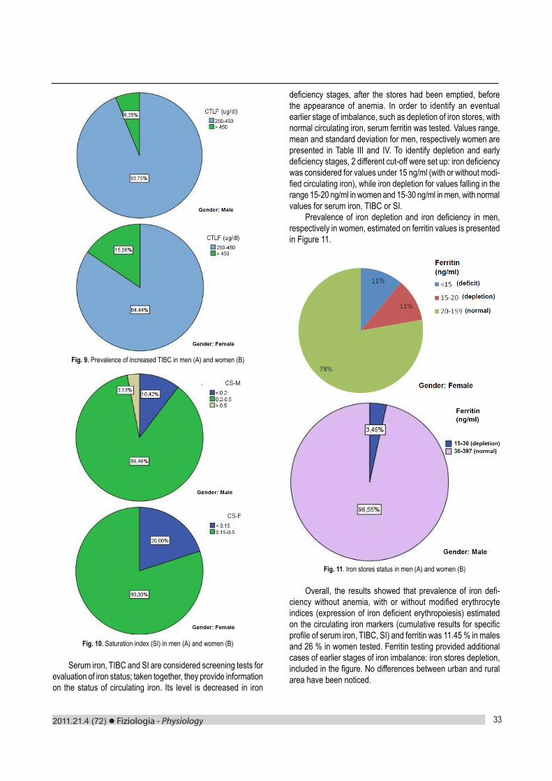
2011.21.4 (72) Fiziologia - Physiology 33
Fig. 9. Prevalence of increased TIBC in men (A) and women (B)
Fig. 10. Saturation index (SI) in men (A) and women (B)
Serum iron, TIBC and SI are considered screening tests for evaluation of iron status; taken together, they provide information on the status of circulating iron. Its level is decreased in iron
deficiency stages, after the stores had been emptied, before the appearance of anemia. In order to identify an eventual earlier stage of imbalance, such as depletion of iron stores, with normal circulating iron, serum ferritin was tested. Values range, mean and standard deviation for men, respectively women are presented in Table III and IV. To identify depletion and early deficiency stages, 2 different cut-off were set up: iron deficiency was considered for values under 15 ng/ml (with or without modi-fied circulating iron), while iron depletion for values falling in the range 15-20 ng/ml in women and 15-30 ng/ml in men, with normal values for serum iron, TIBC or SI.
Prevalence of iron depletion and iron deficiency in men, respectively in women, estimated on ferritin values is presented in Figure 11.
Fig. 11. Iron stores status in men (A) and women (B)
Overall, the results showed that prevalence of iron defi-ciency without anemia, with or without modified erythrocyte indices (expression of iron deficient erythropoiesis) estimated on the circulating iron markers (cumulative results for specific profile of serum iron, TIBC, SI) and ferritin was 11.45 % in males and 26 % in women tested. Ferritin testing provided additional cases of earlier stages of iron imbalance: iron stores depletion, included in the figure. No differences between urban and rural area have been noticed.

Fiziologia - Physiology 2011.21.4 (72)34
DISCUSSIONAll 141 subjects included in the study were eligible first time
donors, selected according to the standards and study precondi-tions set up. It would had been expected that all these persons present normal values for the evaluated biological markers, as long as they all fitted with criteria, declared no relevant history or behavior at risk for an imbalanced iron status and a good health appearance was identified by the responsible physician. Still, various abnormalities were found in 17 % of cases. The range of modifications went from isolated modified erythrocyte indices (MCV- 1 man) to different associations of modified markers, out of the considered reference range. Clear profiles for different stages of iron imbalance, from iron stores depletion to iron deficiency, with or without signs of iron deficient erythropoiesis, were found.
Thus, the study showed the insensitivity of hemoglobin as iron status marker. Eligible donors identified with different stages of iron imbalance, might evolve to iron deficiency anemia even without any other future donations because of their status, without influence of the first donation. Moreover, they would reach a lower hemoglobin level soon, considering the iron loss – approximately 200 – 250 mg – caused by each donation, if they came back to give blood, without any previous iron supplementation. This category of first time donors is not appropriate for giving blood regularly, as frequently as each 8 weeks. Without testing iron status with the occasion of first donation, they would believe that blood donation is the cause of anemia reached, and maybe they will never come back to give blood. This might affect the adher-ence of general population to regular or repeat blood donation programs. Combined screening with both hemoglobin, required by the standard, and iron status markers very effectively predicts first time donors at risk of subsequent deferral. This approach may support the donor management strategy, identifying those who might benefit from iron supplementation, increased donation intervals and changing from whole blood donation to apheresis; all these measures are meant to keep the recruited first time donors and prevent the anemia. Thus, the combined testing, performed in the frame of donor selection procedure, allow the physician to personalize the donation schedule according to the prospective donor characteristics. It is up to the institution
management to decide which iron status markers to use, based on the available technical and financial resources. Cost-efficiency is also a constraint. Appropriate information on healthy nutrition, life style and attitude after donation, with regards to diet and paying attention to any symptoms and signs potentially related to blood donation should be provided by the physician in charge with donors’ surveillance.
The responsibility to ensure donor well-being while facing continue pressure to prevent shortage of the appropriate blood supply has to be kept as priority by all the professionals involved in donor selection.
REFERENCES1. Directive 2002/98/EC of The European Parliament and of The Council of January 2003 setting standards of quality and safety for the collection, testing, processing, storage and distribution of human blood and blood components and amending Directive 2001/83/EC. Official Journal of the European Union, L 33, 8.2.2003: 30-40.2. Commission Directive 2004/33/EC of 22 March 2004 implementing Directive 2002/98/EC of the European Parliament and of the Council as regards certain technical requirements for blood and blood components. Official Journal of the European Union, L 91, 30.03.2004: 25-39.3. European Directorate for the Quality of Medicines & HealthCare, Council of Europe - Guide to the Preparation, Use and Quality Assurance of Blood Components, 16th edition 2010: 85-102, 199-212.4. Ministry of Health - Order number 1193 of 7 July 2007 endorsing regulations regarding information to be provided to blood donors, information to be provided by blood donors with the occasion of each donation and eligibility criteria for blood donation. Official Journal no. 540 of 8 August 2007.5. Klein HG, Anstee DJ. Blood Transfusion in Clinical Medicine- edition 11th, 2005: 1-12.6. Famulener LW. Anemia in professional donors: a study in a new phase of public health. The American Journal of Public Health, (N Y) 1922 May; 12(5): 376-377. 7. Boulton F. Donor selection. Towards evidence-based donor selection. Transfusion Today, 2007; 71: 4-5.8. De Sousa G. Ferro e dadores- ABO, 36 Out/Dez 2008: 5-11.9. Simon LT, Gerry PJ, Hooper EM. Iron stores in blood donors. JAMA, 1981; 245(20): 22-29. 10. Cable RG, Glynn SA, Kiss JE, Mast AE, Steele WR, Murphy EL, Wright DJ, Sacher RA, Gottschall JL, Tobler L, Simon TL. Iron deficiency in blood donors: the REDS-II Donor Iron Status Evaluation (RISE) study. Transfusion, 2011; Oct 24. doi: 10.1111/j.1537-2995.2011.03401.x.

2011.21.4 (72) Fiziologia - Physiology 35
STRATEGIE PENTRU ESTIMAREA POTENŢIALULUI DONATORILOR DE SÂNGE INIŢIALI PENTRU O CARIERĂ DE „DONATOR CU DONĂRI REGULATE”
REZUMATAnemia feriprivă este unul dintre cele mai frecvente motive pentru excluderea de la donare a donatorilor de sânge. Propunem o strategie pentru a identifica donatorii iniţiali cu risc de excludere la prezentări ulterioare şi metode de a-i păstra ca donatori eligibili.Au fost recrutaţi 141 donatori iniţiali, eligibili în conformitate cu standardele, (96 bărbaţi şi 45 femei). Au fost efectu-ate următoarele teste: hemoleucogramă, fierul seric (Fe), capacitatea totală de legare a fierului (CTLF), coeficientul de saturare a transferinei (CS) şi feritina. Datele au fost analizate pentru a identifica profilul donatorilor şi prevalenţa stadiilor deficitului de fier, fără anemie.În ansamblu, rezultatele au arătat că prevalenţa deficitului de fier, fără anemie, cu sau fără indici eritrocitari modificaţi (expresie a eritropoezei cu deficit de fier), estimată pe baza markerilor de evaluare a fierului cir-culant (Fe, CTLF, CS) şi a fierului de depozit (feritina) a fost de 11,45% la bărbaţi şi 26% la femeile testate. Screening-ul donatorilor iniţiali prin asocierea hemoglobinei, impusă de standard, cu markeri de evaluare a statusului fierului , la prima prezentare, poate prezice în mod eficient care dintre donatorii iniţiali admişi prezintă un risc de a fi exclus temporar la prezentări ulterioare, din cauza unei valori neconforme a hemoglobinei, cu sau fără anemie. Această strategie permite medicului să personalizeze programul de donare în funcţie de caracteristicile potenţialului donator şi să-i furnizeze consiliere adecvată.Cuvinte cheie: donator de sange, deficit de fier, strategie, rezerva de componente sanguine
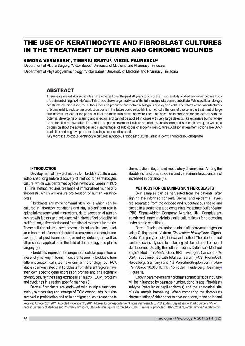
Fiziologia - Physiology 2011.21.4 (72)36
INTRODUCTIONDevelopment of new techniques for fibroblasts culture was
established long before discovery of method for keratinocytes culture, which was performed by Rheinwald and Green in 1975 (1). This method requires presence of immortalized murine 3T3 fibroblasts, which will ensure proliferation of human keratino-cytes.
Fibroblasts are mesenchymal stem cells which can be cultured in laboratory conditions and play a significant role in epithelial-mesenchymal interactions, de to secretion of numer-ous growth factors and cytokines with direct effect on epithelial proliferation, differentiation and formation of extracellular matrix. These cellular cultures have several clinical applications, such as in treatment of chronic decubital ulcers, venous ulcers, burns, coverage of post-traumatic tegumentary defects, as well as other clinical application in the field of dermatology and plastic surgery (2).
Fibroblasts represent heterogenous cellular population of mesenchymal origin, found in several tissues. Fibroblasts from different anatomical sites have similar morphology, but PCA studies demonstrated that fibroblasts from different regions have their own specific gene expression profiles and characteristic phenotypes, synthesizing extracellular matrix (ECM) proteins and cytokines in a region specific manner (3).
Dermal fibroblasts are endowed with multiple functions, mainly synthesizing and storage of ECM compounds, but also involved in proliferation and cellular migration, as a response to
chemotactic, mitogen and modulatory chemokines. Among the fibroblasts functions, autocrine and paracrine interactions are of increased importance (4).
METHODS FOR OBTAINING SKIN FIBROBLASTSSkin samples can be harvested from the patients, after
signing the informed consent. Dermal and epidermal layers are separated from the adipose and subcutaneous tissue and placed in a sterile test tube containing Phosphate Buffer Saline (PBS; Sigma-Aldrich Company, Ayrshire, UK). Samples are transferred immediately into sterile culture flasks for processing under sterile conditions.
Dermal fibroblasts can be obtained after enzymatic digestion using Collagenase IV (from Clostridium histolyticum; Sigma-Aldrich Company) or using the explant method. The latest method can be successfully used for obtaining cellular cultures from small skin biopsies. Usually, the culture media is Dulbecco’s Modified Eagle’s Medium (DMEM; Gibco BRL, Invitrogen, Carlsbad, CA, USA), supplemented with fetal calf serum (FCS; PromoCell, Heidelberg, Germany) and 1% Penicillin/Streptomycin mixture (Pen/Strep, 10,000 IU/ml; PromoCell, Heidelberg, Germany) (Figure 1).
Growth parameters and fibroblasts characteristics in culture will be influenced by passage number, donor’s age, fibroblasts subtype (reticular or papillar dermis) and the anatomical site of skin sample harvesting. When comparing the fibroblasts characteristics of older donor to a younger one, these cells tend
THE USE OF KERATINOCYTE AND FIBROBLAST CULTURES IN THE TREATMENT OF BURNS AND CHRONIC WOUNDS SIMONA VERMESAN1, TIBERIU BRATU1, VIRGIL PAUNESCU2
1Department of Plastic Surgery, “Victor Babes” University of Medicine and Pharmacy Timisoara2Department of Physiology-Immunology, “Victor Babes” University of Medicine and Pharmacy Timisoara
ABSTRACTTissue-engineered skin substitutes have emerged over the past 20 years to one of the most carefully studied and advanced methods of treatment of large skin defects. This article shows a general view of the full structure of a dermic substitute. While acellular biologic constructs are discussed, the authors focus on products that contain autologous or allogenic cells. The efforts of the manufacturers of biomaterial to reduce the production costs in the future could establish this method a the one of choice in the treatment of large skin defects, instead of the partial or total thickness skin grafts that were used until now. These create donor site defects with the potential developing of scarring and infection and cannot be applied in cases with very large defects, like extensive burns, where no donor sites are available. This article compares several cell-culture protocols, some aspects of tissue-engineering, as well as a discussion about the advantages and disadvantages of autologous or allogenic skin cultures. Additional treatment options, like UV-C irradiation and negative pressure dressings are also discussed.Key words: autologous keratinocyte cultures; autologous fibroblast cultures; artificial derm; chondroitin-6-phosphate
Received October 20th, 2011. Accepted November 3rd, 2011. Address for correspondence: Simona Vermesan, MD, PhD student, Department of Plastic Surgery, “Victor Babes” University of Medicine and Pharmacy Timisoara, Eftimie Murgu Square No. 2A, RO-300041, Timisoara, phone/fax: +40256220479, e-mail: [email protected]
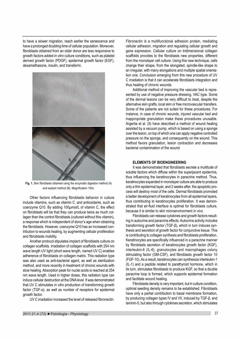
2011.21.4 (72) Fiziologia - Physiology 37
to have a slower migration, reach earlier the senescence and have a prolonged doubling time of cellular population. Moreover, fibroblasts obtained from an older donor are less responsive to growth factors added in vitro culture conditions, such as platelet derived growth factor (PDGF), epidermal growth factor (EGF), dexamethasone, insulin, and transferrin.
Fig. 1. Skin fibroblasts obtained using the enzymatic digestion method (A) and explant method (B). Magnification 100x.
Other factors influencing fibroblasts behavior in culture
include vitamins, such as vitamin C, and antioxidants, such as coenzyme Q10. By adding 100µmol/L of vitamin C, the effect on fibroblasts will be that they can produce twice as much col-lagen than the control fibroblasts (cultured without this vitamin), a response which is independent of donor’s age when obtaining the fibroblasts. However, coenzyme Q10 has an increased con-tribution to wounds healing, by augmenting cellular proliferation and fibroblasts mobility.
Another protocol stipulates implant of fibroblasts culture on collagen scaffolds. Irradiation of collagen scaffolds with 254 nm wave length UV light (short wave length, named UV C) enables adherence of fibroblasts on collagen matrix. This radiation type was also used as anti-bacterial agent, as well as sterilization method, and more recently in treatment of chronic wounds with slow healing. Absorption peak for nuclei acids is reached at 254 nm wave length. Used in higher doses, this radiation type can induce cellular destruction at the DNA level. It was demonstrated that UV C stimulates in vitro production of transforming growth factor (TGF-α), as well as number of receptors for epidermal growth factor.
UV C irradiation increased the level of released fibronectin.
Fibronectin is a multifunctional adhesion protein, mediating cellular adhesion, migration and regulating cellular growth and gene expression. Cellular culture on tridimensional collagen scaffolds provides to the fibroblasts new properties, different from the monolayer cell culture. Using this new technique, cells change their shape, from the elongated, spindle-like shape to an irregular, with many elongations and multiple spatial orienta-tion one. Conclusion emerging from this new procedure of UV C irradiation is that it can accelerate fibroblasts integration and thus healing of chronic wounds.
Additional method of improving the vascular bed is repre-sented by use of negative pressure dressing, VAC type. Some of the dermal lesions can be very difficult to treat, despite the alternative skin grafts, local skin or free microvascular transfers. Some of the patients are not suited for these procedures. For instance, in case of chronic wounds, injured vascular bed and inappropriate granulation make these procedures unusable. Argenta et al. (9) have described a method of wound healing assisted by a vacuum pump, which is based on using a sponge over the lesion, on top of which one can apply negative controlled pressure on the sponge, and consequently on the wound. This method favors granulation, lesion contraction and decreases bacterial contamination of the wound
ELEMENTS OF BIOENGINEERINGIt was demonstrated that fibroblasts secrete a multitude of
soluble factors which diffuse within the superjacent epidermis, thus influencing the keratinocytes in paracrine method. Thus, keratinocytes expanded in monolayer culture are able to produce only a thin epidermal layer, and 2 weeks after, the apoptotic pro-cess will destroy most of the cells. Dermal fibroblasts promoted a better development of keratinocytes from all epidermal layers, thus contributing to keratinocytes proliferation. It was demon-strated that air-fluid interface is optimal for fibroblasts culture, because it is similar to skin microenvironment in vivo.
Fibroblasts can release cytokines and growth factors result-ing in autocrine and paracrine effects. Autocrine activity includes transforming growth factor (TGF-β), which in turn induces syn-thesis and secretion of growth factor for conjunctive tissue. This is contributing to collagen synthesis and fibroblasts proliferation. Keratinocytes are specifically influenced in a paracrine manner by fibroblasts secretion of keratinocytes growth factor (KGF), interleukin-6 (IL-6), granulocytes and macrophages colony stimulating factor (GM-CSF), and fibroblasts growth factor 10 (FGF-10). As a result, keratinocytes can synthesize interleukin-1 (IL-1) and a peptide related to parathyroid hormone, which in its turn, stimulates fibroblasts to produce KGF, so that a double paracrine loop is formed, which supports epidermal formation and facilitate wound healing.
Fibroblasts density is very important, but in culture condition, optimal seeding density remains to be established. Fibroblasts have only a partial contribution to basal membrane formation, by producing collagen types IV and VII, induced by TGF-β, and laminin-5, but also through cytokines secretion, which stimulates

Fiziologia - Physiology 2011.21.4 (72)38
keratinocytes to produce components of basal membrane. Neovascularization and lymphangiogenesis are also impor-
tant processes for maintaining normal homeostasis of the skin. Members of vascular endothelial growth factor family (VEGF), such as VEGF-A, B, C, and D are produced by normal human fibroblasts and are important for regulation of endothelial cells proliferation, acting on specific receptors. It is well known that VEGF-A is involved in activation of resident endothelial cells and progenitor endothelial cells capable of vasculogenesis process. VEGF-B is less mitogenic for endothelial cells, while VEGF-C and -D have receptor specificity, binding to VEGF-R2 which is mediating angiogenesis and VEGF-R3, influencing lymphangiogenesis. A recent study analyzed dermal fibroblasts culture, overexpressing VEGF-C, together with human dermic microvascular endothelial cells. The result was that endothelial cells were activated and had and increased expression of matrix metalloproteinase 1 (MMP-1), which allowed the cells to digest surrounding collagen, invade the supportive gel, and form tubular connections between cells, similar to capillaries, suggesting their ability for neoangiogenesis.
Another important aspect which might be considered in treatment of chronic wounds using fibroblasts culture is that applying local heat can induce acceleration of integration and healing process. Wound microenvironment, comprising the exu-date generated by wound can have both beneficial, as well as inhibitory effects on wound healing process. It was demonstrated that the fluid surrounding acute wound favored in vitro prolifera-tion of fibroblasts, while the fluid resulting during a chronic wound, aminly venous ulcers, had inhibitory effects on wound healing process. Locally applied heat induces vasodilation, increases local blood flow and tissue oxygenation. Thus, this method can be useful for diminishing inhibitory activity of fluid secreted by a chronic, atonic wound. Up to present, it is not elucidated which of these factors is responsible of inhibitory activity exerted by chronic exudate, but an hypothesis suggest that the proteases can be involved, thus explaining the beneficial effect of local heat, which induces denaturation of these enzymes.
CONCLUSION AND DISCUSSIONFibroblasts used in cell cultures can be autologous or al-
logenic. The advantages of autologous fibroblasts are that there are no risks of developing graft versus host disease or intercur-rent infections. Also, use of autologous fibroblasts results in better therapeutic outcomes, such as dermal reconstruction and more esthetic scar, when compared to use of allogenic fibroblasts. However, allogenic fibroblasts present the advantage of time factor. While the autologous fibroblasts culture requires a lot of time for establishing the culture, and expanding the cells, the allogenic fibroblasts can be cryopreserved and are readily avail-able. They can also be used as biological dressing, with the role of optimization of healing process until the permanent grafting. Nevertheless, permanent grafting requires use of autologous fibroblasts (5-8).
The immunological impact of allogenic fibroblasts was also studied using the PCR method. Even when using skin substitutes
composed of keratinocytes and allogenic fibroblasts, foreign cells were not detectable 6 weeks after the grafting. Starting from these studies, a new hypothesis emerged, according to which allogeneic cells are replaced by host cells. Clinical studies results showed that there is no graft versus host disease in these cases, explained by many mechanisms. Neither induction of antibodies specific for class I HLA antigens expressed on allogenic cells was observed, nor proliferation of T cells in patients exposed to such antigens. Dermal fibroblasts lack class II HLA antigens, required for antigen presenting to immune cells. Moreover, it was observed that the in vitro-cultured fibroblasts and keratinocytes gradually lose their ability as antigen presenting cells (similar to Langerhans cells), along cellular passages, and they will not be present in cultured skin substitutes (9).
Presently, cell culture-based therapies have a proven benefic role on faster and better epithelization, in accelerating healing of skin defects and closure of atonic wounds. However, potential improvement of technical skills can increase in the future the effectiveness of this therapeutic procedure (10-12). For instance, improvement of cellular preservation techniques can increase the cellular life span, as well as possible duration of storage. In case of cryopreserved products, simplifying the tawing and washing techniques would make these cells more available for use. Moreover, studies need to be conducted concerning the optimal application regimen of these therapies, including number of useful application, and time interval between two consecutive procedures. Application field of these cellular therapies can be considerably enlarged if it is demonstrated that they can improve certain aspects of wounds, such as cosmetic, scar thickness, recurrence, etc (13). Even the studies regarding the mix allogenic keratinocytes culture deserve much consideration because they seem to be a more rapid and cheap alternative for autologous keratinocytes (14-16).
In the future, a marked development of present techniques for obtaining the cellular cultures can be achieved. However, we think that use of these methods in the entire world, and especially in our country, should be increased, due to the fact that they should not be regarded as the last alternative, after patients suffered for a long time of pain and non-closure of wounds. It is strongly recommended to establish methods for identification of patients which could benefit most of traditional or modern therapeutic approaches.
REFERENCES1. Rheinwald J, Green H. Serial cultivation of strains of human epidermal keratinocytes: formation of keratinizing colonies from single cells. Cell 1975; 331-44.2. Pianigiani E, Cherubini Di Simplicio F, Ierardi F. Dermal Allograft Plus Autologous Epidermal Graft: Evaluation of Two Different Techniques in Chronic, Nonhealing Leg Ulcer. Wounds 2004; 16(3):91-96.3. Eisenbud D, Huang NF, Luke S, Silberklang M. Skin Substitutes and Wound Healing: Current Status and Challenges. Wounds 2004; 16(1):2-17.4. Hansbrough JF, Franco ES. Skin replacements. Clin Plast Surg 1998; 3: 407-23.5. Greenleaf G, Hansbrough JF. Current trends in the use of allograft
2011.21.4 (72) Fiziologia - Physiology 39
skin for patients with burns and reflections on the future of skin banking in the United States. J Burn Care Rahabil 1994; 14: 428-31.6. Bravo D, Rigley TH, Gibran N, et al. Effect of storage and preserva-tion methods on viability in transplantable human skin allografts. Burns 2000; 26: 367-78.7. Flores F, Eaglstein WA, Kirsner RS. Hydroxyurea-induced leg ulcers treated with Apligraf. Ann Int Med 2000; 132: 417-18.8. Martin LK, Kirsner RS. Ulcers caused by bullous morphoea treated with tissue-engineered skin. Int J Dermatol 2003; 42: 402-4.9. Wong SL, Defranzo AJ, Morykwas MJ, Argenta LC. Loxoscelism and negative pressure wound therapy (vacuum-assisted closure): a clinical case series. Am Surg. 2009;75(11):1128-31.10. Streit M, Bohlen LM, Braathen LR. Ulcerative sarcoidosis success-Ulcerative sarcoidosis success-fully treated with Apligraf. Dermatology 2001; 202:367-70.11. Gath HJ, Hell B, Zarrinbal R, et al. Regeneration of intraoral defects after tumour resection with a bioengineered human dermal replacement
(Dermagraft). Plast Reconstruct Surg 2002; 109: 889-93.12. Gohari S, Gambla C, Healey M, et al. Evaluation of tissue-engi-neered skin (human skin substitute) and secondary intention healing in the treatment of full thickness wounds after Mohs micrographic or excisional surgery. Dermatol Surg 2002; 28:1107-14.13. Oka H. Changes in quantity of fibronectin from human skin fibroblasts with cellular aging. Ann Plast Surg 1985; 14: 248-57.14. Gerstein AD, Phillips TJ, Rogers GS, et al. Wound healing and aging. Dermatol Clin 1993; 11: 749-57.15. Krejci-Papa NC, Hoang A, Hansbrough JF. Fibroblast sheets enable epithelialization of wounds that do not support keratinocyte migration. Tissue Eng 1999; 5: 555-62.Marston WA, Hanft J, Norwood P, Pollak R. The efficacy and safety of Dermagraft in improving the healing of chronic diabetic foot ulcers: Results of a prospective randomized trial. Diabetes Care 2003; 26:1701-5.
FOLOSIREA CULTURILOR AUTOLOGE DE KERATINOCITE SI FIBROBLASTE IN TRATAMENTUL ARSURILOR SI PLAGILOR CRONICE
REZUMATSubstituentii dermici obtinuti prin bioinginerie au evoluat mult in ultimii 20 de ani si au devenit una dintre cele intens studiate si avansate metode de tratament ale unor defecte mari de substanta la nivelul pielii. In cadrul acestui articol se face o prezentare generala a structurii complete a unui substituent dermic, sunt discutate si avantajele substituentilor dermici acelulari, dar accentul este pus pe realizarea culturilor autologe de fibroblaste si keratinocite. Eforturile producătorilor de biomateriale de a reduce in viitor costurile de productie ale acestora ar putea implementa aceasta metoda drept cea de electie in tratamentul defectelor mari de substanta, in detrimentul grefelor de piele liberă sau despicata. Acestea din urma implică si crearea unui defect la zona donatoare, cresc riscul de infectie si nu pot fi aplicate in cazurile cu defecte foarte mari, ca si arsurile extinse, unde nu mai exista zone donatoare. Acest articol prezinta in mod comparativ mai multe protocoale de obtinere a culturilor celulare, anumite elemente de bioinginerie, cat si o discutie asupra avantajelor si dezanvantajelor de culturi autologe sau alogene. Sunt prezentate de asemenea si modalitati aditionale benefice de tratament, precum iradierea cu lumina UV C sau bandajele cu presiune negativa.Cuvinte cheie: culturi de keratinocite autologe, cuturi de fibroblaste dermice autologe, substituent dermic, condroitin-6-sulfat
Fiziologia - Physiology 2011.21.4 (72)40
MARKERS OF OXIDATIVE STRESS IN CHRONIC RENAL FAILURE LAVINIA VOINEAGU1, CECILIA ADUMITRESI1, ILEANA ION1, ALINA DOBROTĂ2, VICTORIA BADEA3, LILIANA TUTĂ1
1Faculty of Medicine - ”Ovidius” University, Constanta, Romania2Regional Center of Blood Transfusion Constanta, Romania3Faculty of Dental Medicine - ”Ovidius” University, Constanta, Romania
ABSTRACTOxidative stress is defined as an imbalance of oxidant and antioxidant mechanisms and is generally thought to contribute to the development of many pathological conditions such as hypertension, heart diseases, Parkinson’s disease and even cancer. There are different markers which can be used to assess oxidative stress. The reduced glutathione (GSH), a major antioxidant, the determination of the GSH/GSSG ratio and the quantification of GSSG are useful indicators of oxidative stress in cells and tissues but more stable molecules like F2-isoprostane and 8OHdeoxiguanosine (8OHdG) might present some advantages in assessing this condition In our study we have chosen the renal pathology to show the changes in oxidative markers concentrations on a group of 52 patients with chronic renal failure from Constanta County Hospital. We tried to establish whether there is a significant correlation between the concentrations of reduced glutathione (GSH), its oxidized form (GSSG) and GSH/GSSG ratio) and, on the other side, F2-isoprostane and 8OhdG concentrations. Tests were performed from whole blood and urine, using ELISA kits of reagents.A significant difference between patients with renal failure and healthy controls was found in the levels of GSH, GSSG, and GSH/GSSG ratio. Compared with controls, the patients with renal insufficiency had a significantly higher level of urinary 8OHdG and isoprostane. Increased concentrations suggest an excess of free radicals and oxidant injury. A significant correlation was found between GSH, GSSG, GSH/GSSG ratio, isoprostane and 8OHdG.Key words: oxidative stress, chronic renal failure, F2-isoprostane, 8OHdeoxiguanosine.
Received August 15th, 2011. Accepted September 5th, 2011. Address for correspondence: Lavinia Voineagu, University ”Ovidius”, Constanta, Faculty of Medicine, Universitătii Street, C2, Constanta 900527, phone/fax: +40241-672899, e-mail: [email protected]
INTRODUCTIONThe broad definition of reactive oxygen species (ROS) in-
cludes oxygen-containing species that are all capable of reacting with proteins and lipids to produce abnormal cellular responses. ROS include molecules such as superoxide, hydroxyl radical, H2O2, peroxynitrite and nitric oxide. Oxidative stress is defined as an imbalance of oxidant and antioxidant mechanisms and is generally thought to contribute to the development of many pathological conditions such as hypertension, chronic renal failure, heart disease, Parkinson’s disease and even cancer.
There are several key enzymes and cytokines in vascular and tubular cells within the kidney that produce increased amounts of reactive oxygen species (NADPH oxidase, xanthine oxidase, cyclo-oxygenase, lipoxygenase). Renal sources for ROS are activated macrophages, vascular cells, and various glomerular cells. ROS may affect cells of the host organism, especially at sites of inflammation, in addition to playing a role in the defense system against other agents. This effect plays a role in a variety of renal diseases such as glomerulonephritis and tubulointerstitial nephritis, which can contribute to proteinuria and other conditions. ROS are also thought to contribute to the patho-genesis of ischemia reperfusion injury in the kidney. This sug-gests that the kidney may be particularly susceptible to oxidative stress. Additional uremia-related metabolic aberrations such as
intravenous iron exposure, biocompatibility changes associated with dialysis, and hyper homocystinemia may also contribute to increased oxidative stress. Renal anemia is another contributor to oxidative stress in patients with chronic renal failure.
Reduced glutathione (γ-glutamyl-cysteine-glycine) has been identified as the prominent, highly effective radical-eliminating system. When this is inhibited, there is significant delay in the elimination of free radicals, illustrating a defect in the antioxi-dant forces. GSH is a major antioxidant that provides reducing equivalents for the glutathione peroxidase (GPx). In the GPx catalyzed reaction, the formation of a disulfide bond between two GSH molecules gives rise to oxidized glutathione (GSSG). When cells are exposed to increased levels of oxidative stress, GSSG will accumulate and the ratio of GSH to GSSG will decrease. Therefore, the determination of the GSH/GSSG ratio and the quantification of GSSG are useful indicators of oxidative stress in cells and tissues.
Evaluation of oxidative stress in a clinical setting is difficult because free radicals have very short half-lives (in seconds). In contrast, there are actual, more stable marker molecules that have longer half-lives, ranging from hours to weeks, which can be used to assess oxidative stress.
For example, F2-isoprostane, which is a nonenzymatic, free radical-catalyzed isomer of cyclooxygenase-derived enzymatic

2011.21.4 (72) Fiziologia - Physiology 41
products of arachidonic acid, can be found in both urine and plasma. It is a stable end product of arachidonic acid oxidation and can be measured with high sensitivity and specificity. Iso-prostane increased level was associated with hepatorenal syn-drome, rheumatoid arthritis, atherogenesis and carcinogenesis. Several of these compounds possess potent biological activity, as evidenced mainly through their renal vasoconstrictive effects, and have short half-lives. Both human and experimental studies have indicated associations of isoprostanes and severe inflammatory conditions, ischemia-reperfusion, diabetes and atherosclerosis. Bioactive 15-isoprostane F2t are continuously formed in various tissues and large amounts of these potent compounds are found unmetabolized in their free acid form in the urine in normal basal conditions with a wide inter-individual variation.
8-OH-dG (8-hydroxy-2-deoxy Guanosine) is produced by the oxidative damage of DNA by reactive oxygen and nitrogen species. Although more than 20 base lesions have been identi-fied, only a fraction of these have received appreciable study, most notably 8-oxo-2’deoxyguanosine. In complex samples such as plasma, cell lysates, and tissues, 8-OH-dG can exist as either the free nucleoside or incorporated in DNA. Once the blood enters the kidney, free 8-OH-dG is readily filtered into the urine, while larger DNA fragments remain in the bloodstream. Its levels are increased by smocking and excessive physical exercise
MATERIALS AND METHODSThe analyses were conducted in two groups of individuals:
Group of healthy controls – HC (n = 40) recruited from among the patients with biochemical parameters in normal range and without any history of renal disease (age: 56.56 ± 4.95 years). This control group comprises individuals who have not been exposed to the main exogenous factors of ROS production. The individuals in the control group were not on any prescribed medications (including anti-oxidants), no subject was an active smoker, the individuals did not participate in regular intense exercise, and all had moderate exposure to the sun; Group of patients with chronic renal failure – CRF (n = 52) were recruited from the Constanta County Hospital, (age: 65.57 ± 9.36 years). The CRF group comprised a variety of disease etiologies, and with glomerular filtration rate between 30 and 89 ml/min/1.73 m2. All participants gave fully informed consent to participation in the study.
Samples we have used are whole blood and urine. Blood was obtained by venous puncture and collected in EDTA tubes for GSH and GSSG measurement. Urine was collected as the first morning void. It was obtained for 8-OH-dG and isoprostane detection.
Freezing and thawing will lyse red blood cells and maximize the concentration of GSSG in the sample. Blood samples that have been frozen without prior treatment with the scavenger are not ideal for GSSG assay, although the scavenger can be added to frozen samples before they thaw. This will result in a reduction of GSSG and an increase in GSH in the sample. GSH and GSSG are relatively stable in intact ”resting” cells for up 24 hr at 4°C. The stability of ”elevated” GSSG in intact red blood cell has not been
determined. It is recommended that blood samples are treated with scavenger as soon as possible and frozen immediately. Upon disruption of the cell, GSH is rapidly oxidized.
Quantitative and sensitive detection of oxidative stress markers have been carried out using ELISA both for blood and urine sample. ELISA conventional method is excellent in light of its sensitivity and great convenience. It is a competitive assay that can be used for the quantification of isoprostane in urine. The plates are coated with specific polyclonal antibodies. The tracer consists in an enzyme linked with the isoprostane. The analytes in the samples are competes with tracer for binding to antibodies coated on the microplate. The activity of the enzyme results in color development when substrate is added, with intensity of the color inversely proportional to the amount of unconjugated analyte in the samples.
Specific ELISA kits were used for measuring serum levels of isoprostane, according to the manufacturer’s protocol. Each sample was tested in duplicate. Regarding the reproducibility, we measured isoprostane levels more than twice in seven serum samples; as a result, the mean percentage coefficient of variation was 3.6%.
Using the SOCR program we perform statistical analysis of the data, searching for correlations between the GSH, GSSG, GSH/GSSG ratio and isoprostane, 8OHdG. A p value less than 0.05 was considered to indicate statistical significance.
RESULTS AND DISCUSSIONSA significant difference between patients with renal failure
and healthy controls was found in the levels of GSH, GSSG, and GSH/GSSG ratio. The renal patients had significantly lower levels of GSH (p<0.001) (Figure 1). GSH is an important low-molecular-weight antioxidant and, as such, low values would indicate oxidative stress. Healthy people are protected against free radicals by several defense mechanisms. Reduced GSH is the most important intracellular scavengers of free radicals. GSH serves as a reductant in oxidation reactions resulting in the formation of GSSG. High values of GSSG therefore indicate oxidative stress (p<0.04) (Figure 2). A decrease in GSH/GSSG ratio (p<0.001) was taken as indicative of oxidative damage. The GSH/GSSG ratio is considered one of the best ways to determine the antioxidant capacity of cells, and any decreases suggest a strong oxidant effect (Figure 3).
Fig. 1. Average level, standard deviation, and standard error of GSH for healthy control and patients with chronic renal failure

Fiziologia - Physiology 2011.21.4 (72)42
Fig. 2. Average level, standard deviation, and standard error of GSSG for healthy control and patients with chronic renal failure
Fig. 3. Average level, standard deviation, and standard error of GSH/GSSG ratio for healthy control and patients with chronic renal failure
Compared with controls, the patients with renal insufficiency had a significantly higher level of urinary 8-OH-dG (p<0.002) and isoprostane (p<0.001). Increased F2-Isoprostanes concentra-tion suggests an excess of free radicals and oxidant injury. Remarkably, 90% (51/52) of patients with CRF exhibited elevated 8-isoprostane levels and isoprostane, while none of the healthy individuals showed increased levels. Thus, almost all patients exhibited elevated levels of markers that could discriminate patients from normal controls.
A significant negatively correlation was found between GSH and isoprostane for controls (r = -0.67) and patients with CRF too (r = -0.85). In addition, positive correlation were found between GSSG and isoprostane (r = 0.75). We found a significant negatively correlation between GSH/GSSG ratio and isoprostane (-0.78) (Figure 5). These may be attributable to the inflammatory process, but vascular endothelial dysfunction is a very important event in CRF, and oxidative stress induced by vasospasm is the most characteristic sign that reflects this dysfunction. Ischaemia and reperfusion injury during renal disease can generate reactive oxygen species that may result in vascular endothelial damage. In addition, isoprostane itself is a potent vasoconstrictor, has platelet pro-aggregate functions and stimulates endothelial cells to bind monocytes, which may promote vascular oblitera-tion, inflammation and spasm. Therefore, the finding that high
isoprostane levels correlated with low GSH/GSSG ratio and high GSSG suggests that excessive oxidative stress is related to vascular damage in CRF.
It has already been shown that patients with diabetes mel-litus and renal insufficiency undergo oxidative stress, as indicated by decreased plasma superoxide dismutase, increased serum 8-OHdG or comet assay parameters [9]. For 8OHdG we found a negatively correlation between GSH/GSSG ratio and 8OHdG (r = -0.75) (Figure 4). In this study, we confirmed that blood 8-OHdG in CRF patients was significantly higher than in general subjects. These findings suggest that circulating 8-OHdG might reflect oxidative stress in renal tissue.
Fig. 4. Linear regression curve of GSH versus isoprostane
Fig. 5. Linear regression curve of GSH/GSSg versus 8OhdG
CONCLUSIONS
1. Patients with chronic renal failure have diminished re-sponse to oxidative stress due, probably, to a decrease in the antioxidant capacity or to a continued consumption of GSH in oxidative processes; the mechanisms underlying this decrease, however, are not well established.
2. A significant difference between patients with renal failure and healthy controls was found in the levels of GSSG, and GSH/GSSG ratio.
3. Patients with renal insufficiency had a significantly higher level of urinary 8-OH-dG.
4. Patients with chronic renal failure have an increased isoprostane concentration which suggests an excess of free

2011.21.4 (72) Fiziologia - Physiology 43
radicals and oxidant injury. 5. A progressive reduction of GSH/GSSG ratio and a
progressive increase in lipid peroxidation products such as isoprostane provide a strong evidence of increasing oxidative stress in chronic renal failure.
6. For 8OHdG we found a negatively correlation between GSH/GSSG ratio and 8OhdG (r = -0.75) (Figure 4).
7. Isoprostane is authentic biomarkers of lipid peroxidation and can be used as potential in vivo indicators of oxidant stress in renal failure.
8. The urinary excretion of DNA repair product 8OHdG has been proposed as a noninvasive biomarker as oxidative DNA damage in humans in vivo.
9. A larger number of biomarkers bring more information on the extent of tissular damage in oxidative stress.
REFERENCES1. Beswick RA, Dorrance AM, Leite R, et al. NADH/NADPH oxidase and enhanced superoxide production in the mineralocorticoid hypertensive rat. Hypertension, 2001; 38: 1107-11.2. Dobashi K, Ghosh B, Orak JK, et al. Kidney ischemia-reperfusion: modulation of antioxidant defenses. Mol Cell Biochem., 2000; 205(1-2): 1-11.3. Elmarakby AA, Loomis ED, Pollock JS, et al. NADPH oxidase in-hibition attenuates oxidative stress but not hypertension produced by chronic ET-1. Hypertension, 2005; 45: 283-7.4. Fassett RG, Driver R. Comparison of markers of oxidative stress, inflammation and arterial stiffness between incident hemodialysis and peritoneal dialysis patients – an observational study. BMC Nephrology, 2009; 10: 8.5. Li L, Fink GD, Watts SW, et al. Endothelin-1 increases vascular
superoxide via endothelin A-NADPH oxidase pathway in low-renin hypertension. Circulation, 2003; 107: 1053-58.6. Kolagal V, Karnam S. Determination of oxidative stress markers and their importance in early diagnosis of uremia-related complications. Indian Journal of Nephrology, 2009; 19(1):8-12.7. Loomis ED, Sullivan JC, Osmond DA, et al. Endothelin mediates superoxide production and vasoconstriction through activation of NADPH oxidase and uncoupled nitric-oxide synthase in the rat aorta. J. Pharmacol. Exp. Ther., 2005; 315: 1058-64.8. Lykkesfeldt J. Malondialdehyde as biomarker of oxidative damage to lipids caused by smoking. Clin Chim Acta, 2007; 380: 50-58. 9. Pollock DM. How does endothelin induce vascular oxidative stress in mineralocorticoid hypertension? Clinical Science, 2006; 110: 205-206.10. Montuschi P, Barnes P, Roberts J. Isoprostanes: markers and media-tors of oxidative stress. The FASEB Journal, 2004; 18: 1517-19.11. Naito Y, Masaichi C, Kato Y, et al. Oxidative stress markers. Anti-aging Medicine, 2010; 25: 3.12. Reed MC, Thomas RL, Pavisic J, et al. A mathematical model of glutathione metabolism. Theor Biol Med Model, 2008; 28: 5-8.13. Romeu M, Nogues R, Marcas L, Sánchez-Martos V, Mulero M, Martinez-Vea A, Mallol J, Giralt M. Evaluation of oxidative stress biomarkers in patients with chronic renal failure: a case control study. BMC Research Notes, 2010; 3: 20.14. Wu LL, Chiou CC, Chang PY, Wu JT. Urinary 8-OHdG: a marker of oxidative stress to DNA and a risk factor for cancer, atherosclero-sis and diabetics. Clin Chim Acta., 2004; 339(1-2): 1-9.15. Mastalerz-Migas A, Steciwko A, Pokorski M, et al. What infl u-What influ-ences the level of oxidative stress as measured by 8-hydroxy-2’-deoxyguanosine in patients on hemodialysis? J Physiol Pharmacol., 2006; 57(4): 199-205. 16. Yoshihisa T, Yukihiko H, Masayo I. No inhibitory effects of (−)-epi-gallocatechin gallate and lycopene on spontaneous hepatotumorigene-sis in c3h/hen mice. Fukushima J Medical Science., 2010; 56(1): 7-27.
MARKERI AI STRESULUI OXIDATIV IN INSUFICIENTA RENALA CRONICA
REZUMATStresul oxidativ, consecinţă a dezechilibrului între intensitatea acţiunii factorilor oxidanţi si cea a mecanismelor protective antoixidante, s-a dovedit a fi implicat în patogeneza şi evoluţia unui număr semnificativ de afecţiuni cum ar fi hipertensiunea, bolile cardio-vasculare, boala Parkinson, cancerul, etc. Există un număr considerabil de markeri destinaţi studiului stresului oxidativ. Măsurarea concentraţiei glutationului redus (GSH), unul dintre cei mai importanţi apărători intracelulari impotriva acţiunii speciilor reactive ale oxigenului, cuantificarea formei sale oxidate (GSSG), determinarea raportului GSH/GSSG reprezintă printre markerii cei mai utilizaţi în acest scop. Au fost descrise însă şi molecule mai stabile, cum ar fi F2-isoprostanul si 8OHdeoxiguanozina (8OHdG) ce pot aduce o serie de avantaje în aprecierea intensităţii acţunii factorilor oxidanţi.Studiul nostru a fost efectuat pe un lot alcătuit din pacienţi aflaţi în insuficienţă relală cronică, internaţi în secţia de nefrologie a Spitalului Clinic Judetean Constanta. Am urmărit să stabilim dacă există o corelatie între evoluţia concentraţiei markerilor de stres oxidativ reprezentaţi prin: GSH, GSSG, raport GSH/GSSG, F2-isoprostan, respectiv 8OHdG. Testele s-au realizat din sânge integral si urină, folosind truse de reactivi pentru metode ELISA. Am găsit diferente semnificative între pacientii cu insuficientă renală cronică si lotul de control privind concentraţiile GSH, GSSG si raportul GSH/GSSG. Pacientii cu insuficientă renală cronică au prezentat nivele de 8OHdG si isoprostan semnificativ mai ridicate decât cele ale lotului de control. Concentratiile crescute ale acestora sugerează existenţa unui exces de radicali liberi si atac oxidativ şi/sau o diminuare semnificativă a puterii de apărare antioxidantă. Valorile GSH, GSSG si ale raportului GSH/GSSG s-au corelat cu cele ale F2-isoprostanului si ale 8OHdG.Cuvinte cheie: stres oxidativ, insuficientă renală cronică, F2-isoprostan, 8OHdeoxiguanozină

Fiziologia - Physiology 2011.21.4 (72)44