Fiziologia nr 1 (95) 2018 A4 46 pag cu 6 art pentru...
46
FIZIOLOGIA physiology FOUNDING EDITOR FRANCISC SCHNEIDER CHIEF EDITOR CARMEN PANAITESCU CO-CHIEF EDITORS CARMEN TATU FLORINA BOJIN ASSOCIATE EDITORS MIHAI NECHIFOR SORIN RIGA EXECUTIVE EDITORS GABRIELA TANASIE DACIANA NISTOR CALIN MUNTEAN EDITORIAL BOARD ARDELEAN AUREL (Arad) BĂDĂRĂU ANCA (Bucureşti) BENEDEK GYORGY (Szeged) BENGA GHEORGHE (Cluj) COJOCARU MANOLE (Bucureşti) GĂLUȘCAN ATENA (Timișoara) IANCAU MARIA (Craiova) MIHALAŞ GEORGETA (Timişoara) MUNTEAN DANINA (Timişoara) MUREŞAN ADRIANA (Cluj) NESTIANU VALERIU (Craiova) OPREA TUDOR (New Mexico) PANAITESCU CARMEN (Timişoara) PĂUNESCU VIRGIL (Timişoara) PETROIU ANA (Timişoara) PODARIU ANGELA CODRUTA (Timișoara) RÂCZ OLIVER (Kosice) RIGA DAN (Bucureşti) SABĂU MARIUS (Tg. Mureş) SAULEA I. AUREL (Chişinău) SIMIONESCU MAIA (Bucureşti) SWYNGHEDAUW BERNARD (Paris) TANGUAY M. ROBERT (Canada) TATU ROMULUS FABIAN (Timişoara) VLAD AURELIAN (Timişoara) VOICU VICTOR (Bucureşti) ZĂGREAN LEON (Bucureşti) ACCREDITED BY CNCSIS - B+CATEGORY ■ CODE 240 http://www.ebscohost.com/titleLists/a9h-journals.pdf Fiziologia (Physiology) is issued quarterly Printed at Editura EUROSTAMPA www.eurostampa.ro Bd. Revoluţiei din 1989 nr. 26, Timişoara Tel/fax: 0256-204816 ISSN 1223-2076
Transcript of Fiziologia nr 1 (95) 2018 A4 46 pag cu 6 art pentru...
FIZIOLOGIA p h y s i o lo g y
FOUNDING EDITOR FRANCISC SCHNEIDER CHIEF EDITOR CARMEN PANAITESCU CO-CHIEF EDITORS CARMEN TATU FLORINA BOJIN ASSOCIATE EDITORS MIHAI NECHIFOR SORIN RIGA EXECUTIVE EDITORS GABRIELA TANASIE
DACIANA NISTOR CALIN MUNTEAN
E D I T O R I A L B O A R D
ARDELEAN AUREL (Arad) BĂDĂRĂU ANCA (Bucureşti) BENEDEK GYORGY (Szeged) BENGA GHEORGHE (Cluj) COJOCARU MANOLE (Bucureşti) GĂLUȘCAN ATENA (Timișoara) IANCAU MARIA (Craiova) MIHALAŞ GEORGETA (Timişoara) MUNTEAN DANINA (Timişoara) MUREŞAN ADRIANA (Cluj) NESTIANU VALERIU (Craiova) OPREA TUDOR (New Mexico) PANAITESCU CARMEN (Timişoara)
PĂUNESCU VIRGIL (Timişoara) PETROIU ANA (Timişoara) PODARIU ANGELA CODRUTA (Timișoara) RÂCZ OLIVER (Kosice) RIGA DAN (Bucureşti) SABĂU MARIUS (Tg. Mureş) SAULEA I. AUREL (Chişinău) SIMIONESCU MAIA (Bucureşti) SWYNGHEDAUW BERNARD (Paris) TANGUAY M. ROBERT (Canada) TATU ROMULUS FABIAN (Timişoara) VLAD AURELIAN (Timişoara) VOICU VICTOR (Bucureşti) ZĂGREAN LEON (Bucureşti)
ACCREDITED BY CNCSIS - B+CATEGORY ■ CODE 240
http://www.ebscohost.com/titleLists/a9h-journals.pdf
Fiziologia (Physiology) is issued quarterly
Printed at Editura EUROSTAMPA www.eurostampa.ro
Bd. Revoluţiei din 1989 nr. 26, Timişoara Tel/fax: 0256-204816
ISSN 1223-2076
2 Fiziologia - Physiology • 2018.28.1(95)
Instructions to Authors
Submission: Only original papers in English are considered and should be sent to the following address: [email protected] Manuscripts should be submitted by e-mail only, written in Microsoft Word 97 or later versions. Conditions: AII manuscripts are subject to editorial review. Manuscripts are received with the explicit understanding that they are not under simultaneous consideration by any other publication. Submission of an article for publication implies the transfer of the Copyright from the author the publisher upon acceptance. Accepted papers become the permanent property of "Fiziologia" (Physiology) and may not be reproduced by any means, in-whole or in part, without the written consent of the publisher. It is the author's responsibility to obtain permission to reproduce illustrations, tables, etc. from other publications. Arrangement:
Title page: The first of each paper should indicate the title, the authors' names and their affiliation(s). A short title for use as running head is also required.
Keywords: for indexing purposes, a list of 3-5 keywords in English and Romanian is essential.
Corresponding author: Indicate the full name, the email address and the phone number.
Abstract: Each paper needs abstract and title in Romanian and English language, fonts size 9, Arial Narrow.
Body text: fonts size 10, Arial Narrow. Small type: Paragraphs which can or must be set
in smaller type (case histories, test methods, etc.) should be indicated with a „p" (petit) in the margin on the left-hand side.
Footnotes: Avoid footnotes. When essential, they are numbered consecutively and typed at the foot of the appropriate page, fonts size 8, Arial Narrow.
Tables and illustrations: Tables (numbered in Roman numerals) and illustrations (numbered in Arabic numerals) should be prepared on separate sheets, fonts size 9, Arial Narrow. Tables require a heading, and figures a legend, also prepared on a separate sheet. For the reproduction of illustrations, only good drawings and original photographs can be accepted; negatives or photocopies cannot be used. When possible, group several illustrations on one block for reproduction (max. size 140x188 mm) or
provide crop marks. On the back of each illustration indicate its number, the author's name, and article title.
References: In the text identify references by Arabic figures, (in brackets), fonts size 9, Arial Narrow. Material submitted for publication but not yet accepted should be noted as "unpublished data" and not be included in the reference list. The list of references should include only those publications which are cited in the text. The references should be numbered and arranged alphabetically by the authors' names. The surnames of the authors followed by initials should be given. There should be no punctuation signs other than a comma to separate the authors. When there are more than 3 authors, the names of the 3 only are used, followed by "et al" abbreviate journal names according to the Index Medicus system. (also see International Committee of Medical Journal Editors: Uniform Requirements for manuscripts submitted to biomedical journals. Ann Intern Med 1982; 96: 766-771).
Examples: (a) Papers published in periodicals: Kauffman
HF, van der Heide S, Beaumont F, et al: Class-specific antibody determination against Aspergillus fumigatus by mean of the enzyme-linked immunosorbent assay. III. Comparative study: IgG, IgA, IgM, ELISA titers, precipitating antibodies and IGE biding after fractionation of the antigen. Int Arch Allergy Appl Immunol 1986; 80:300 - 306.
(b) Monographs; Matthews DE, Farewell VT: Using and Understanding Medical Statistics. Basel, Karger, 1985.
(c) Edited books: Hardy WD Jr, Essex M:.FeLV-inducted feline acquired immune deficiency syndrome: A model for human AIDS; in Klein E(ed): Acquired Immunodeficiency Syndrome. Prag Allergy, Busel, Karger, 1986, vol 37,353 - 376.
Galley proofs: unless indicated otherwise, galley proofs are sent to the first-named author and should be returned with the least possible delay. Alternations made in galley proofs, other than the corrections of printer's errors, are charged to the author. No page proofs are supplied.

Fiziologia - Physiology • 2018.28.1(95) 3
CONTENTS
1. CAR T Cells Therapy vs. TILs Therapy: The Future of Cancer Immunotherapy ........................................ 4
Szekely FAE, Zogorean R, Anghel S, Paunescu V 2. Faculty Perception for the Need of Change in I Year MBBS Indian Physiology Practical Curriculum ....... 16
Smita R Sorte, Sachin B Rathod 3. Impact of Isotonic Hand Grip Exercise on Blood Pressure and Sympathetic Response
in Prehypertensives ..................................................................................................................................... 21 Saravanan Murugan, Singh S K
4. Misdiagnosis of Aspirated Bronchial Foreign Body. A Case Report ......................................................... 31
Cioboata R, Bazavan I, Nitu M, Georgescu M, Olteanu M 5. Clinical and Etiological Aspects of the Confusional Syndrome ................................................................ 35
Albu VC, Sandu RE, Pirscoveanu D, Bumbea AM, Parvulescu OC, Bogdan C, Florescu AO, Balseanu TA, Enescu A, Mirea CS, Barbulescu AL
6. Clinical Patterns of Neuropsychiatric Systemic Lupus Erythematosus ................................................... 40
Sandu RE, Barbulescu AL, Florescu AO, Pirscoveanu D, Bumbea AM, Parvulescu OC, Tartea AE, Bogdan C, Balseanu TA, Enescu A, Mirea CS, Albu VC, Burada E
CUPRINS
1. Terapia cu celule CAR-T vs. TIL: Viitorul imunoterapiei în cancer .............................................................. 4
Szekely FAE, Zogorean R, Anghel S, Paunescu V 2. Percepția academică referitoare la nevoia de schimbare a curriculum de fiziologie practică
pentru studenții MBBs din anul I ................................................................................................................ 16 Smita R Sorte, Sachin B Rathod
3. Impactul exercițiilor isotonice hand grip privind presiunea de sânge și răspunsul simpatic
la pre-hipertensivi ........................................................................................................................................ 21 Saravanan Murugan, Singh S K
4. Diagnostic greșit al unui corp strain aspirat bronșic. Raport de caz ....................................................... 31
Cioboata R, Bazavan I, Nitu M, Georgescu M, Olteanu M 5. Aspecte clinico-etiologice ale sindromului confuzional ............................................................................ 35
Albu VC, Sandu RE, Pirscoveanu D, Bumbea AM, Parvulescu OC, Bogdan C, Florescu AO, Balseanu TA, Enescu A, Mirea CS, Barbulescu AL
6. Afectarea neuropsihiatrică în lupus eritematos sistemic - aspecte clinice .............................................. 40
Sandu RE, Barbulescu AL, Florescu AO, Pirscoveanu D, Bumbea AM, Parvulescu OC, Tartea AE, Bogdan C, Balseanu TA, Enescu A, Mirea CS, Albu VC, Burada E
4 Fiziologia - Physiology • 2018.28.1(95)
CAR T CELLS THERAPY VERSUS TILs THERAPY: THE FUTURE OF CANCER IMMUNOTHERAPY SZEKELY FLAVIA ANNE-ELISE1,2, ZOGOREAN ROXANA1, ANGHEL SIMONA1,3, GAVRILIUC OANA1,3, BOJIN FLORINA1,3, PĂUNESCU VIRGIL1,3 1Clinical Emergency County Hospital “Pius Brînzeu” Timișoara, Centre for Cellular and Gene Therapies in the Treatment of Cancer – OncoGen 2Emergency Hospital for Children Louis Țurcanu Timișoara 3”Victor Babes” University of Medicine and Pharmacy Timisoara, Department of Functional Sciences, Immunology Discipline
ABSTRACT Worldwide, cancers are one of the main leading causes of death and the conventional treatment has its own limits. Researches from immunotherapy field are focusing on discovering feasible alternative therapeutic approaches for treating this disorder. In cancers, immunotherapy has the role to enhance anti-tumor effect of the immune cells involved in the process. Depending on the type of targeted antigens, side effects of this therapy may vary from local and mild to systemic and more severe reactions. The principle of CAR T cells and TILs immunotherapies is based on using T cells that are specific for a certain antigen and re-infusing back to the patient after those cells were expended ex vivo, in order to cause tumor destruction. Association of these therapies with a lymphodepleting regime and T cell growth factor IL-2 leads to a potent effect on patients. Within this article, we make an overview of the CAR T cell therapy and TILs therapy, by describing the principles of this immunotherapies, their clinical applications, as well as their advantages, disadvantages and future perspectives. Key words: CAR T cells, TILs, immunotherapies, cancer
INTRODUCTION
An overview of cancer immunotherapy strategies During the last decades, cancer has become one of the
primary diseases that threaten human lives, metastatic cancers remaining an incurable disease for the most patients. Each year, approximately 8,8 million people are dying of cancer, this disease having the highest frequency in low-income and middle-income countries (LMICs), where the number of cancer cases is rising most rapidly. By 2035, the cancer incidence is estimated to double, especially in LMICs because of the population aging and the increasing exposure to risk factors [1, 2].
The conventional therapy strategies against cancer have incorporated surgery, chemotherapeutic agents and radiotherapy to eliminate the tumor mass. Although many of these therapies offered substantial benefits, the high relapse rate associated with poor prognosis, continues to be a major challenge [3, 4]. Because of the limited effectiveness of
these conventional antitumor treatments, there is a need for cancer treatments with favorable benefits and toxicity profiles that can potentially result in long-term survival [5].
In last years, researchers and clinicians have focused on discovering therapeutic alternatives designed to induce a potent anti-tumor response and to eliminate the resistant tumor cells, with fewer side effects. One alternative strategy that opened the door to new cancer therapies is immunotherapy [6]. Cancer immunotherapy is becoming an attractive strategy to induce anti-tumor response, focusing on the host’s immune system to recognize and eliminate the cancer cells [1].
Even though the idea of using immune system in fighting cancer was novel in the 1980s, its practice wasn’t. The first successful immunotherapy to treat cancer was developed by William B. Coley, using toxins from Streptococcus erysipelas and Bacillus prodigious. Two decades later, after the Coley’s death in 1936, was the advent of the
Received April 16th 2018. Accepted May 20th 2018. Address for correspondence: Gavriliuc Oana, PhD, ”Victor Babes” University of Medicine and Pharmacy Timisoara, Department of Functional Sciences, Immunology Discipline, Clinical Emergency County Hospital “Pius Brînzeu” Timișoara, Centre for Cellular and Gene Therapies in the Treatment of Cancer – OncoGen; Eftimie Murgu Square No. 2A, RO-300041, Timisoara, Romania; phone: +40728301083; e-mail: [email protected]
Fiziologia - Physiology • 2018.28.1(95) 5
immunology modern era. This new era in medicine began with discovering of interferon in 1957 and continued with: publishing the first ever cancer vaccine study, a 114 patient cohort of gynecologic cancer patients in 1959, discovery and characterization of dendritic cells in 1973, description of MHC restriction in 1974 and continued with many more other discoveries. Since our understanding of immune mechanisms has expended, there have been identified a wide array of immune pathways useful to promote an antitumor response in cancer patients [6,7].
Due to the research of the last decades, could have been developed different immunotherapy strategies aiming to enhance the immune system for a potent anti-tumor response. For this, a wide range of immunotherapy approaches have been developed and we are going to discuss them in the following [8].
Monoclonal antibodies-based treatment, which is one of the most successful immunotherapy used for solid tumor and hematologic malignancies. Monoclonal antibodies are modified antibodies used to target in different ways tumor cell. There are four types of monoclonal antibodies: murine, chimeric, humanized and human chemotherapeutic monoclonal antibodies. Once the monoclonal antibodies are attached to the targeted antigen tumor, cell destruction can be caused through three main mechanisms:
x induction of apoptosis, inhibition of cell survival signaling, or through the direct delivery of cytotoxic drugs.
x immune mediated tumor cell killing by activation of cellular phagocytosis and engaging antibody-dependent-cell-mediated-cytotoxicity, complement-mediated-cytotoxicity.
x vascular ablation and disruption of stromal interaction with cancer cells [9].
Cytokines are immune modulators of the innate and adaptive immune system which helps cells of the immune system to communicate with one another. These immune regulators, such as interleukin (IL)-2, IL-12, IL-15, IL-21, granulocyte macrophage colony-stimulating factor (GM-CSF), interferon (IFN)- α have been explored in the treatment of cancer, showing their effectiveness in preclinical murine cancer models [10,11].
Cancer vaccines are designed to strength patient’s own immune system in order to eradicate cancer cells. During the last decades, several therapeutic vaccination strategies have been developed or are currently evaluated in clinical trials. Based on their format, they are classified into several major categories, presented below:
- autologous cell vaccines, one of the first types of vaccines to be tasted, are using patient-derived tumor cells. Once these types of cells are obtained, they irradiated and then administrated to the patient alone, or in combination with an adjuvant.
- allogenic tumor cell vaccines would be a better option because may overcome some limitations of autologous tumor cell vaccines such as a varied source of antigens of cancer cells, easier expression manipulation of immunostimulatory molecules, large scale production of this vaccine type.
- dendritic cells are cells specialized in presenting the antigen, having an important role in taking, processing, presenting tumor antigens, inducing an immune system. This type of therapy is based on injecting patients with in vitro matured autologous monocytes, treated with antigen right before administration.
- protein based cancer vaccines are based on generation an immune- mediated response against a single cell tumor antigen, associated with an HLA complex from its surface.
- genetic vaccines are used to trigger on immune-mediated response by injecting the patient with vectors that carries genetic information of the antigen. Thereby, dendritic and somatic cells are transfected, generating an immune-response [8,12].
Cell based immunotherapy is an adoptive cell therapy (ACT) used for hematologic malignancies and solid cancers. This treatment involves use of autologous lymphocytes with antitumor activity, or lymphocytes genetically engineered to express antitumor TCRs [8].
With this review, our goal is to present the molecular basis of adoptive T cell therapy, discuss the clinical application in different types of cancers, with presentation of the latest result from clinical trials. Using all this data, we hope to identify the best immunotherapy solution in compliance with different types of cancers.
CAR T CELLS THERAPY
Adoptive T cell therapy (ACT) is one of the main treatment approaches, using genetically modified lymphocytes in order to fight against cancer cells from hematological malignancies and solid tumors. There are three main adaptive T cell therapies such as chimeric antigen receptors (CAR-T), T cell receptor engineered T cells (TCR) and tumor infiltrating lymphocytes (TILs). In the following we will focus on adaptive T cell transfer using genetically modified lymphocytes [3].
1. Chimeric antigen receptors: production,
structure, generations of CARs
Chimeric antigen receptor (CAR-T) is an immunotherapy based on using genetically engineered T cells in order to recognize and eliminate specific tumor cells [13].

6 Fiziologia - Physiology • 2018.28.1(95)
Production of CARs Autologous T-cells are collected from patients’
peripheral blood, isolated through leukapheresis and followed by apheresis to obtain the T-cells. After the purification, T-cells are genetically modified using a viral (retroviral or lentiviral) or non-viral vector. The viral vector used (which encode the CAR), will integrate its genetic material into the genome of the patient cells. Thus, CARs-expressing lymphocytes will be produced and will be expended in culture. When the cell expansion process is finished and passed all the quality control testing, the final cell product will be infused into the patient. In order to have a better T cell expansion, a lymphodepleting treatment should be done two days prior the CAR-T cells administration [13, 14].
Structure of CARs Genetically modifications of T lymphocytes used to
produce CAR T-cells, determine reconstruction of the receptor region to recognize specific antigens of the cancer cells that are to be destroyed.
The CAR T receptor has three primary domains: extracellular domain, a transmembrane domain (TMD) and a signal transduction domain (STD) (Figure 1).
The extracellular domain is represented by antigen-binding single-chain variable fragment domain (scFv) from a monoclonal antibody, which is designed to recognize a specific surface molecule on B cells. It consists of signal peptide, antigen recognition region and spacer.
The TMD is the linkage between scFv region and intracellular signaling/activation domain and its structure is represented by a hydrophobic alpha helix that spans the membrane.
The functional end of the receptor is represented by the endodomain, containing a CD3ζ domain and a costimulatory domain (usually 4-1BB or CD28) [15,16].
Fig. 1. Structure of CAR T receptor
Generations of CARs In 1989 was developed this concept therapy,
represented by the first generation of CART cells. First generation of CARs had the extracellular domain (scFV) attached to a cytoplasmic domain represented by zeta chain CD3 complex (CD3 ζ). Because of a low proliferation rate, short life duration and insufficient cytokines secretion, has led to an improvement of the CAR-T cells. A new type of cells was developed represented by the second generation of CARs. This new type of CARs had incorporated a co-stimulatory protein receptor (CD28 or CD137) attached to the cytoplasmic tail. This co-stimulatory protein receptor has the role to provide a supplementary signal to the T cell, in order to improve the CAR-T cells features and responses. Third generation of CARs is characterized by combining sequences of multiple stimulatory signals (OX40 (CD134), CD28, 4-1BB (CD137), CD27, DAP10 or other molecules) with CD3ζ. The role of multiple co-stimulatory signals is to increase the cytokine production, T-cell proliferation rate and to improve killing ability [3,17].
The desire to broaden the anti-tumor response by recruiting other immune cells due to the cancer cells heterogeneity from solid tumors, has led to creation of the fourth generation of CAR-T cells, called TRUCKs (universal cytokine-mediated killing). TRUCK cells are CAR-T cells modified to produce a cytokine (IL-12), using as base the second-generation construct. The role of the IL-12 is to enhance T-cell function, stimulate additional immune cells to fight against tumor cells and to influence the immunological and vascular tumor environment (Figure 2) [18].
Fig. 2. Generations of CARs
2. Risk factors of CAR T cells therapy
Even though there are and will be introduced new targets for CAR-T cells immunotherapy, for achieving better results in treating a larger variety of cancers, there are some challenges to overcome such as factors that may affect in a negative way safety and efficiency of this therapy (Table I) [16,17; 19-21].

Fiziologia - Physiology • 2018.28.1(95) 7
Table I. Risk factors that may affect therapy with CAR T cells.
Factors that may affect efficacy
Factors that may affect safety
lymphodepletion (because it can reduce the number of circulating T cells)
on target off-tumor activity
cell dose of CAR-T cells
off-target reactivity
tumor microenvironment (tumor cells, vasculature, immune-cells)
cytokine release syndrome (CRS-which is a major side effect observed after CAR-T cell therapy)
administration of a defined number of CAR-T cells
sterility of the vector (potential for insertional mutagenesis caused by the integration of vector DNA into the host cells)
long term safety of viral vectors used
types and concentration of cytokines released during the CAR T cell immunotherapy
autoimmune disease (caused by elimination of potential healthy cellular antigens)
CAR-T cell-related encephalopathy syndrome (CRES)
Legend: adapted from Sharma P et al. Cell. 2017;168(4):707-723. (1)
3. TCR and UCART therapies
TCR therapy T cell receptors (TCR) engineering technologies are
based on producing tumor antigen-specific TCR through altering specificity of lymphocytes T by modifying expression of alpha and beta chains. Thereby this type of cells will be able to target specific cancer antigen presented by the major histocompatibility complex proteins. In order to that, first, T cells must be isolated from the patient blood or tissue affected by the tumor. Clones of TCR α and β chain genes are inserted into a vector (lentivirus or retrovirus) and then transduced into the T cells. All this process allows gene modification in order to obtain modified T cells with a specific TCR sequences. After the new T cells are expended, in vitro, enough to have a sufficient number of cells, will be re-infused to the patient [22, 23].
Normally, a T-cell receptor is a protein of the adaptive immune response and has in its structure α and β chains. Each chain has two extracellular domains, a variable domain (VR – which binds to the peptide/MHC complex) and a constant region (C). The constant region is associated transmembrane with a CD3 complex with three dimers
(CD3γ and CD3ε; CD3δ and CD3ε; CD3ζ). For an efficient activation of the cell, also there is required an accessory adhesion molecules expressed by T cell (CD4 for MHC class II and CD8 for MHC class I) (Figure 3) [19,24].
Fig. 3. T cell receptor
Even though TCRs and CARs therapies obtained promising results in cancer treatment, CARs therapy is preferred because those type of cells recognize antigens structure which are not associated with major histocompatibility complex (MHC) [4].
UCART therapy Universal Chimeric Antigen Receptor T-cells are a new
generation of modified CAR T cells, using cells from allogenic healthy donor. UCARTS are considered to be the future of cancer immunotherapy, even though there are some issues that need to be fixed in the practical utilization.
The common base of this new system of UCARTS is to modify allogenic T cells using genome-editing techniques and generating universal T cells, in order to obtain universal T cells for a specific antigen.
The principle of genome-editing methods used to produce universal T cells, is to eliminate graft-versus-host disease (GVHD) by abrogating at the genomic level the TCR expression or antigen of HLA class 1. Therefore, T cells are not able to recognize and target allogenic antigens and GVHD is abolished. Gene-editing methods used to generate universal T cells are [25]:
- zinc finger nuclease (ZNF): is a type of enzyme which has two main domains. A DNA – binding domain has the role to target the DNA sequence we want to replace/modify. Cleavage domain is used for double-strand breaking of the DNA molecule [26].
- transcription activator like effector nuclease (TALEN), another gene-editing tool, may be engineered to target any desired sequence. Like
8 Fiziologia - Physiology • 2018.28.1(95)
ZFN, TALEN has in its structure two domains, a DNA-cleaving nuclease which is bind to a DNA-binding domain. Attaching of the TALEN molecule to the desired DNA sequence, will induce double-strand-breaks (DSBs). Although TALENs and ZFNs are similar and equally effective, TALEN is a more suitable editing tool because of the wide range of targeting genes and its simplicity [27].
- clustered regularly interspaced short palindromic repeat – associated protein 9 (CRISPR/CAS 9) is the newest generation of gene-editing tool, used to inactivate the target gene, through deletion or insertion of DNA. CRISPR/CAS9 uses an RNA guide which hybridize to the target sequence and Cas 9 nucleases that generates a double-strand break. This new technology was successfully used in production of the universal T cells and have been initiated clinical trials in order to see the efficiency of this new method [25, 28].
4. CAR T cells therapy in blood cancer
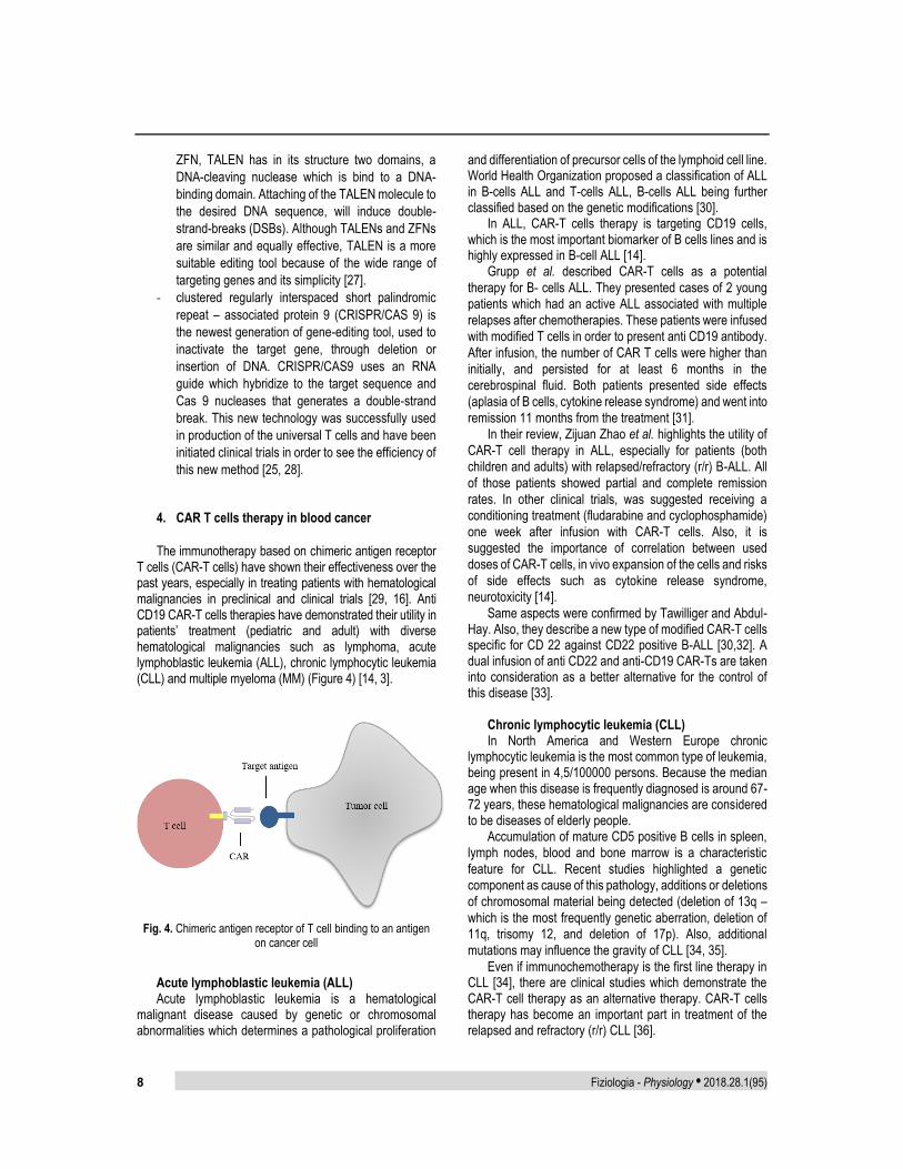
The immunotherapy based on chimeric antigen receptor T cells (CAR-T cells) have shown their effectiveness over the past years, especially in treating patients with hematological malignancies in preclinical and clinical trials [29, 16]. Anti CD19 CAR-T cells therapies have demonstrated their utility in patients’ treatment (pediatric and adult) with diverse hematological malignancies such as lymphoma, acute lymphoblastic leukemia (ALL), chronic lymphocytic leukemia (CLL) and multiple myeloma (MM) (Figure 4) [14, 3].
Fig. 4. Chimeric antigen receptor of T cell binding to an antigen
on cancer cell
Acute lymphoblastic leukemia (ALL) Acute lymphoblastic leukemia is a hematological
malignant disease caused by genetic or chromosomal abnormalities which determines a pathological proliferation
and differentiation of precursor cells of the lymphoid cell line. World Health Organization proposed a classification of ALL in B-cells ALL and T-cells ALL, B-cells ALL being further classified based on the genetic modifications [30].
In ALL, CAR-T cells therapy is targeting CD19 cells, which is the most important biomarker of B cells lines and is highly expressed in B-cell ALL [14].
Grupp et al. described CAR-T cells as a potential therapy for B- cells ALL. They presented cases of 2 young patients which had an active ALL associated with multiple relapses after chemotherapies. These patients were infused with modified T cells in order to present anti CD19 antibody. After infusion, the number of CAR T cells were higher than initially, and persisted for at least 6 months in the cerebrospinal fluid. Both patients presented side effects (aplasia of B cells, cytokine release syndrome) and went into remission 11 months from the treatment [31].
In their review, Zijuan Zhao et al. highlights the utility of CAR-T cell therapy in ALL, especially for patients (both children and adults) with relapsed/refractory (r/r) B-ALL. All of those patients showed partial and complete remission rates. In other clinical trials, was suggested receiving a conditioning treatment (fludarabine and cyclophosphamide) one week after infusion with CAR-T cells. Also, it is suggested the importance of correlation between used doses of CAR-T cells, in vivo expansion of the cells and risks of side effects such as cytokine release syndrome, neurotoxicity [14].
Same aspects were confirmed by Tawilliger and Abdul-Hay. Also, they describe a new type of modified CAR-T cells specific for CD 22 against CD22 positive B-ALL [30,32]. A dual infusion of anti CD22 and anti-CD19 CAR-Ts are taken into consideration as a better alternative for the control of this disease [33].
Chronic lymphocytic leukemia (CLL) In North America and Western Europe chronic
lymphocytic leukemia is the most common type of leukemia, being present in 4,5/100000 persons. Because the median age when this disease is frequently diagnosed is around 67-72 years, these hematological malignancies are considered to be diseases of elderly people.
Accumulation of mature CD5 positive B cells in spleen, lymph nodes, blood and bone marrow is a characteristic feature for CLL. Recent studies highlighted a genetic component as cause of this pathology, additions or deletions of chromosomal material being detected (deletion of 13q – which is the most frequently genetic aberration, deletion of 11q, trisomy 12, and deletion of 17p). Also, additional mutations may influence the gravity of CLL [34, 35].
Even if immunochemotherapy is the first line therapy in CLL [34], there are clinical studies which demonstrate the CAR-T cell therapy as an alternative therapy. CAR-T cells therapy has become an important part in treatment of the relapsed and refractory (r/r) CLL [36].
Fiziologia - Physiology • 2018.28.1(95) 9
Zou Y et al. presented different clinical trials which confirmed the potential of CAR-T cells as a valid therapy for treatment of chronic lymphocytic leukemia [36].
In a study published in 2018, Fraiette and his team have tried to use this novel immunotherapy in order to overcome tumor mediated rejection and tolerance of the immune system, for patients diagnosed with chronic lymphocytic leukemia (CLL). 41 patients with advanced CLL were included in the study and some of them responded to the CD19-targeted T cells (CTL019) treatment, with a dramatic expansion of the number of CAR T cell, associated with B cell aplasia. Other patients displayed limited response. Also, there were a small group of patients that had a high number of active T cells but later relapsed into an aggressive form of B cell Lymphoma. Efficiency of the treatment wasn’t related to the age of patient, therapies followed prior CART cells, or any other factors. All these different patients’ responses to the treatment have been attributed to the innate immune system [37].
Porter and his colleagues present a clinical trial where 14 patients were infused with CAR-modified T cell, with monitoring the evolutions of patients. All patients who took part of this trial presented a refractory-relapsed (r/r) form of CLL. From all those 14 patients, just 8 of them presented different rates of response towards the treatment. Half of those cases presented a complete remission, while the other four patients presented a partial remission. All the patients that responded to the treatment developed side effects such as B cell aplasia and cytokine release syndrome [38].
Multiple myeloma (MM) In the Western World, multiple myeloma represents 10%
from all of hematological cancers, being presents in 5,6/100000 persons. It is defined by an accumulation inside the bone marrow of plasmatic clonal cells.
Conventional therapies are focused to prolong the survival rate of the patient with a number reduction of malignant plasmatic cells, followed by a therapy in order to maintain therapeutic effectiveness. During the last years, have been developed novel immunotherapies in order to improve the curative potential and disease management, CAR-T cells therapy being one of the most recent therapeutic alternatives, with successful results in ALL and CLL treatment [49,40].
With their review article, Ghosh et al focuses on CD 19 CAR T cells therapy which targets multiple myeloma, putting on spot other CAR T cells that target different tumor antigen.
CD 19 CAR T cells therapy has been studied as an alternative treatment for patients with relapse-refractory disease, without satisfactory results. They also proposed N CAR T cells, CD 138 CAR T cells and BCMA CAR T cells. Usage N CAR T cells is a new strategy developed to eliminate in a more selective way malignant B cells, by targeting kappa
light chain in order to kill tumor cells. The clinical trial result showed the potential of N CAR T cells as an alternative therapy.
Because of the highly expressed CD 138 on MM cells, a new series of anti CD138-CAR T cells were developed. In the presented clinical trial, 4 of 5 patients had a stable disease, while the other patients presented a number reduction of MM cells.
Last, but not least, CAR-T cells targeting B cell maturation antigen, which is highly expressed by all of tumor plasma cells, is another therapeutic strategy in order to treat patients with MM. All trials conducted to see the efficiency of this new treatment, showed a dramatic response toward this new treatment. Besides CD 138 CAR-T cells, patients received a treat also received a conditioning chemotherapy like fludarabine, cyclophosphamide prior the infusion [40].
Berahovich and his team showed the potential anti-tumor activity of BCMA 4C8A CAR T cells. BCMA CAR T cells are a novel type of CAR T cells in order to be used for patients with MM. For creation of this cells, a BCMA specific mAb, clone 4C8A was generated. Clone 4C8A was used in construction of single chain variable fragments (scFv), which was inserted into a second generation of CARs. These modified CAR T cells were infused to the mice with xenograft tumor model, to measure their reaction against the tumor [41]. In order to establish the utility of this novel treatment, is necessary to test their utility in clinical trials. Some studies have showed or are about to presents their results regarding the potential anti-tumor function of BCMA CAR T cells [42,43].
Lymphoma In 1832, this malignancy was described for the first time
by Thomas Hodgkin which described six cases, of which only three were proven to be right later [44].
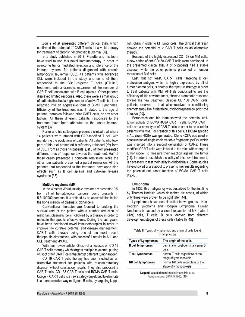
Lymphomas have been classified in two groups: Non-Hodgkin lymphoma and Hodgkin Lymphoma. Human lymphoma is caused by a clonal expansion of NK (natural killer) cells, T cells, B cells, derived from different development stages of these cells (Table II) [45].
Table II. Types of lymphomas and origin of cells found in lymphomas
Types of Lymphomas The origin of the cells B cell lymphomas germinal or post-germinal center B
cells T cell lymphomas normal T cells regardless of the
stage of lymphopoiesis NK cell lymphomas normal NK cells regardless of the
stage of lymphopoiesis
Legend: adapted from Knochelmann HM et al. Front Immunol. 2018; 9:1740. (46)

10 Fiziologia - Physiology • 2018.28.1(95)
Organs which are rich in lymphoid immune tissue, represent the starting point for Hodgkin (HL) and Non-Hodgkin (NHL) lymphomas and represents a very easy access for antibodies. Because of that, immunotherapy represent a useful tool in lymphoma treatment [45].
Besides anti CD 19 CAR-T cells, have been studied the utility of other potential target antigens like CD 20, CD 30, ĸ light chain. Ramos et al. [45] reviewed the results of clinical trials in order to identify a better treatment alternative for patients with lymphoma. Even though, the rate of disease remissions from the reviewed clinical trials was not as expected, CAR T cells still remain a valid therapy as the “one and done” treatment. I order to increase the efficiency of CAR T cells, it is necessary to adopt alternative solutions to improve CAR T cells function. Targeting checkpoint inhibition, inhibitory cytokines or cells from tumor stroma may enhance are the alternative strategies used to inhibit the tumor immune-suppression.
Zhao, Chen et al. highlights the superior qualities of second and third generations of T cells, associated with CD28 or with a cytoplasmic signaling domain 4-1BB; this association causing a higher proliferation rate and persistence of T cells. Also, CD20 CAR T cells usage for patients with DLBCC (diffuse large B cell lymphoma), showed promising results. In regard to CD30 CAR T cells, there is clinical trial that reports a regression of disease after receiving a conditioning chemotherapy prior CD30 CAR T cells infusion. CD30 is an antigen expressed in DLBCL, in lymphoma with anaplastic large cells, primary mediastinal B cell and peripheral T cell lymphoma, HL [44].
5. CAR T cells therapy in solid tumor
Even if adoptive cell therapy using chimeric antigen
receptor has encouraging results in treating hematological malignancies, especially B cell malignancies, we cannot talk about the same results when it comes to CAR T cells treatment of solid tumor [46].
Because desired antigens aren’t often expressed just in tumors, CAR T cells therapy determine severe toxicities. Knochelmann and his colleagues described several clinical trials and case reports regarding this type of immunotherapy for solid tumor, focusing on side effects like liver toxicity in renal cell carcinoma, the multi organ failure in CAR T cell treatment against ERB2. Also, poor results were obtained in a trial with CEACAM5- CAR T cells for gastrointestinal cancer, EGFR VIII in glioblastoma, HER2-based CAR in sarcoma. Despite these challenging cases, recently has been recorded success in treatment of glioblastoma with CAR T cell therapy [46].
Schmidts and Maus present in their review, article studies that shows promising results in treatment of phase I clinical trial of pediatric neuroblastoma with CART T cells specific for GD2, phase II/III clinical studies for treatment
with CAR T cells specific for HER2. Currently there are more than 270 CAR T cells clinical trials registered at U.S. National Library of Medicine, third of these studies researching the use of solid tumors in solid tumor of CAR T cells therapies [47].
Same results as Knochelmann, Schmidts and Maus was described by Newick and his team [48].
Because of this poor results of CAR T cells therapy in solid tumor treatment, was identified some berriers involved in failure of CAR T cells, such as structure of CARs, choice of desired antigen, doses, frequency and way of administration of CARs, survival on long-term in the environment of tumor tissue, lymphodepletion treatment [49].
TILs THERAPY
1. What is TIL therapy?
Tumor infiltrating lymphocytes therapy is an adoptive cell therapy, being an alternative approach to the CAR T cells therapy [50].
For the first time, in 1863, was described the presence of lymphoid cells in the structure of neoplastic tissue by Rudolf Virchow, who hypothesized a link between tumor and the presence of inflammation [51]. Years later, in 1969, the notion of tumor infiltrating lymphocytes was introduce for the first time by Wallace H. Clark Jr, to describe the immune response of the host against cancer [50,52]. Tumor infiltrating lymphocytes are considered to be lymphocytes cells that are leaving the bloodstream and infiltrate the tumor, being into a direct contact with cancer cells. TILs which are involved into this “defensive response” are formed by T cells, B cells, natural killer cells, T regulatory cells, dendritic cells, myeloid-derived suppressor cells and macrophages [52,53]. Even though TILs are presented inside the tumor, they are inactivated by the immunosuppressive tumor environment. In vivo, immunosuppressive factors such as cytotoxic T lymphocyte antigen 4 (CTLA4), lymphocyte activation gene 3 protein (LAG 3), programmed cell death protein 1 (PD1), T cell immunoglobulin and mucin domain-containing protein 3 (LAG3), enables TILs expansion and antitumor activity [54].
Because of their property of recognizing tumor cells antigens, TILs are used as an alternative immunotherapy, especially in treating melanoma [24]. Doctor Steven Rosenberg and his colleagues from Surgery Branch of the National Cancer Institute were the pioneers who introduced the benefit of TILs as adoptive cell therapy, especially in treatment of melanoma. During years of clinical trials coordinated by Rosenberg and his team, have been highlighted the importance of this novel immunotherapy for the treatment of patients with melanoma, which will be detailed later [55].
Fiziologia - Physiology • 2018.28.1(95) 11
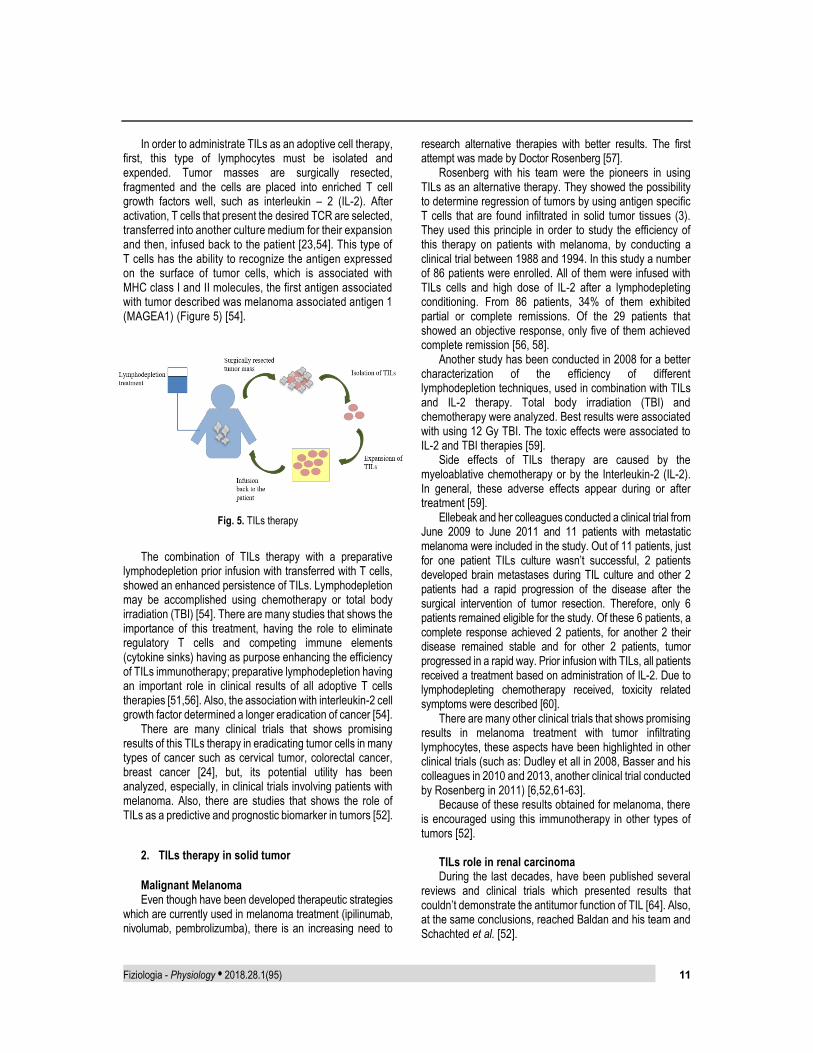
In order to administrate TILs as an adoptive cell therapy, first, this type of lymphocytes must be isolated and expended. Tumor masses are surgically resected, fragmented and the cells are placed into enriched T cell growth factors well, such as interleukin – 2 (IL-2). After activation, T cells that present the desired TCR are selected, transferred into another culture medium for their expansion and then, infused back to the patient [23,54]. This type of T cells has the ability to recognize the antigen expressed on the surface of tumor cells, which is associated with MHC class I and II molecules, the first antigen associated with tumor described was melanoma associated antigen 1 (MAGEA1) (Figure 5) [54].
Fig. 5. TILs therapy
The combination of TILs therapy with a preparative lymphodepletion prior infusion with transferred with T cells, showed an enhanced persistence of TILs. Lymphodepletion may be accomplished using chemotherapy or total body irradiation (TBI) [54]. There are many studies that shows the importance of this treatment, having the role to eliminate regulatory T cells and competing immune elements (cytokine sinks) having as purpose enhancing the efficiency of TILs immunotherapy; preparative lymphodepletion having an important role in clinical results of all adoptive T cells therapies [51,56]. Also, the association with interleukin-2 cell growth factor determined a longer eradication of cancer [54].
There are many clinical trials that shows promising results of this TILs therapy in eradicating tumor cells in many types of cancer such as cervical tumor, colorectal cancer, breast cancer [24], but, its potential utility has been analyzed, especially, in clinical trials involving patients with melanoma. Also, there are studies that shows the role of TILs as a predictive and prognostic biomarker in tumors [52].
2. TILs therapy in solid tumor
Malignant Melanoma Even though have been developed therapeutic strategies
which are currently used in melanoma treatment (ipilinumab, nivolumab, pembrolizumba), there is an increasing need to
research alternative therapies with better results. The first attempt was made by Doctor Rosenberg [57].
Rosenberg with his team were the pioneers in using TILs as an alternative therapy. They showed the possibility to determine regression of tumors by using antigen specific T cells that are found infiltrated in solid tumor tissues (3). They used this principle in order to study the efficiency of this therapy on patients with melanoma, by conducting a clinical trial between 1988 and 1994. In this study a number of 86 patients were enrolled. All of them were infused with TILs cells and high dose of IL-2 after a lymphodepleting conditioning. From 86 patients, 34% of them exhibited partial or complete remissions. Of the 29 patients that showed an objective response, only five of them achieved complete remission [56, 58].
Another study has been conducted in 2008 for a better characterization of the efficiency of different lymphodepletion techniques, used in combination with TILs and IL-2 therapy. Total body irradiation (TBI) and chemotherapy were analyzed. Best results were associated with using 12 Gy TBI. The toxic effects were associated to IL-2 and TBI therapies [59].
Side effects of TILs therapy are caused by the myeloablative chemotherapy or by the Interleukin-2 (IL-2). In general, these adverse effects appear during or after treatment [59].
Ellebeak and her colleagues conducted a clinical trial from June 2009 to June 2011 and 11 patients with metastatic melanoma were included in the study. Out of 11 patients, just for one patient TILs culture wasn’t successful, 2 patients developed brain metastases during TIL culture and other 2 patients had a rapid progression of the disease after the surgical intervention of tumor resection. Therefore, only 6 patients remained eligible for the study. Of these 6 patients, a complete response achieved 2 patients, for another 2 their disease remained stable and for other 2 patients, tumor progressed in a rapid way. Prior infusion with TILs, all patients received a treatment based on administration of IL-2. Due to lymphodepleting chemotherapy received, toxicity related symptoms were described [60].
There are many other clinical trials that shows promising results in melanoma treatment with tumor infiltrating lymphocytes, these aspects have been highlighted in other clinical trials (such as: Dudley et all in 2008, Basser and his colleagues in 2010 and 2013, another clinical trial conducted by Rosenberg in 2011) [6,52,61-63].
Because of these results obtained for melanoma, there is encouraged using this immunotherapy in other types of tumors [52].
TILs role in renal carcinoma During the last decades, have been published several
reviews and clinical trials which presented results that couldn’t demonstrate the antitumor function of TIL [64]. Also, at the same conclusions, reached Baldan and his team and Schachted et al. [52].
12 Fiziologia - Physiology • 2018.28.1(95)
Even though it has been difficult to prove the efficiency anti-tumor activity of TILs, there are studies that shows the utility of TIL in prediction of the disease evolution, by studying the immune profile of immune cells which infiltrated the tumor [65,66].
Overall, TIL therapy can be take into consideration as an alternative therapy, but more clinical trials are necessary in order to prove its success in treatment of renal cell carcinoma [52].
TILs in breast cancer In their study, Lee et al., successful isolated and expended,
ex vivo, TIL cells which derived from all types of breast cancer tissues. Also, expended TIL cells have been shown anti-tumor reaction, in vivo, in a xenograft mouse model [53].
Many other studies presented by Badalamenti in his review, showed the potential role of TIL as a predictive biomarker in breast cancers [66].
TILs and gastrointestinal cancers Even though gastrointestinal cancer is a rare type of
cancer, several studies have been conducted in order to obtain more biological information for a better prognosis, diagnosis of this tumor [66].
Also, Turcotte et al. demonstrated in their studies the achievement of a successful TILs culture and expansion to a sufficient number of cells which can be used in clinical trials [67, 68].
3. TILs immunotherapy limitations
Even though all studies have shown promising results in
using TIL as an alternative immunotherapy, there are some issues (Table III) to overcome in order to make this treatment a feasible one [69].
Table III. Limitations of TILs immunotherapy
Variable successful rates Relatively high costs for production of this treatment because it is personalized for every patient Too long production time (> 1 month), especially when there are patients with a rapidly progressive type of tumor Highly specialized facilities that requires highly trained specialists and extensive investments
Legend: adapted from Rohaan MW et al. J Immunother Cancer. 2018;6(1):102. (71)
CONCLUSIONS AND FUTURE PERSPECTIVES
Cancer is one of the most complex, frequent disease and, because of this, there are under development a series of novel immunotherapies in order to improve patient’s condition [58].
Because of the limited effect of the classical therapies, there is a great desire in searching alternatives treatments with a better outcome. One of the alternative strategies that prove its efficiency is immunotherapy, is using the patient immune system in order to make fight against tumor cells. Adoptive T cells therapy, cancer vaccines, immune checkpoint blockade, immunomodulatory agents may represent an alternative in cancer treatment and their uses in clinical trials [55].
In order to obtain the desired results using immunotherapy as main treatment it is important to take in consideration the patient status, type of tumor, the purpose of the therapy, as well as tumor classification that dictates the available time to obtain desired clinical results [58].
From all those immunological therapeutic strategies, we tried to highlight the importance of adoptive cell therapy (CAR T cells, TILs) in treating patients with blood malignancies and solid tumor.
Evan if CAR T cells therapy is making steps in changing the management of blood malignancies, there are some issues in applying successfully this therapy to solid tumors [24].
In 2018, have been successful approved by USA FDA two types of CAR-T cell therapies: YESCARTA and KYMRIAH. Both of them have the role to attack B – cells malignancies (leukemia and lymphoma) [70].
In conclusion, with this review article, we wanted to focus on the potential benefits of treating various types of tumors using novel immunotherapies such as adoptive cell transfer and, also, we expect a faster improvement of these technologies in order to solve the problems we are now facing in clinical trials.
In future, it will be important to explore other methods to determine an improvement of the ablation of immune system in order to obtain promised results in cancer studies, by using ACT technologies. Also, the ability to identify patient specific tumor antigens, through DNA sequencing techniques, will revolutionize the immunotherapy in cancer [50].
Acknowledgement: This work was supported by the grant “Chimeric Antigen Receptor Targeted Oncoimmunotherapy with Natural Killer Cells (CAR-NK)”, POC 92/09/09/2016, ID: P_37_786, MySMIS code: 103662.
REFERENCES
1. Sharma P, Hu-Lieskovan S, Wargo JA, Ribas A. Primary, Adaptive, and Acquired Resistance to Cancer Immunotherapy. Cell. 2017;168(4):707-723.
2. Prager GW, Braga S, Bystricky B, et al. Global cancer control: responding to the growing burden, rising costs and inequalities in access. ESMO Open. 2018;3(2):e000285.
3. Miliotou AN, Papadopoulou LC. CAR T-cell Therapy: A New Era in Cancer Immunotherapy. Curr. Pharm. Biotechnol. 2018;19:5-18.
Fiziologia - Physiology • 2018.28.1(95) 13
4. Zhang H, Chen J. Current status and future directions of cancer immunotherapy. J Cancer. 2018;9(10):1773-81.
5. Ascierto PA, Daniele B, Hammers H, et al. Perspectives in immunotherapy: meeting report from the "Immunotherapy Bridge", Napoli, November 30th 2016. J Transl Med. 2017;15(1):205.
6. Borghaei H, Smith MR, Campbell KS. Immunotherapy of cancer. Eur J Pharmacol. 2009;625(1-3):41-54.
7. Decker WK, da Silva RF, Sanabria MH, et al. Cancer Immunotherapy: Historical Perspective of a Clinical Revolution and Emerging Preclinical Animal Models. Front Immunol. 2017;8:829.
8. Ventola CL. Cancer Immunotherapy, Part 1: Current Strategies and Agents. P&T. 2017;42(6):375-383.
9. Coulson A, Levy A, Gossell-Williams M. Monoclonal Antibodies in Cancer Therapy: Mechanisms, Successes and Limitations. West Indian Med J. 2015;63(6):650-4.
10. Waldmann TA. Cytokines in Cancer Immunotherapy. Cold Spring Harb Perspect Biol. 2018 Dec 3;10(12).
11. Lee S, Margolin K. Cytokines in cancer immunotherapy. Cancers (Basel). 2011;3(4):3856-93.
12. Guo C, Manjili MH, Subjeck JR, Sarkar D, Fisher PB, Wang XY. Therapeutic cancer vaccines: past, present, and future. Adv Cancer Res. 2013;119:421-75.
13. Levine BL, Miskin J, Wonnacott K, Keir C. Global Manufacturing of CAR T Cell Therapy. Mol Ther Methods Clin Dev. 2016;4:92-101.
14. Zijun Z, Yu C, Ngiambudulu MF, Yuanqing Z, Minhao W. The application of CAR-T cell therapy in hematological malignancies: advantages and challenges. Acta Pharm Sin B. 2018; 8(4):539-551.
15. Xia AL, Wang XC, Lu YJ, et al. Chimeric-antigen receptor T (CAR-T) cell therapy for solid tumors: challenges and opportunities. Oncotarget. 2017;8(52):90521-90531.
16. Aaron J. Smith, John Oertle, Dan Warren, Dino Prato. Chimeric antigen receptor (CAR) T cell therapy for malignant cancers: Summary and perspective. Journal of Cellular Immunotherapy. 2016; 2(2):59-68.
17. Zhang C, Liu J, Zhong JF, Zhang X. Engineering CAR-T cells. Biomark Res. 2017; 5:22.
18. Chmielewski M, Abken H. TRUCKs: the fourth generation of CARs. Expert Opin Biol Ther. 2015; 15(8):1145-1154.
19. Sharpe M, Mount N. Genetically modified T cells in cancer therapy: opportunities and challenges. Dis Model Mech. 2015;8(4):337-50.
20. Graham C, Hewitson R, Pagliuca A, Benjamin R. Cancer immunotherapy with CAR-T cells – behold the future. Clin Med. 2018;18(4):324-328.
21. Chu F, Cao J, Neelalpu SS. Versatile CAR T-cells for cancer immunotherapy. Contemp Oncol (Pozn). 2018;22(1A):73-80.
22. Ping Y, Liu C, Zhang Y. T-cell receptor-engineered T cells for cancer treatment: current status and future directions. Protein Cell. 2017;9(3):254-266.
23. Rosenberg SA. Cell transfer immunotherapy for metastatic solid cancer--what clinicians need to know. Nat Rev Clin Oncol. 2011;8(10):577-85.
24. Eisenberg V, Hoogi S, Shamul A, et al. T-cells “à la CAR-T(e)” – Genetically engineering T-cell response against cancer. Adv Drug Deliv Rev. 2019.
25. Zhao J, Lin Q, Song Y, Liu D. Universal CARs, universal T cells, and universal CAR T cells. J Hematol Oncol. 2018; 11:132.
26. Carroll D. Genome engineering with zinc-finger nucleases. Genetics. 2011;188(4):773-82.
27. Joung JK, Sander JD. TALENs: a widely applicable technology for targeted genome editing. Nat Rev Mol Cell Biol. 2012;14(1):49-55.
28. Tsai SQ, Joung JK. Defining and improving the genome-wide specificities of CRISPR–Cas9 nucleases. Nat Rev Genet. 2016;17(5):300-12.
29. Tomuleasa C, Fuji S, Berce C, et al. Chimeric Antigen Receptor T-Cells for the Treatment of B-Cell Acute Lymphoblastic Leukemia. Front Immunol. 2018;9:239.
30. Terwilliger T, Abdul-Hay M. Acute lymphoblastic leukemia: a comprehensive review and 2017 update. Blood Cancer J. 2017;7(6):e577.
31. Grupp SA, Kalos M, Barrett D, et al. Chimeric antigen receptor-modified T cells for acute lymphoid leukemia. N Engl J Med. 2013;368(16):1509-1518.
32. Shah NN, Stetler-Stevenson M, Yuan CM, et al. Minimal Residual Disease Negative Complete Remissions Following Anti-CD22 Chimeric Antigen Receptor (CAR) in Children and Young Adults with Relapsed/Refractory Acute Lymphoblastic Leukemia (ALL). Blood. 2016;128(22).
33. Huang L, Wang N, Li C, CaoY, et al. Sequential Infusion of Anti-CD22 and Anti-CD19 Chimeric Antigen Receptor T Cells for Adult Patients with Refractory/Relapsed B-Cell Acute Lymphoblastic Leukemia. Blood. 2017;130(1).
34. Hus I, Roliński J. Current concepts in diagnosis and treatment of chronic lymphocytic leukemia. Contemp Oncol (Pozn). 2015;19(5):361-7.
35. Hallek M. Chronic lymphocytic leukemia: 2017 update on diagnosis, risk stratification, and treatment. Am J Hematol. 2017;92:946–965.
36. Zou Y, Xu W, Li J. Chimeric antigen receptor-modified T cell therapy in chronic lymphocytic leukemia. J Hematol Oncol. 2018;11(1):130.
37. Fraietta JA, Lacey SF, Orlando EJ, et al. Determinants of response and resistance to CD19 chimeric antigen receptor (CAR) T cell therapy of chronic lymphocytic leukemia. Nat Med. 2018;24(5):563-571.
38. Porter DL, Hwang WT, Frey NV, et al. Chimeric antigen receptor T cells persist and induce sustained remissions in relapsed refractory chronic lymphocytic leukemia. Sci Transl Med. 2015;7(303):303ra139.
39. Ormhøj M, Bedoya F, Frigault MJ, Maus MV. CARs in the Lead Against Multiple Myeloma. Curr Hematol Malig Rep. 2017;12(2):119-125.
40. Ghosh A, Mailankody S, Giralt SA, et al. CAR T cell therapy for multiple myeloma: where are we now and where are we headed?. Leuk Lymphoma. 2018;59(9):2056-2067.
41. Berahovich R, Zhou H, Xu S, et al. CAR-T Cells Based on Novel BCMA Monoclonal Antibody Block Multiple Myeloma Cell Growth. Cancers (Basel). 2018;10(9):323.

14 Fiziologia - Physiology • 2018.28.1(95)
42. Xu J, Wang Q, Xu H, et al. Anti-BCMA CAR-T cells for treatment of plasma cell dyscrasia: case report on POEMS syndrome and multiple myeloma. J Hematol Oncol. 2018;11(1):128.
43. Cho SF, Anderson KC, Tai YT. Targeting B Cell Maturation Antigen (BCMA) in Multiple Myeloma: Potential Uses of BCMA-Based Immunotherapy. Front Immunol. 2018;9:1821.
44. Aisenberg, A. C. Historical review of lymphomas. Br J Haematol. 2000;109(3):466-476.
45. Ramos CA, Heslop HE, Brenner MK. CAR-T Cell Therapy for Lymphoma. Annu Rev Med. 2015;67:165-83.
46. Knochelmann HM, Smith AS, Dwyer CJ, et al. CAR T Cells in Solid Tumors: Blueprints for Building Effective Therapies. Front Immunol. 2018;9:1740.
47. Schmidts A, Maus MV. Making CAR T Cells a Solid Option for Solid Tumors. Front Immunol. 2018;9:2593.
48. Newick K, O'Brien S, Moon E, Albelda SM. CAR T Cell Therapy for Solid Tumors. Annu Rev Med. 2017;68(1):139-152.
49. D'Aloia MM, Zizzari IG, Sacchetti B, et al. CAR-T cells: the long and winding road to solid tumors. Cell Death Dis. 2018;9(3):282.
50. Sackstein R, Schatton T, Barthel SR. T-lymphocyte homing: an underappreciated yet critical hurdle for successful cancer immunotherapy. Lab Invest. 2017;97(6):669-697.
51. Geukes Foppen MH, Donia M, Svane IM, Haanen JB. Tumor-infiltrating lymphocytes for the treatment of metastatic cancer. Mol Oncol. 2015;9(10):1918-35.
52. Lee N, Zakka LR, Mihm MC, Jr, Schatton T. Tumour-infiltrating lymphocytes in melanoma prognosis and cancer immunotherapy. Pathology. 2016;48(2):177–87.
53. Lee HJ, Kim YA, Sim CK, et al. Expansion of tumor-infiltrating lymphocytes and their potential for application as adoptive cell transfer therapy in human breast cancer. Oncotarget. 2017;8(69):113345-59.
54. Restifo NP, Dudley ME, Rosenberg SA. Adoptive immunotherapy for cancer: harnessing the T cell response. Nat Rev Immunol. 2012;12(4):269-81.
55. Lizée G, Overwijk WW, Radvanyi L, et al. Harnessing the Power of the Immune System to Target Cancer. Annu Rev Med. 2013;64(1):71-90.
56. Muranski P, Boni A, Wrzesinski C, et al. Increased intensity lymphodepletion and adoptive immunotherapy--how far can we go?. Nat Clin Pract Oncol. 2006;3(12):668-81.
57. Lee S, Margolin K. Tumor-infiltrating lymphocytes in melanoma. Curr Oncol Rep. 2012;14(5):468-74.
58. Alatrash G, Jakher H, Stafford PD, Mittendorf EA. Cancer immunotherapies, their safety and toxicity. Expert Opin Drug Saf. 2013;12(5):631-645.
59. Dudley ME, Yang JC, Sherry R, et al. Adoptive cell therapy for patients with metastatic melanoma: evaluation of intensive myeloablative chemoradiation preparative regimens. J Clin Oncol. 2008;26(32):5233-9.
60. Ellebaek E, Iversen TZ, Junker N, et al. Adoptive cell therapy with autologous tumor infiltrating lymphocytes and low-dose Interleukin-2 in metastatic melanoma patients. J Transl Med. 2012;10:169.
61. Besser MJ, Shapira-Frommer R, Treves AJ, et al. Clinical responses in a phase II study using adoptive transfer of short-term cultured tumor infiltration lymphocytes in metastatic melanoma patients. Clin Cancer Res. 2010;16(9):2646–2655.
62. Rosenberg SA, Yang JC, Sherry RM, et al. Durable complete responses in heavily pretreated patients with metastatic melanoma using T-cell transfer immunotherapy. Clin Cancer Res. 2011;17(13):4550–4557.
63. Besser MJ, Shapira-Frommer R, Itzhaki O, Treves AJ, Zippel DB, Levy D, Kubi A, Shoshani N, Zikich D, Ohayon Y, Ohayon D, Shalmon B, et al. Adoptive transfer of tumor-infiltrating lymphocytes in patients with metastatic melanoma: intent-to-treat analysis and efficacy after failure to prior immunotherapies. Clin Cancer Res. 2013;19(17):4792–4800.
64. Shablak A, Hawkins RE, Rothwell DG, Elkord E. T cell-based immunotherapy of metastatic renal cell carcinoma: modest success and future perspective. Clin. Cancer Res. 2009;15(21):6503–6510.
65. Geissler K, Fornara P, Lautenschläger C, et al. Immune signature of tumor infiltrating immune cells in renal cancer. Oncoimmunology. 2015;4(1):e985082.
66. Badalamenti G, Fanale D, Incorvaia L, et al. Role of tumor-infiltrating lymphocytes in patients with solid tumors: Can a drop dig a stone?. Cell Immunol. 2018.
67. Turcotte S, Gros A, Hogan K, et al. Phenotype and function of T cells infiltrating visceral metastases from gastrointestinal cancers and melanoma: implications for adoptive cell transfer therapy. J Immunol. 2013;191(5):2217-25.
68. Turcotte S, Gros A, Tran E, et al. Tumor-reactive CD8+ T cells in metastatic gastrointestinal cancer refractory to chemotherapy. Clin Cancer Res. 2014;20(2):331–343.
69. Rohaan MW, van den Berg JH, Kvistborg P, Haanen JBAG. Adoptive transfer of tumor-infiltrating lymphocytes in melanoma: a viable treatment option. J Immunother Cancer. 2018;6(1):102.
70. Salmikangas P, Kinsella N, Chamberlain P. Chimeric Antigen Receptor T-Cells (CAR T-Cells) for Cancer Immunotherapy - Moving Target for Industry?. Pharm Res. 2018;35(8):152.
Fiziologia - Physiology • 2018.28.1(95) 15
TERAPIA CU CELULE CAR-T VS. TIL: VIITORUL IMUNOTERAPIEI ÎN CANCER REZUMAT La nivel mondial, cancerele sunt unele dintre principalele cauze de deces, iar tratamentul convențional are propriile limitări. Cercetările din domeniul imunoterapiei se concentrează pe descoperirea unor alternative terapeutice fezabile pentru tratarea acestei patologii. În cancere, imunoterapia are rolul de a spori efectul antitumoral al celulelor imune implicate în acest proces. În funcție de antigenul țintă, efectele adverse ale acestei terapii pot varia de la reacții locale și ușoare, la cele sistemice și mai severe. Principiul imunoterapiilor cu celule CAR T și TILs se bazează pe folosirea celulelor T, care sunt specifice pentru un anumit antigen și reinfuzarea înapoi în pacient dupa ce aceste celule au fost multiplicate ex vivo, pentru a determina distrugerea tumorii. Asocierea acestor terapii cu un regim de depleție limfoci tară și cu administrarea factorului IL-2 de creștere a celulelor T determină un efect puternic asupra pacienților. În cadrul acestui articol, vom face o prezentare generală a terapiei cu celule CAR T și a terapiei cu TIL, descriind principiile acestor imunoterapii, aplicațiile clinice, precum și avantajele, dezavantajele și perspectivele acestora. Key words: celule CAR-T, TILs, imunoterapii, cancer

16 Fiziologia - Physiology • 2018.28.1(95)
FACULTY PERCEPTION FOR THE NEED OF CHANGE IN I YEAR MBBS INDIAN PHYSIOLOGY PRACTICAL CURRICULUM SMITA R SORTE1, SACHIN B RATHOD2 1Assistant Professor, Department of Physiology, Shri Shankaracharya Institute of Medical Sciences, Bhilai, Chhattisgarh, India 2Assistant Professor, Department of Physiology, Shri Shankaracharya Institute of Medical Sciences, Bhilai, Chhattisgarh, India.
ABSTRACT Indian medical curriculum differs from other foreign universities. In India, Bachelor of Medicine and Bachelor of Surgery (MBBS) is 5½ year professional course, in which physiology is 1st-year MBBS subject. Physiology deals with teaching the basic principles of functioning of various system of body. Indian physiology Practical syllabus includes four sections -Haematology, Experimental-Amphibian practical, human experiments and basic clinical examination. This syllabus was formulated many years back which has not been revised. Physiology is the base of medicine, so physiology curriculum should be well-designed to build basic clinical concept that can be applied in medicine practice. Graduate medical education regulation, long-term goal is to get the qualified doctor for the better patient care health system. But alignment is not seen in curriculum and the physiology practical syllabus. So this study was conducted on 330 teaching faculty of India to know their perspective about the need of change in I year MBBS Indian physiology practical curriculum. Our study found that many topics have no clinical relevance. Student gained irrelevant knowledge which has no future clinical use. Those topics should be replaced by newer practicals and demonstration. Teaching faculty staff suggested early clinical exposure, simulation experiments, basic of research and healthy lifestyle practice should be inculcated in the newer syllabus. Short Running Title: Faculty perception for the need of change in I year MBBS Indian physiology practical curriculum Keywords: curriculum, physiology syllabus, medical education, MBBS.
INTRODUCTION
Curricula” is two wheeled chariot drawn by horse and curriculum is the path or the course. The curriculum is planned educational experience [1]. Indian medical curriculum differs from other foreign universities. Indian medical council act was established in 1933 under which Medical Council of India was established in 1934 [2]. The act was reconstituted in 1956 [3] and its first, second and third schedule was formed later on. The amendment was added in 1993 [4], 2011 [5], 2012 [6] and 2016 [7]. In 1997, Graduate medical education regulation (GMER) was published in part III, section- 4 [8] which was modified many times, and the latest modification was done in May 2018.
In India, Bachelor of Medicine and Bachelor of Surgery (MBBS) is a 5½ year professional course. In the first year, the preclinical subjects – Anatomy, Physiology, and Biochemistry are taught for the duration of 1 year. The
Second year includes – Pathology, Microbiology, Pharmacology and Forensic Medicine and toxicology covered in the duration of 1 and ½ year. The third year includes PSM, Ophthalmology, and ENT in the duration of 1 year and final year includes Medicine, Surgery, Pediatrics and Obstetrics & Gynecology in the duration of 1 year. It is followed by 1 year of compulsory clinical rotator internship.
Physiology is basic science subject taught in 1st MBBS. It teaches the basic principles of functioning of the various system of the body. Graduate medical curriculum is oriented towards training students to undertake the responsibilities of a physician of first contact who is capable of looking after the preventive, promotive, curative & rehabilitative aspect of medicine. According to GME regulation goal of teaching physiology is to providing the student comprehensive knowledge of the normal functions of the organ systems of the body to facilitate an understanding of the physiological basis of health and disease [8].
Received April 15th 2018. Accepted May 21st 2018. Address for correspondence: Dr Sachin B Rathod, Department of Physiology, Shri Shankaracharya Institute of Medical Sciences, Junwani, Bhilai, India; Phone: +91-9407796105, +91-9423409931, e-mail: drsuchinrat10@gmail. Name of department to which work is attributed: Department of Physiology, Rural Medical College, Loni, PIMS, Ahmednagar, Maharashtra, India
Fiziologia - Physiology • 2018.28.1(95) 17
Process objectives of teaching physiology are - at the end of the course, the student will be able to: (1) Explain the normal functioning of all the organ systems and their interactions for well-coordinated total body function. (2) Assess the relative contribution of each organ system to the maintenance of the milieu interior. (3) Elucidate the physiological aspects of normal growth and development. (4) Describe the physiological response and adaptations to environmental stresses. (5) List the physiological principles underlying pathogenesis and treatment of disease [8].
Indian physiology Practical syllabus includes four sections - Haematology, Experimental-Amphibian practical, human experiments and basic clinical examination. This syllabus was formulated many years back which has not been revised. Curriculum hypertrophy or curriculomegaly is the disease of curriculum described by Stephen Abrahamson in 1978 [9]. It is caused due to the addition of newer knowledge in pre-existing knowledge. Ever expanding syllabus has to be taught in limited duration of teaching hours. So some part of older outdated syllabus must be removed while newer syllabus should be regularly added in the curriculum at some time intervals.
The curriculum should be aligned with the specific learning objectives, the syllabus, teaching-learning methods, and assessment. But this alignment is not seen in physiology practical part of the curriculum. According to GMER, learners objectives in practical skills are at the end of the course the student will be able to i) conduct experiments designed for the study of physiological phenomena; ii) Interpret experimental/investigative data; iii) Distinguish between normal and abnormal data derived as a result of tests which he/she has performed and observed in the laboratory.
The government of India had banned animal experiments in 2012 [10] due to which animal dissection is not conducted in the teaching of experimental amphibian practicals. The amphibian graphs are taught theoretically on basis of experiments conducted in past. This government rule leads to misalignment of the curriculum with the skill learning objective (that students should be able to conduct experiments designed for the study of physiological phenomena). This makes it necessary to formulate new a skill learning objective to fulfill the physiology curriculum need.
Physiology is the base of medicine, so physiology curriculum should be well-designed to build basic clinical concept that can be applied in medical practice. Some studies were conducted from students’ perspective of MBBS curriculum which found many topics of physiology curriculum to be irrelevant and obsolete [11, 12]. Another study was conducted on faculty perception of only experimental physiology curriculum which also found that amphibian experimental physiology is outdated and need to be replaced by newer practicals. Our study was conducted to find faculty perception about the need to change I year MBBS physiology practical curriculum in India.
METHOD AND MATERIAL
A descriptive cross-sectional study was conducted in the Department of Physiology, RMC Loni, Ahmednagar, Maharashtra, India. The study was approved by IEC. The structured organized online survey was conducted on website- Survey Monkey. The questionnaire was validated by experts. Qualitative questions were evaluated by 3 points Likert scale. Internal reliability of question was evaluated by Cronbach’s alpha. Open-ended question to obtain the opinion of faculty regarding new physiology topics to be added in practical syllabus was also asked.
Physiology faculty of different cadre from various colleges of India was informed about the survey by telephones and emails, where the link of the survey was attached. Consent of participant was taken as the first question and then they were allowed to participate in the survey. Data was collected from 330 physiology faculty in the duration of 6 months.
Statistical analysis Data was analyzed and the result was calculated as
percentage. No statistical test was applied.
RESULTS
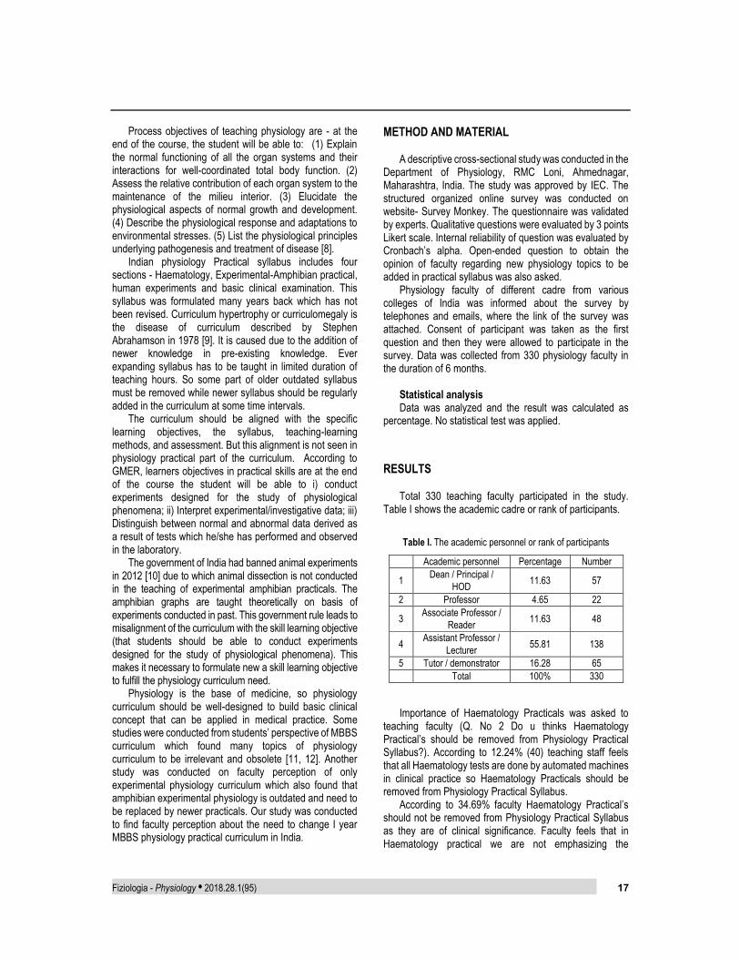
Total 330 teaching faculty participated in the study. Table I shows the academic cadre or rank of participants.
Table I. The academic personnel or rank of participants
Academic personnel Percentage Number
1 Dean / Principal / HOD 11.63 57
2 Professor 4.65 22
3 Associate Professor / Reader 11.63 48
4 Assistant Professor / Lecturer 55.81 138
5 Tutor / demonstrator 16.28 65 Total 100% 330
Importance of Haematology Practicals was asked to
teaching faculty (Q. No 2 Do u thinks Haematology Practical’s should be removed from Physiology Practical Syllabus?). According to 12.24% (40) teaching staff feels that all Haematology tests are done by automated machines in clinical practice so Haematology Practicals should be removed from Physiology Practical Syllabus.
According to 34.69% faculty Haematology Practical’s should not be removed from Physiology Practical Syllabus as they are of clinical significance. Faculty feels that in Haematology practical we are not emphasizing the

18 Fiziologia - Physiology • 2018.28.1(95)
techniques but we teach the students to correlate the haematological finding with clinical scenario. In Haematology, the student can appreciate blood cell which can be utilized to understand and diagnose the abnormality of blood cell in pathology. So the applied aspect of Haematology should be the part of physiology practicals. When student (future doctor) practice in rural areas where automated machines are not available, these old haematological test and techniques are useful for diagnosis of certain diseases.
While 53.06% teaching faculty thinks that, newer Haematology techniques should replace older techniques. They suggested the addition of platelet count and genetic practical’s like extraction of chromosomes from WBC for mapping. The principle of research and curiosity should be inculcated by Haematology practical’s.
The significance of amphibian experimental physiology was asked in Question no 3 (Do u think Experimental Physiology (Amphibian Graphs) should be removed from Syllabus?)
40.63 % teaching staff consider that Experimental Physiology (Amphibian Graphs) should be removed from syllabus as it is of no clinical significance. The relevance of these practicals is decreased as the dissection of animal is banned. 31.25 % teaching staffs think that Experimental Physiology (Amphibian Graphs) should not be removed from syllabus but its marks weight-age should be decreased, or it should be integrated with Pharmacology.
28.13% teaching staff think that Experimental Physiology (Amphibian Graphs) should not be removed from the practical syllabus. Amphibian experiments show the basic invented of Physiology subject. “I hear and I forget. I see and I remember. I do and I understand.” Is a common Chinese proverb, which can be also applicable in experimental Physiology. Unless animal (frog) dissections are done, mere graphs are not understood by I year MBBS students. As animal dissection is banned by the government of India, Physiology society should seriously consult with animal protection organization and get permission to dissection animal for medical and research centers. Though animal killing should not be practiced, Frogs may be replaced with other easily replaceable lab animals. As it forms the basis of the physiology, newer animation and soft copy of the experiment can be shown to the students or it can be replaced with computer simulations. Good software’s like COMPU FROG can be used and faculties should be trained to use such software.
The implication of human experiments was asked in question no 3.(Are human practical’s like Ergography/ stethography/ Pulmonary function test etc required in Human physiology practical’s?)
11 % teaching staff thinks that Human physiology practical’s syllabus requires no modifications. 5% teaching staff thinks that Human physiology practicals are outdated practical’s of no clinical significance. Some experiments like perimetry, spirometry should be removed. But 84% faculty
appreciates the significance of human experiments but suggest modification in experiments, like the use of computerized / simulation techniques. Some faculty suggests the addition of exercise physiology related experiments in this section.
Opinion regarding addition of Neurophysiology experiments was asked in question no 4 (Do u think that Neurophysiology experiments like NCV, BERA, VEP, EMG etc should be included in physiology practical?)
79.79% teaching staff thinks that Neurophysiology experiments like NCV, BERA, VEP, EMG etc should be included in physiology practicals. With the progressive scope of research for neurophysiology, yoga, and meditation, it becomes essential to learn basics in I MBBS. Some staff suggested it should be taught in form of demonstration or integrated with theory teachings. Demonstration in the clinical setting would expose students to the patients which will help to understand their clinical relevance. 13.83% teaching staff thinks that Neurophysiology experiments like NCV, BERA, VEP, EMG etc should not be included in physiology practical’s, whereas 6.38% teaching staff did not express their view in this context.
The open-ended question was asked (What other experiments can be added to Physiology practical syllabus for first yr MBBS students?). Results were tabulated in Table II.
Table II. The answers of open ended questions
Topics Opinions for adding topics in physiology practical curriculum
1 Hematology Clinically oriented experiments should be added. Discussion of Computerized haemogram should be done. Visit to blood banks and pathology labs should be done.
2 Amphibian Replace amphibian experiments by other experiments
3 Human Computerized perimetry, Computerized PFT, pulse oximetry, Blood pressure alteration with stress, cold etc. Vision test, Memory/cognitive test, Tonometry, Endoscopy, effect of hypoxia on body functions, and other environmental practical’s should be considered
4 Clinical More emphasis should be given to clinical examination and early exposure to patients. It must be given more marks distribution. In Early clinical exposure students can learn to differentiate normal from abnormal. Comprehensive clinical history taking and clinical examination of clinical cases should include. They should be master of clinical examinations e.g. General, CVS, RS, CNS and abdomen

Fiziologia - Physiology • 2018.28.1(95) 19
examination. Examination of heart sound by electronic stethoscope, Phonocardiogram, Murmurs should be added. Communication skills should be included.
5 Experiments on yoga and meditation
Compulsion of yoga and meditation to students for thirty days to establish the benefits and implementation among students to better citizens of our country
6 Radiology Brief introduction and principles of x rays, ultrasound, Doppler, Echocardiography, CT scan, Radionucleotide imaging and MRI. There demonstration should be considered.
7 Exercise physiology
Treadmill test, bicycle Ergography, cardiorepiratory fitness assessment, exercise stress test.
7 Neuro physiology
Demonstration of Reaction time, ECG, EEG, EMG, NCV, Autonomic function testing- HRV and sleep study.
8 Simulation experiments
Simulation lab with latest equipments for clinical skills, First aid techniques, Cardio-Pulmonary Resuscitation, simulators for artificial respiration. Simulation lab for renal physiology, nerve muscle physiology, transport across cell membrane.
9 Biochemical experiments
Experiments based on Resting membrane potentials e.g voltage-clamp technique. Biochemical Estimations using standard kits comparing normal and patient samples. Automated ELISA, colorimetric, PCR, electrophoresis Hormonal assay, tests based on Antigen-antibody reactions may be included in the Physiology curriculum
10 Diet and Healthy lifestyle
Basal Metabolic Rate calculation, Estimation nutrition /diet prescriptions, caloric requirement and daily calories burn.
11 Endocrine Endocrine photos in form of cases to find pathophysiology could increase the application of their knowledge
12 Biophysics Calculation based biophysics logical graph/chart/photograph
13 Reproductive Semen analysis 14 Assessment More problems based clinical learning;
less weightage should be given on memorizing facts than learning concepts, and increase viva and objective questions marks while decreasing theory marks.
DISCUSSION Physiology curriculum must be designed with a long-
term goal of better patient care and health practice system. Even graduate medical education regulation aims to make students better doctors of tomorrow. But alignment is not seen in physiology curriculum and its syllabus which is also perceived by the faculty members in our study. Due to the daily addition of newer information and techniques, there is curriculomegaly or hypertrophy which should be included in the syllabus by time to time amendments.
The cadre of most of the faculty in our study is Assistant professor> tutors> HOD> Associate professor> Professor. So we can consider the results are the opinion of young budding teachers and researchers generation. According to our study, most part of physiology curriculum is outdated and obsolete, which should be replaced by newer practicals and demonstration. Study on faculty perspective by Dr. paralikar [13], Dr. Narlawar [11] suggested parallel findings whereas study on student’s perspective by Dr. Goyal [14] and Dr. Chawla [15] also suggested similar findings, thus supports our study. Students complained that though they have gained basic knowledge they had also learned too much irrelevant knowledge in some areas.
The government of India had banned animal experiments, due to which amphibian experimental syllabus has lost its charm, making it a dull and bore section of the practical syllabus. Certain topics have no clinical relevance. Physiology societies should find an alternative (simulation/videos) or remove this section from the practical syllabus.
Majority of teaching faculty (53.06%) thinks that newer hematology techniques should replace older techniques. The principle of research should be inculcated by hematology practicals. A good number of teaching faculties (84%) appreciates the significance of human experiments but suggest modification in experiments, like the use of computerized / simulation techniques. 79.79% teaching staff feels that Neurophysiology experiments like NCV, BERA, VEP, EMG etc should be included in physiology practical’s.
Teaching faculty staff suggested early clinical exposure and many newer topics that should be added in the newer curriculum. These topics are mostly clinically oriented experiments. They are computerized perimetry, computerized PFT, pulse oximetry, Blood pressure alteration with stress, vision test, Memory/cognitive test, Tonometry, Endoscopy, effect of hypoxia on body functions, and other environmental practical’s, simulators for first aid techniques, Cardio-Pulmonary Resuscitation, Brief introduction of x rays, ultrasound, Doppler, Echocardiography, CT scan and MRI. Treadmill test, bicycle ergography cardiorespiratory fitness assessment, exercise stress test, Reaction time, ECG, EEG, EMG, NCV, Autonomic function testing- HRV and sleep study.
As lifestyle diseases are one of the major causes of mortality and morbidity, one should also focus on practicing a healthy lifestyle and healthy diet. The basic principle and
20 Fiziologia - Physiology • 2018.28.1(95)
practice should be included in I year MBBS. Yoga and meditation should also be taught and practiced. The doctor is a researcher and so the seed of research should also be sowed in IST year MBBS.
CONCLUSION
In fact practical’s help in understanding the underlying physiological phenomenon. We learn faster and better by performing the experiments. Thus the entire physiological phenomenon must be taught with practical demonstration. Many of physiology practicals are outdated and useless. We should focus on the current clinical scenario to teach students. SPICES model of curriculum organization should be used in formulating the newer curriculum. It should be Student-centered, Problem based learning, Integrated teaching, Community based education, Elective and Systematic. It should facilitate horizontal and vertical integration to link disparity between theory and practical. Clinical training, research and principles of a healthy lifestyle and diet should start in first MBBS focusing on professionalism, basic clinical skills and communication skills. It is the responsibility of physiological bodies and society to consider the opinion of young teaching faculty and develop a new modified physiology practical curriculum for better future doctors.
REFERENCES
1. Curriculum Development for Medical Education [Internet]. Available from: https://www.ncbi.nlm.nih.gov/pmc/articles/PMC3074744/
2. Introduction | MCI India [Internet]. Available from: https://www.mciindia.org/CMS/about-mci/introduction
3. The Indian Medical Council Act, 1956 | MCI India [Internet]. Available from: https://www.mciindia.org/CMS/acts-amendments/ the-indian-medical-council-act-1956
4. Amendment to the Act | MCI India [Internet]. Available from: https://www.mciindia.org/CMS/acts-amendments/amendment-to-the-act
5. IMC Amendment Act 2011 | MCI India [Internet]. Available from: https://www.mciindia.org/CMS/acts-amendments/imc-amendment-act-2011
6. IMC Amendment Act 2012 | MCI India [Internet]. Available from: https://www.mciindia.org/CMS/acts-amendments/imc-amendment-act-2012
7. IMC Amendment Act 2016 | MCI India [Internet]. Available from: https://www.mciindia.org/CMS/acts-amendments/imc-amendment-act-2016
8. Graduate Medical Education Regulations, 1997 | MCI India [Internet]. Available from: https://www.mciindia.org/ CMS/rules-regulations/graduate-medical-education-regulations-1997
9. Abrahamson S. Diseases of the curriculum. J Med Educ. 1978 Dec; 53(12):951–7.
10. Govt bans use of live animals for education, research | India News - Times of India [Internet]. Available from: https://timesofindia.indiatimes.com/india/Govt-bans-use-of-live-animals-for-education-research / articleshow / 12696452.cms
11. Perception of medical teachers towards existing graduate medical education regulations 1997 | Narlawar | International Journal of Community Medicine and Public Health [Internet]. Available from: http://www.ijcmph.com/index.php/ijcmph/article/view/499
12. Need for Changes in the Practical Physiology Curriculum of Medical Undergraduates [Internet]. Available from: https://www.ncbi.nlm.nih.gov/pmc/articles/PMC5535342/
13. Paralikar S, Shah C. Faculty perceptions of the strengths, weaknesses and future prospects of the current medical undergraduate experimental physiology curriculum in Gujarat, India. Indian J Physiol Pharmacol. 2015 Mar; 59(1):109–16.
14. TS H. Need for changes in the practical pharmacology curriculum of medical undergraduates. Indian J Pharmacol. 2004 May 1;36(3):181.
15. Chawla O, Bhattacharjee M, Kansal NK. Students’ perspective of the MBBS Physiology curriculum. 2012;6:5.
Fiziologia - Physiology • 2018.28.1(95) 21
IMPACT OF ISOTONIC HAND GRIP EXERCISE ON BLOOD PRESSURE AND SYMPATHETIC RESPONSE IN PREHYPERTENSIVES SARAVANAN MURUGAN1, SINGH SK2
1Associate Professor, The Sarvajanik College of Physiotherapy, Rampura, Surat, Gujarat, India 2Head, Department of Physiology, Pramukh Swami Medical College, Gokulnagar, Karamsad, Gujarat, India
ABSTRACT Background of the study: Physical exercise is one of the essential elements for controlling blood pressure. However, certain constraints like feasibility of exercise equipment and time have reduced the compliance of people towards exercising. Isotonic hand grip (IHG) exercise can be considered an alternative to other forms of exercise if it could be studied for the effects it has on blood pressure (BP). Hence, this study was conducted to determine and compare the short term effect of IHG exercise versus brisk walking (BW) on BP and cardiovascular reactivity in prehypertensives. Methods: 60 volunteers with prehypertension (male–30; female–30) participated in this study. The study was conducted on three consecutive days: Day 1- Control or non-exercise, Day 2 –IHG exercise and Day 3 – BW. Baseline pre exercise systolic (SBP) and diastolic blood pressure (DBP) and pulse rate (PR) of the participants were recorded on arrival followed by cold pressor test (CPT). This was followed by no exercise, isotonic hand grip exercise or brisk walking for 20 minutes on day 1, 2 and 3 respectively. BP was recorded immediately after 20 minutes followed by CPT. Measurements were again recorded after 1 hour in the same sequence on all three days. Repeated measures ANOVA (p<0.05) was used for analyzing differences between pre exercise, immediate post exercise and 60 minutes post exercise BP, PR, mean arterial pressure (MAP) and Pulse pressure (PP). CPT measurements were also analyzed to find out the differences in response of cardiovascular system to IHG exercise and BW. Results: Among the female participants, significant differences were found in BP and PR measurements immediately after exercise. IHG exercise showed significant reduction in PR (mean ±SD; -0.36±5.09), SBP (-0.33±4.87), DBP (-0.53±4.49), MAP (-0.24±3.74) and PP (-0.86±5.75) as compared to BW which showed an increase in PR (20.20±11.75), SBP (12.00±5.88), DBP (3.76±4.85), MAP (6.51±4.01) and PP (8.23±7.06). However, after 60 minutes of exercise, no significant differences in SBP, DBP, MAP and PP were observed to IHG exercise and BW, with only PR showing a significant reduction to IHG exercise (-4.26±5.34) than BW (0.33±4.45). CPT response measured at baseline and 60 minutes post exercises were not statistically significant on the two experiment days. However, immediate post exercise CPT response to BW was significantly higher (15.33±4.42) than IHG exercise (11.10±4.07) Significant differences were found in PR, SBP, MAP and PP measurements immediately after exercise and that of pre exercise baseline in male participants. IHG exercise showed significant reductions in SBP (-1.93±4.65), MAP (-0.82±4.07) and PP (-1.66±4.43) as compared to BW which showed an increase in SBP (18.16±5.47), MAP (7.52±3.18) and PP (15.96±7.60). An increase in PR was observed on both exercise conditions with BW (14.73±4.51), significantly higher than IHG exercise (2.20±3.77). No significant difference was found in DBP on both exercise days. Similarly, after 60 minutes of exercise, no significant differences were observed in DBP and MAP to IHG exercise and BW. PR showed a significant reduction to IHG exercise (-2.70±4.00) than BW (2.23±4.36). Significant reduction was also observed in SBP (-6.10±4.18) and PP (-3.83±4.44) to IHG exercise than SBP (-2.96±3.47) and PP (-0.43±5.69) to BW. CPT response measured at baseline and 60 minutes post exercises were not statistically significant on the two experiment days. However, immediate post exercise CPT response to BW was significantly higher (17.00±4.26) than IHG exercise (12.03±4.27). Conclusions: The findings suggest that 20 minutes of IHG exercise can be used as a cost effective and less time consuming method for reducing BP levels in both male and female prehypertensive adolescents as compared to BW. It is also found that acute IHG exercise appears to have a significant impact on BP responses to cold stress. Further studies to investigate the long term training effects of IHG exercise on BP are required. Short Title: Blood pressure and sympathetic response to Hand grip exercise in Prehypertensives Key words: Isotonic hand grip exercise, Cold pressor test, Prehypertension
Received April 26th 2018. Accepted May 22nd 2018. Address for correspondence: Dr. Saravanan Murugan, Associate Professor, The Sarvajanik College of Physiotherapy, Rampura, Surat, Gujarat, India; phone: + 91-937 598 5609; e-mail: [email protected]
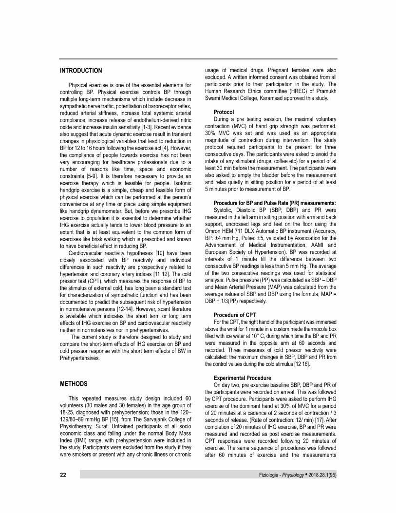
22 Fiziologia - Physiology • 2018.28.1(95)
INTRODUCTION
Physical exercise is one of the essential elements for controlling BP. Physical exercise controls BP through multiple long-term mechanisms which include decrease in sympathetic nerve traffic, potentiation of baroreceptor reflex, reduced arterial stiffness, increase total systemic arterial compliance, increase release of endothelium-derived nitric oxide and increase insulin sensitivity [1-3]. Recent evidence also suggest that acute dynamic exercise result in transient changes in physiological variables that lead to reduction in BP for 12 to 16 hours following the exercise act [4]. However, the compliance of people towards exercise has not been very encouraging for healthcare professionals due to a number of reasons like time, space and economic constraints [5-9]. It is therefore necessary to provide an exercise therapy which is feasible for people. Isotonic handgrip exercise is a simple, cheap and feasible form of physical exercise which can be performed at the person’s convenience at any time or place using simple equipment like handgrip dynamometer. But, before we prescribe IHG exercise to population it is essential to determine whether IHG exercise actually tends to lower blood pressure to an extent that is at least equivalent to the common form of exercises like brisk walking which is prescribed and known to have beneficial effect in reducing BP.
Cardiovascular reactivity hypotheses [10] have been closely associated with BP reactivity and individual differences in such reactivity are prospectively related to hypertension and coronary artery indices [11 12]. The cold pressor test (CPT), which measures the response of BP to the stimulus of external cold, has long been a standard test for characterization of sympathetic function and has been documented to predict the subsequent risk of hypertension in normotensive persons [12-14]. However, scant literature is available which indicates the short term or long term effects of IHG exercise on BP and cardiovascular reactivity neither in normotensives nor in prehypertensives.
The current study is therefore designed to study and compare the short-term effects of IHG exercise on BP and cold pressor response with the short term effects of BW in Prehypertensives.
METHODS
This repeated measures study design included 60 volunteers (30 males and 30 females) in the age group of 18-25, diagnosed with prehypertension; those in the 120–139/80–89 mmHg BP [15], from The Sarvajanik College of Physiotherapy, Surat. Untrained participants of all socio economic class and falling under the normal Body Mass Index (BMI) range, with prehypertension were included in the study. Participants were excluded from the study if they were smokers or present with any chronic illness or chronic
usage of medical drugs. Pregnant females were also excluded. A written informed consent was obtained from all participants prior to their participation in the study. The Human Research Ethics committee (HREC) of Pramukh Swami Medical College, Karamsad approved this study.
Protocol During a pre testing session, the maximal voluntary
contraction (MVC) of hand grip strength was performed. 30% MVC was set and was used as an appropriate magnitude of contraction during intervention. The study protocol required participants to be present for three consecutive days. The participants were asked to avoid the intake of any stimulant (drugs, coffee etc) for a period of at least 30 min before the measurement. The participants were also asked to empty the bladder before the measurement and relax quietly in sitting position for a period of at least 5 minutes prior to measurement of BP.
Procedure for BP and Pulse Rate (PR) measurements: Systolic, Diastolic BP (SBP, DBP) and PR were
measured in the left arm in sitting position with arm and back support, uncrossed legs and feet on the floor using the Omron HEM 711 DLX Automatic BP instrument (Accuracy, BP: ±4 mm Hg, Pulse: ±5, validated by Association for the Advancement of Medical Instrumentation, AAMI and European Society of Hypertension). BP was recorded at intervals of 1 minute till the difference between two consecutive BP readings is less than 5 mm Hg. The average of the two consecutive readings was used for statistical analysis. Pulse pressure (PP) was calculated as SBP – DBP and Mean Arterial Pressure (MAP) was calculated from the average values of SBP and DBP using the formula, MAP = DBP + 1/3(PP) respectively.
Procedure of CPT For the CPT, the right hand of the participant was immersed
above the wrist for 1 minute in a custom made thermocole box filled with ice water at 10° C, during which time the BP and PR were measured in the opposite arm at 60 seconds and recorded. Three measures of cold pressor reactivity were calculated: the maximum changes in SBP, DBP and PR from the control values during the cold stimulus [12 16].
Experimental Procedure On day two, pre exercise baseline SBP, DBP and PR of
the participants were recorded on arrival. This was followed by CPT procedure. Participants were asked to perform IHG exercise of the dominant hand at 30% of MVC for a period of 20 minutes at a cadence of 2 seconds of contraction / 3 seconds of release. (Rate of contraction: 12/ min) [17]. After completion of 20 minutes of IHG exercise, BP and PR were measured and recorded as post exercise measurements. CPT responses were recorded following 20 minutes of exercise. The same sequence of procedures was followed after 60 minutes of exercise and the measurements
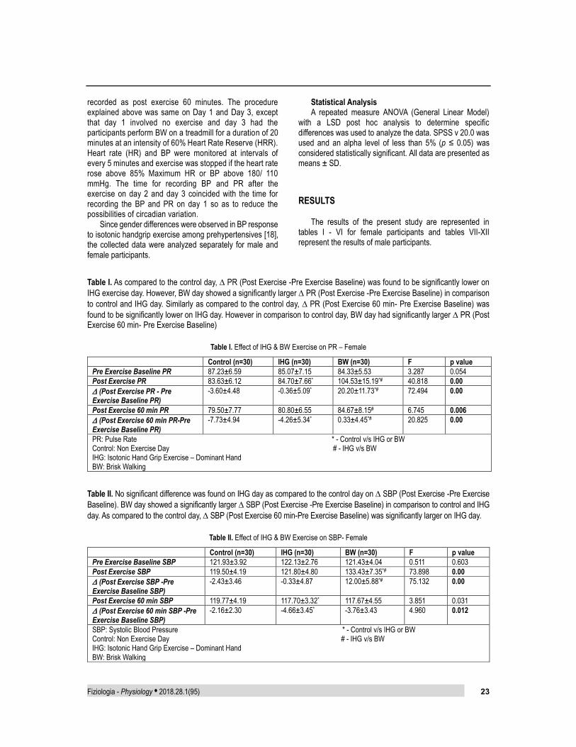
Fiziologia - Physiology • 2018.28.1(95) 23
recorded as post exercise 60 minutes. The procedure explained above was same on Day 1 and Day 3, except that day 1 involved no exercise and day 3 had the participants perform BW on a treadmill for a duration of 20 minutes at an intensity of 60% Heart Rate Reserve (HRR). Heart rate (HR) and BP were monitored at intervals of every 5 minutes and exercise was stopped if the heart rate rose above 85% Maximum HR or BP above 180/ 110 mmHg. The time for recording BP and PR after the exercise on day 2 and day 3 coincided with the time for recording the BP and PR on day 1 so as to reduce the possibilities of circadian variation.
Since gender differences were observed in BP response to isotonic handgrip exercise among prehypertensives [18], the collected data were analyzed separately for male and female participants.
Statistical Analysis A repeated measure ANOVA (General Linear Model)
with a LSD post hoc analysis to determine specific differences was used to analyze the data. SPSS v 20.0 was used and an alpha level of less than 5% (p ≤ 0.05) was considered statistically significant. All data are presented as means ± SD.
RESULTS
The results of the present study are represented in tables I - VI for female participants and tables VII-XII represent the results of male participants.
Table I. As compared to the control day, ' PR (Post Exercise -Pre Exercise Baseline) was found to be significantly lower on IHG exercise day. However, BW day showed a significantly larger ' PR (Post Exercise -Pre Exercise Baseline) in comparison to control and IHG day. Similarly as compared to the control day, ' PR (Post Exercise 60 min- Pre Exercise Baseline) was found to be significantly lower on IHG day. However in comparison to control day, BW day had significantly larger ' PR (Post Exercise 60 min- Pre Exercise Baseline)
Table I. Effect of IHG & BW Exercise on PR – Female
Control (n=30) IHG (n=30) BW (n=30) F p value Pre Exercise Baseline PR 87.23±6.59 85.07±7.15 84.33±5.53 3.287 0.054 Post Exercise PR 83.63±6.12 84.70±7.66* 104.53±15.19*# 40.818 0.00
' (Post Exercise PR - Pre Exercise Baseline PR)
-3.60±4.48 -0.36±5.09* 20.20±11.73*# 72.494 0.00
Post Exercise 60 min PR 79.50±7.77 80.80±6.55 84.67±8.15# 6.745 0.006
' (Post Exercise 60 min PR-Pre Exercise Baseline PR)
-7.73±4.94 -4.26±5.34* 0.33±4.45*# 20.825 0.00
PR: Pulse Rate * - Control v/s IHG or BW Control: Non Exercise Day # - IHG v/s BW IHG: Isotonic Hand Grip Exercise – Dominant Hand BW: Brisk Walking
Table II. No significant difference was found on IHG day as compared to the control day on ' SBP (Post Exercise -Pre Exercise Baseline). BW day showed a significantly larger ' SBP (Post Exercise -Pre Exercise Baseline) in comparison to control and IHG day. As compared to the control day, ' SBP (Post Exercise 60 min-Pre Exercise Baseline) was significantly larger on IHG day.
Table II. Effect of IHG & BW Exercise on SBP- Female
Control (n=30) IHG (n=30) BW (n=30) F p value Pre Exercise Baseline SBP 121.93±3.92 122.13±2.76 121.43±4.04 0.511 0.603 Post Exercise SBP 119.50±4.19 121.80±4.80 133.43±7.35*# 73.898 0.00
' (Post Exercise SBP -Pre Exercise Baseline SBP)
-2.43±3.46 -0.33±4.87 12.00±5.88*# 75.132 0.00
Post Exercise 60 min SBP 119.77±4.19 117.70±3.32* 117.67±4.55 3.851 0.031
' (Post Exercise 60 min SBP -Pre Exercise Baseline SBP)
-2.16±2.30 -4.66±3.45* -3.76±3.43 4.960 0.012
SBP: Systolic Blood Pressure * - Control v/s IHG or BW Control: Non Exercise Day # - IHG v/s BW IHG: Isotonic Hand Grip Exercise – Dominant Hand BW: Brisk Walking
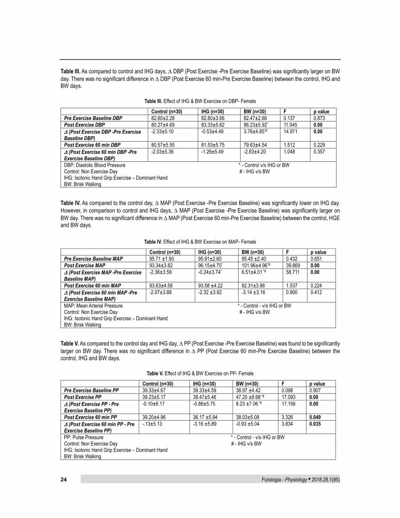
24 Fiziologia - Physiology • 2018.28.1(95)
Table III. As compared to control and IHG days, ' DBP (Post Exercise -Pre Exercise Baseline) was significantly larger on BW day. There was no significant difference in ' DBP (Post Exercise 60 min-Pre Exercise Baseline) between the control, IHG and BW days.
Table III. Effect of IHG & BW Exercise on DBP- Female
Control (n=30) IHG (n=30) BW (n=30) F p value Pre Exercise Baseline DBP 82.60±2.28 82.80±3.66 82.47±2.66 0.137 0.873 Post Exercise DBP 80.27±4.69 83.33±5.62 86.23±5.92* 11.045 0.00
' (Post Exercise DBP -Pre Exercise Baseline DBP)
-2.33±5.10 -0.53±4.49 3.76±4.85*# 14.971 0.00
Post Exercise 60 min DBP 80.57±5.55 81.53±5.75 79.63±4.54 1.512 0.229
' (Post Exercise 60 min DBP -Pre Exercise Baseline DBP)
-2.03±5.36 -1.26±5.49 -2.83±4.20 1.048 0.357
DBP: Diastolic Blood Pressure * - Control v/s IHG or BW Control: Non Exercise Day # - IHG v/s BW IHG: Isotonic Hand Grip Exercise – Dominant Hand BW: Brisk Walking
Table IV. As compared to the control day, ' MAP (Post Exercise -Pre Exercise Baseline) was significantly lower on IHG day. However, in comparison to control and IHG days, ' MAP (Post Exercise -Pre Exercise Baseline) was significantly larger on BW day. There was no significant difference in ' MAP (Post Exercise 60 min-Pre Exercise Baseline) between the control, HGE and BW days.
Table IV. Effect of IHG & BW Exercise on MAP- Female
Control (n=30) IHG (n=30) BW (n=30) F p value Pre Exercise Baseline MAP 95.71 ±1.93 95.91±2.60 95.45 ±2.40 0.432 0.651 Post Exercise MAP 93.34±3.82 96.15±4.70* 101.96±4.96*# 39.869 0.00
' (Post Exercise MAP -Pre Exercise Baseline MAP)
-2.36±3.59 -0.24±3.74* 6.51±4.01 *# 58.711 0.00
Post Exercise 60 min MAP 93.63±4.58 93.58 ±4.22 92.31±3.86 1.537 0.224
' (Post Exercise 60 min MAP -Pre Exercise Baseline MAP)
-2.07±3.88 -2.32 ±3.92 -3.14 ±3.16 0.900 0.412
MAP: Mean Arterial Pressure * - Control - v/s IHG or BW Control: Non Exercise Day # - IHG v/s BW IHG: Isotonic Hand Grip Exercise – Dominant Hand BW: Brisk Walking
Table V. As compared to the control day and IHG day, ' PP (Post Exercise -Pre Exercise Baseline) was found to be significantly larger on BW day. There was no significant difference in ' PP (Post Exercise 60 min-Pre Exercise Baseline) between the control, IHG and BW days.
Table V. Effect of IHG & BW Exercise on PP- Female
Control (n=30) IHG (n=30) BW (n=30) F p value Pre Exercise Baseline PP 39.33±4.67 39.33±4.59 38.97 ±4.42 0.098 0.907 Post Exercise PP 39.23±5.17 38.47±5.46 47.20 ±8.68 *# 17.093 0.00
' (Post Exercise PP - Pre Exercise Baseline PP)
-0.10±6.17 -0.86±5.75 8.23 ±7.06 *# 17.156 0.00
Post Exercise 60 min PP 39.20±4.96 36.17 ±5.94 38.03±5.08 3.326 0.049
' (Post Exercise 60 min PP - Pre Exercise Baseline PP)
-.13±5.13 -3.16 ±5.89 -0.93 ±5.04 3.834 0.035
PP: Pulse Pressure * - Control - v/s IHG or BW Control: Non Exercise Day # - IHG v/s BW IHG: Isotonic Hand Grip Exercise – Dominant Hand BW: Brisk Walking
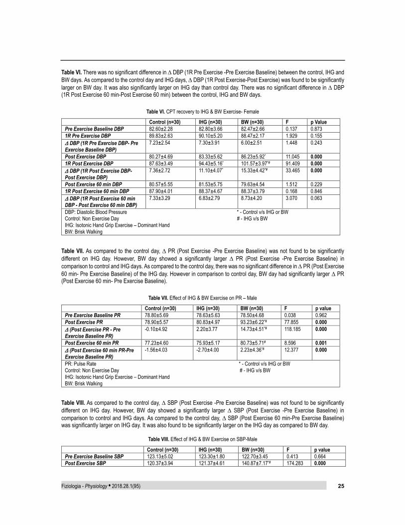
Fiziologia - Physiology • 2018.28.1(95) 25
Table VI. There was no significant difference in ' DBP (1R Pre Exercise -Pre Exercise Baseline) between the control, IHG and BW days. As compared to the control day and IHG days, ' DBP (1R Post Exercise-Post Exercise) was found to be significantly larger on BW day. It was also significantly larger on IHG day than control day. There was no significant difference in ' DBP (1R Post Exercise 60 min-Post Exercise 60 min) between the control, IHG and BW days.
Table VI. CPT recovery to IHG & BW Exercise- Female
Control (n=30) IHG (n=30) BW (n=30) F p Value Pre Exercise Baseline DBP 82.60±2.28 82.80±3.66 82.47±2.66 0.137 0.873 1R Pre Exercise DBP 89.83±2.63 90.10±5.20 88.47±2.17 1.929 0.155 ' DBP (1R Pre Exercise DBP- Pre Exercise Baseline DBP)
7.23±2.54 7.30±3.91 6.00±2.51 1.448 0.243
Post Exercise DBP 80.27±4.69 83.33±5.62 86.23±5.92* 11.045 0.000 1R Post Exercise DBP 87.63±3.49 94.43±5.16* 101.57±3.97*# 91.409 0.000 ' DBP (1R Post Exercise DBP- Post Exercise DBP)
7.36±2.72 11.10±4.07* 15.33±4.42*# 33.465 0.000
Post Exercise 60 min DBP 80.57±5.55 81.53±5.75 79.63±4.54 1.512 0.229 1R Post Exercise 60 min DBP 87.90±4.01 88.37±4.67 88.37±3.79 0.168 0.846 ' DBP (1R Post Exercise 60 min DBP - Post Exercise 60 min DBP)
7.33±3.29 6.83±2.79 8.73±4.20 3.070 0.063
DBP: Diastolic Blood Pressure * - Control v/s IHG or BW Control: Non Exercise Day # - IHG v/s BW IHG: Isotonic Hand Grip Exercise – Dominant Hand BW: Brisk Walking
Table VII. As compared to the control day, ' PR (Post Exercise -Pre Exercise Baseline) was not found to be significantly different on IHG day. However, BW day showed a significantly larger ' PR (Post Exercise -Pre Exercise Baseline) in comparison to control and IHG days. As compared to the control day, there was no significant difference in ' PR (Post Exercise 60 min- Pre Exercise Baseline) of the IHG day. However in comparison to control day, BW day had significantly larger ' PR (Post Exercise 60 min- Pre Exercise Baseline).
Table VII. Effect of IHG & BW Exercise on PR – Male
Control (n=30) IHG (n=30) BW (n=30) F p value Pre Exercise Baseline PR 78.80±5.69 78.63±5.63 78.50±4.68 0.038 0.962 Post Exercise PR 78.90±5.57 80.83±4.97 93.23±6.22*# 77.855 0.000
' (Post Exercise PR - Pre Exercise Baseline PR)
-0.10±4.92 2.20±3.77 14.73±4.51*# 118.185 0.000
Post Exercise 60 min PR 77.23±4.60 75.93±5.17 80.73±5.71# 8.596 0.001
' (Post Exercise 60 min PR-Pre Exercise Baseline PR)
-1.56±4.03 -2.70±4.00 2.23±4.36*# 12.377 0.000
PR: Pulse Rate * - Control v/s IHG or BW Control: Non Exercise Day # - IHG v/s BW IHG: Isotonic Hand Grip Exercise – Dominant Hand BW: Brisk Walking
Table VIII. As compared to the control day, ' SBP (Post Exercise -Pre Exercise Baseline) was not found to be significantly different on IHG day. However, BW day showed a significantly larger ' SBP (Post Exercise -Pre Exercise Baseline) in comparison to control and IHG days. As compared to the control day, ' SBP (Post Exercise 60 min-Pre Exercise Baseline) was significantly larger on IHG day. It was also found to be significantly larger on the IHG day as compared to BW day.
Table VIII. Effect of IHG & BW Exercise on SBP-Male
Control (n=30) IHG (n=30) BW (n=30) F p value Pre Exercise Baseline SBP 123.13±5.02 123.30±1.80 122.70±3.45 0.413 0.664 Post Exercise SBP 120.37±3.94 121.37±4.61 140.87±7.17*# 174.283 0.000
26 Fiziologia - Physiology • 2018.28.1(95)
' (Post Exercise SBP -Pre Exercise Baseline SBP)
-2.76±4.35 -1.93±4.65 18.16±5.47*# 165.435 0.000
Post Exercise 60 min SBP 118.77±2.22 117.20±2.39* 119.73±2.82# 9.256 0.001
' (Post Exercise 60 min SBP -Pre Exercise Baseline SBP)
-4.36±4.42 -6.1±4.18* -2.96±3.47# 29.469 0.000
SBP: Systolic Blood Pressure * - Control v/s IHG or BW Control: Non Exercise Day # - IHG v/s BW IHG: Isotonic Hand Grip Exercise – Dominant Hand BW: Brisk Walking
Table IX. There was no significant difference in ' DBP (Post Exercise -Pre Exercise Baseline) between the control day and IHG day. However, in comparison to control, ' DBP (Post Exercise -Pre Exercise Baseline) was significantly larger on BW day. There was no significant difference in ' DBP (Post Exercise 60 min-Pre Exercise Baseline) between the control, IHG and BW days.
Table IX. Effect of IHG & BW Exercise on DBP-Male
Control (n=30) IHG (n=30) BW (n=30) F p value Pre Exercise Baseline DBP 80.40±2.22 79.97±2.73 80.23±2.81 0.296 0.745 Post Exercise DBP 79.23±3.49 79.70±5.19 82.43±5.42* 4.692 0.013
' (Post Exercise DBP -Pre Exercise Baseline DBP)
-1.16±3.42 -0.26±4.54 2.20±4.41* 5.646 0.006
Post Exercise 60 min DBP 77.53±3.86 77.70±5.30 77.70±4.28 0.018 0.982
' (Post Exercise 60 min DBP -Pre Exercise Baseline DBP)
-2.86±4.03 -2.26±4.07 -2.53±3.83 0.177 0.838
DBP: Diastolic Blood Pressure * - Control v/s IHG or BW Control: Non Exercise Day # - IHG v/s BW IHG: Isotonic Hand Grip Exercise – Dominant Hand BW: Brisk Walking
Table X. There was no significant difference in ' MAP (Post Exercise -Pre Exercise Baseline) between the control days and IHG days. However, in comparison to control and IHG days, ' MAP (Post Exercise -Pre Exercise Baseline) was significantly larger on BW day. There was no significant difference in ' MAP (Post Exercise 60 min-Pre Exercise Baseline) between the control, IHG and BW days.
Table X. Effect of IHG & BW Exercise on MAP-Male
Control (n=30) IHG (n=30) BW (n=30) F p value Pre Exercise Baseline MAP 94.64±2.22 94.41±2.11 94.38±2.47 0.208 0.813 Post Exercise MAP 92.94±2.77 93.58±4.40 101.91±4.36*# 63.077 0.000
' (Post Exercise MAP -Pre Exercise Baseline MAP)
-1.70±3.06 -0.82±4.07 7.52±3.18 *# 70.32 0.000
Post Exercise 60 min MAP 91.27±2.87 90.86 ±3.74 91.71±2.83 0.690 0.506
' (Post Exercise 60 min MAP -Pre Exercise Baseline MAP)
-3.36±2.98 -3.54 ±3.05 -2.67 ±2.58 0.861 0.428
MAP: Mean Arterial Pressure * - Control - v/s IHG or BW Control: Non Exercise Day # - IHG v/s BW IHG: Isotonic Hand Grip Exercise – Dominant Hand BW: Brisk Walking
Table XI. As compared to the control and IHG days, ' PP (Post Exercise -Pre Exercise Baseline) was found to be significantly larger on BW day. As compared to the control day, there was no difference in ' PP (Post Exercise 60 min-Pre Exercise Baseline) on the IHG and BW days. ' PP (Post Exercise 60 min-Pre Exercise Baseline) was found to be significantly lower on the BW day as compared to IHG day.

Fiziologia - Physiology • 2018.28.1(95) 27
Table XI. Effect of IHG & BW Exercise on PP-Male
Control (n=30) IHG (n=30) BW (n=30) F p value Pre Exercise Baseline PP 42.73±5.50 43.33±2.67 42.47±3.73 0.49 0.613 Post Exercise PP 41.13±5.01 41.67±5.03 58.43 ±8.93 *# 75.86 0.000
' (Post Exercise PP - Pre Exercise Baseline PP)
-1.60±4.62 -1.66±4.43 15.96 ±7.60 *# 83.92 0.000
Post Exercise 60 min PP 41.23±3.88 39.50 ±5.46 42.03±5.55 2.65 0.083
' (Post Exercise 60 min PP - Pre Exercise Baseline PP)
-1.50±6.17 -3.83±4.44 -0.43 ±5.69 # 3.50 0.041
PP: Pulse Pressure * - Control - v/s IHG or BW Control: Non Exercise Day # - IHG v/s BW IHG: Isotonic Hand Grip Exercise – Dominant Hand BW: Brisk Walking
Table XII. As compared to the control and IHG days, ' DBP (1R Pre Exercise -Pre Exercise Baseline) was found to be significantly larger on BW day. As compared to the control and IHG days, ' DBP (1R Post Exercise-Post Exercise) was found to be significantly larger on BW day. There was no significant difference in ' DBP (1R Post Exercise 60 min-Post Exercise 60 min) between the control, IHG and BW days.
Table XII. CPT recovery to IHG & BW Exercise – Male
Control (n=30) IHG (n=30) BW (n=30) F p Value Pre Exercise Baseline DBP 80.40±2.22 79.97±2.73 80.23±2.81 0.296 0.745 1R Pre Exercise DBP 85.97±2.87 86.40±4.44 88.10±3.55* 3.547 0.041 ' DBP (1R Pre Exercise DBP- Pre Exercise Baseline DBP)
5.56±2.73 6.43±3.50 7.86±3.21* 4.185 0.022
Post Exercise DBP 79.23±3.49 79.70±5.19 82.43±5.42* 4.692 0.014
1R Post Exercise DBP 88.83±3.38 91.73±4.48* 99.43±3.90*# 61.962 0.000 ' DBP (1R Post Exercise DBP- Post Exercise DBP)
9.60±3.13 12.03±4.27 17.00±4.26*# 32.46 0.000
Post Exercise 60 min DBP 77.53±3.86 77.70±5.30 77.70±4.28 0.018 0.982
1R Post Exercise 60 min DBP 83.93±3.61 84.80±4.38 86.17±4.03 2.947 0.067 ' DBP (1R Post Exercise 60 min DBP - Post Exercise 60 min DBP)
6.40±3.55 7.10±4.02 8.46±4.80 1.705 0.191
DBP: Diastolic Blood Pressure * - Control v/s IHG or BW Control: Non Exercise Day # - IHG v/s BW IHG: Isotonic Hand Grip Exercise – Dominant Hand BW: Brisk Walking
DISCUSSION
Recent studies have proven that small reductions of BP in prehypertensive or hypertensive population could have significant health benefits. Studies suggest that a 2mm Hg drop in DBP would lead to a 17% decrease in Hypertension, 6% reduction in Coronary Heart Disease and 15% reduction in Stroke related events[19]. The results of the present study shows that 20 minutes of IHG exercise reduces BP in prehypertensive young male and females. Immediately after 20 minutes of IHG exercise, mean reduction in PR, SBP, DBP, MAP and PP were observed both in female (Tables: 1-5) and male (Tables: 7-11) participants whereas there was an increase in mean of all the above parameters after BW. The mean reductions of PR, SBP and PP of females were higher in IHG exercise as compared to BW. However, mean DBP (-
2.83±4.20) and MAP (-3.14±3.16) of BW were higher than IHG exercise. Similarly, the mean reductions of PR, SBP, MAP and PP of males were higher in IHG exercise as compared to BW except for reductions in DBP which was higher in BW.
Earlier studies reported variations in reductions of SBP and DBP to various forms of exercise. Quinn TJ [20] observed 4 mm Hg and 5 mm Hg reductions in SBP and DBP to treadmill walking for 30 minutes at 75% VO2 max, whereas Wallace et al [21] observed 8 mm Hg and 6 mm Hg in SBP and DBP respectively, after treadmill exercise of 5 sessions of 10 minutes at 50% VO2 max. Pescatello et al [22 23], Lehmkuhl et al [24], Hara et al [25], Rueckert et al [26], in their studies found no significant changes in BP after various intensities of treadmill walking and running.
Differences in reductions of BP and PR of this present study from past studies can be attributed to the different duration and intensities of the exercise, as the present study

28 Fiziologia - Physiology • 2018.28.1(95)
had focused on the benefits of only 20 minutes of exercising, which was the least of all the above mentioned studies.
Various studies in the past have postulated numerous potential mechanisms responsible for the reduction in BP following handgrip exercises, most of these studies focusing on isometric handgrip exercises [27 28]. Some of the factors include oxidative stress [29], improved endothelium-dependent vasodilatation [30 31], and modulation of Autonomic Nervous system [32 33]. Despite the fact that the present study recommending IHG exercise to reduce BP in prehypertensives, the exact mechanisms responsible for BP reductions following IHG exercise remains elusive.
CPT response immediately after IHG exercise and BW showed a rise in DBP in both female and male participants, however this increase was quite larger on BW day (Tables 6 and 12). At 60 minutes post exercise, DBP response to CPT returned close to baseline resting values on IHG as compared to BW day, where the DBP stayed higher than baseline values.
Once stimulated by a stressor, the sympathetic system sets off a rise in heart rate and blood pressure, but usually these effects return to normal levels within a very short period of time (5 minutes) after the withdrawal of the stressor. Diastolic blood pressure is the index of peripheral resistance. Higher diastolic pressure even after the withdrawal of the cold stimulus indicated that once constricted through sympathetic stimulation, vessels took longer to return to their normal original diameters [34]. Persons with higher cardiovascular reactivity to a stressor show slower rate of recovery after the withdrawal of the stressor responsible for sympathetic stimulation, indicating that the autonomic control system is not competent enough to lower heart rate and blood pressure to the baseline quickly.
LIMITATIONS OF THE STUDY
This study had some limitations: First, our inclusion criteria were purposely restrictive so as to limit the strong confounding effects of age, BMI, and training, but they also limit the generalization of our findings to the population studied. Secondly, being a repeated measures study design, the practice effects on participants on three consecutive days were not controlled. Finally, it focused only on the acute effect of IHG exercise on BP and PR parameters, hence further studies should advance the findings of this research by taking into consideration, the long term training effects of IHG exercise.
CONCLUSION
One of the common barrier to exercise among patient and normal population is time. The present study protocol requires very little adjustments in day to day routine and at
the same time is effective to yield a better reduction in resting SBP and PP in comparison to treadmill walking. This might increase the adherence of prehypertensives to exercise as the IHG exercise can be considered as participation in leisure time. The clinical benefits of IHG exercise found out from this study, which is least explored till date, can be considered as a novel therapy in transcending time and cost and might be provided as an alternative method for prehypertensive population to control BP by improving exercise adherence.
REFERENCES
1. Hamer M. The effects of exercise on haemodynamic function in relation to the familial hypertension risk model. Journal of human hypertension 2006;20(5):313-9.
2. Md. Shahidur Rahman AKMS. Role of Exercise as a Therapeutic Intervention for Hypertension. University Heart Journal 2009;5(1):36-39.
3. Peter F. Kokkinos A, Athanasios Manolis, Andreas Pittaras. Physical Activity in the Prevention and Management of High Blood Pressure. Hellenic J Cardiol 2009;50:52-59.
4. Fagard R, Vanhees L. Twenty-four hour blood pressure after exercise in patients with coronary artery disease. Journal of human hypertension 2000;14(4):231-4.
5. Arikawa AY, O'Dougherty M, Kaufman BC, et al. Attrition and adherence of young women to aerobic exercise: lessons from the WISER study. Contemporary clinical trials 2012;33(2):298-301.
6. Irwin ML TS, Yasui Y, Rajan B, McVarish L, LaCroix K, et al. Influence of demographic, physiologic, and psychosocial variables on adherence to a yearlong moderateintensity exercise trial in postmenopausal women. . Prev Med 2004;39:1080-6.
7. King AC BS, Bild DE, Dishman RK, Dubbert PM, Marcus BH, et al. Determinants of physical activity and interventions in adults. Med Sci Sports Exerc 1992;24:S221-36.
8. Martin KA SA. Who will stay and who will go? A review of older adults’ adherence to randomized controlled trials of exercise. JAPA 2001;9:91-114.
9. Perri MG AS, Durning PE, Ketterson TU, Sydeman SJ, Berlant NE, et al.nAdherence to exercise prescriptions: effects of prescribing moderate versus higher levels of intensity and frequency. Health Psychol 2002;21:452-58.
10. Krantz DS, Manuck SB. Acute psychophysiologic reactivity and risk of cardiovascular disease: a review and methodologic critique. Psychological bulletin 1984;96(3):435-64.
11. Jennings JR, Kamarck TW, Everson-Rose SA, et al. Exaggerated blood pressure responses during mental stress are prospectively related to enhanced carotid atherosclerosis in middle-aged Finnish men. Circulation 2004;110(15):2198-203.
12. Menkes MS, Matthews KA, Krantz DS, et al. Cardiovascular reactivity to the cold pressor test as a predictor of hypertension. Hypertension 1989;14(5):524-30.
13. Victor RG, Leimbach WN, Jr., Seals DR, et al. Effects of the cold pressor test on muscle sympathetic nerve activity in humans. Hypertension 1987;9(5):429-36.
14. Wood DL, Sheps SG, Elveback LR, et al. Cold pressor test as a predictor of hypertension. Hypertension 1984;6(3):301-6.

Fiziologia - Physiology • 2018.28.1(95) 29
15. NM. K. Kaplan’s Clinical Hypertension. 9th ed: Lippincott Williams & Wilkins, 2006.
16. Vivek Verma SKS, S. Ghosh. Identification of susceptibility to hypertension by the cold pressor test. Indian J Physiol Pharmacol 2005;49(1):119-20.
17. Brian A. Batman Jch UAL, Michael B. Smith, Qing X. Yang, Lawrence I. Sinoway. . Sympathetic nerve activity during prolonged rhythmic forearm exercise. The American Physiological Society 1994:1077-81.
18. Saravanan Murugan SKS, Wasim Sheikh. Does gender affect the response of blood pressure to isotonic hand grip exercise in prehypertensives? Sch J App Med Sci 2016;4(5(D)):1693-97.
19. Cook NR, Cohen J, Hebert PR, et al. Implications of small reductions in diastolic blood pressure for primary prevention. Archives of internal medicine 1995;155(7):701-9.
20. TJ Q. Twenty-four hour, ambulatory blood pressure responses following acute exercise: impact of exercise intensity. J Hum Hypertens 2000;14:547-53.
21. Wallace JP, Bogle PG, King BA, et al. A comparison of 24-h average blood pressures and blood pressure load following exercise. American journal of hypertension 1997;10(7 Pt 1):728-34.
22. Pescatello LS, Kulikowich JM. The aftereffects of dynamic exercise on ambulatory blood pressure. Medicine and science in sports and exercise 2001;33(11):1855-61.
23. Thompson PD, Crouse SF, Goodpaster B, et al. The acute versus the chronic response to exercise. Medicine and science in sports and exercise 2001;33(6 Suppl):S438-45; discussion S52-3.
24. Lehmkuhl LA, Park S, Zakutansky D, et al. Reproducibility of postexercise ambulatory blood pressure in Stage I hypertension. Journal of human hypertension 2005;19(8):589-95.
25. Hara K, Floras JS. Influence of naloxone on muscle sympathetic nerve activity, systemic and calf haemodynamics and
ambulatory blood pressure after exercise in mild essential hypertension. Journal of hypertension 1995;13(4):447-61.
26. Rueckert PA, Slane PR, Lillis DL, et al. Hemodynamic patterns and duration of post-dynamic exercise hypotension in hypertensive humans. Medicine and science in sports and exercise 1996;28(1):24-32.
27. Mortimer J, McKune AJ. Effect of short-term isometric handgrip training on blood pressure in middle-aged females. Cardiovascular journal of Africa 2011;22(5):257-60.
28. Millar PJ, Bray SR, McGowan CL, et al. Effects of isometric handgrip training among people medicated for hypertension: a multilevel analysis. Blood pressure monitoring 2007;12(5):307-14.
29. Peters PG, Alessio HM, Hagerman AE, et al. Short-term isometric exercise reduces systolic blood pressure in hypertensive adults: possible role of reactive oxygen species. International journal of cardiology 2006;110(2):199-205.
30. McGowan CL, Visocchi A, Faulkner M, et al. Isometric handgrip training improves local flow-mediated dilation in medicated hypertensives. European journal of applied physiology 2007;99(3):227-34.
31. McGowan CL, Levy AS, Millar PJ, et al. Acute vascular responses to isometric handgrip exercise and effects of training in persons medicated for hypertension. American journal of physiology Heart and circulatory physiology 2006;291(4):H1797-802.
32. Millar PJ, MacDonald MJ, Bray SR, et al. Isometric handgrip exercise improves acute neurocardiac regulation. European journal of applied physiology 2009;107(5):509-15.
33. Taylor AC, McCartney N, Kamath MV, et al. Isometric training lowers resting blood pressure and modulates autonomic control. Medicine and science in sports and exercise 2003;35(2):251-6.
34. LeBlanc J, Dulac S, Cote J, et al. Autonomic nervous system and adaptation to cold in man. J Appl Physiol 1975;39(2):181-6.
IMPACTUL EXERCIȚIILOR ISOTONICE HAND GRIP PRIVIND PRESIUNEA DE SÂNGE ȘI RĂSPUNSUL SIMPATIC LA PRE-HIPERTENSIVI REZUMAT Contextul studiului: Exercițiul fizic este unul dintre elementele esențiale pentru controlul tensiunii arteriale. Cu toate acestea, anumite constrângeri, cum ar fi fezabilitatea echipamentului de exerciții fizice și a timpului, au redus respectarea oamenilor față de exerciții fizice. Exercitarea izotonică a mâinii (IHG) poate fi considerată o alternativă la alte forme de exercițiu dacă ar putea fi studiată pentru efectele pe care le are asupra tensiunii arteriale (BP). Prin urmare, acest studiu a fost realizat pentru a determina și compara efectul pe termen scurt al exercițiului IHG față de mersul brusc (BW) asupra BP și a reactivității cardiovasculare la prehypertensive. Metode: 60 de voluntari cu prehypertensiune (bărbați-30; femei-30) au participat la acest studiu. Studiul a fost efectuat pe trei zile consecutive: Ziua 1- Control sau non-exercițiu, Ziua 2 - exercițiu IGG și Ziua 3 - BW. La sosire s-au înregistrat valorile presiunii sistolice (SBP) și tensiunii arteriale diastolice (DBP) și frecvența pulsului (PR), urmate de testul de presiune la rece (CPT). Acest lucru a fost urmat de nici un exercițiu, exercițiu izotonic de prindere pe mâini sau o plimbare intensă timp de 20 de minute în ziua 1, 2 și respectiv 3. BP a fost înregistrată imediat după această perioadă de 20 de minute urmată de CPT. Măsurătorile au fost înregistrate din nou după o oră în aceeași ordine în toate cele trei zile. Măsuri repetate
30 Fiziologia - Physiology • 2018.28.1(95)
ANOVA (p <0,05) a fost utilizată pentru analiza diferențelor dintre exercițiul prealabil de bază, exercițiul imediat post și 60 de minute după exercițiul BP, PR, presiunea arterială medie (MAP) și presiunea pulsului (PP). Măsurătorile CPT au fost, de asemenea, analizate pentru a afla diferențele de răspuns ale sistemului cardiovascular la exercițiile IHG și BW. Rezultate: În rândul participanților feminini, s-au constatat diferențe semnificative în măsurătorile BP și PR imediat după exercițiu. Exercițiul IHG a evidențiat o reducere semnificativă a PR (media ± SD, -0,36 ± 5,09), SBP (-0,33 ± 4,87), DBP (-0,53 ± 4,49), MAP (-0,24 ± 3,74) și PP (-0,86 ± 5,75) comparativ cu BW care au prezentat o creștere a PR (20,20 ± 11,75), SBP (12,00 ± 5,88), DBP (3,76 ± 4,85), MAP (6,51 ± 4,01) și PP (8,23 ± 7,06). Cu toate acestea, după 60 de minute de exercițiu, nu s-au observat diferențe semnificative în SBP, DBP, MAP și PP la exercițiul IHG și BW, numai PR prezentând o reducere semnificativă a exercițiilor IHG (-4,26 ± 5,34) decât BW (0,33 ± 4,45) . Răspunsul CPT măsurat la momentul inițial și 60 de minute după exerciții nu a fost statistic semnificativ în cele două zile de experiment. Cu toate acestea, răspunsul CPT exercițiu imediat la BW a fost semnificativ mai mare (15,33 ± 4,42) decât exercițiul IHG (11,10 ± 4,07) S-au constatat diferențe semnificative în măsurătorile PR, SBP, MAP și PP imediat după exercițiu și cea a pre-exercițiului fizic la participanții de sex masculin. Exercițiul IHG a arătat o reducere semnificativă a SBP (-1,93 ± 4,65), MAP (-0,82 ± 4,07) și PP (-1,66 ± 4,43) comparativ cu BW care au prezentat o creștere a SBP (18,16 ± 5,47), MAP (7,52 ± 3,18 ) și PP (15,96 ± 7,60). O creștere a PR a fost observată în ambele condiții de efort cu BW (14,73 ± 4,51), semnificativ mai mare decât exercițiul IHG (2,20 ± 3,77). Nu sa constatat nici o diferență semnificativă în DBP în ambele zile de exerciții fizice. În mod similar, după 60 de minute de exercițiu, nu s-au observat diferențe semnificative în exercițiul DBP și MAP la exercițiul IHG și BW. PR a prezentat o reducere semnificativă a exercițiului IHG (-2,70 ± 4,00) decât BW (2,23 ± 4,36). O reducere semnificativă a fost observată și la SBP (-6,10 ± 4,18) și PP (-3,83 ± 4,44) la efortul IHG decât SBP (-2,96 ± 3,47) și PP (-0,43 ± 5,69) la BW. Răspunsul CPT măsurat la momentul inițial și 60 de minute după exerciții nu a fost statistic semnificativ în cele două zile de experiment. Cu toate acestea, răspunsul CPT imediat la exercițiul BT a fost semnificativ mai mare (17,00 ± 4,26) decât exercițiul IHG (12,03 ± 4,27). Concluziile: Constatările sugerează că 20 de minute de exercițiu IHG pot fi utilizate ca o metodă eficientă din punct de vedere al costurilor și mai puțin consumatoare de timp pentru reducerea nivelurilor de BP la adolescenții prehypertensivi masculi și femele în comparație cu BW. De asemenea, sa constatat că exercițiul IHG acut pare să aibă un impact semnificativ asupra răspunsurilor BP la stresul rece. Sunt necesare studii suplimentare pentru a investiga efectele pe termen lung ale antrenamentului IHG asupra BP. Cuvinte cheie: exercițiu isotonic, test de presor rece, pre-hipertensiune

Fiziologia - Physiology • 2018.28.1(95) 31
MISDIAGNOSIS OF ASPIRATED BRONCHIAL FOREIGN BODY. A CASE REPORT RAMONA CIOBOATA1, ILEANA BAZAVAN1, MIMI NITU1, MARIUS GEORGESCU2, MIHAI OLTEANU 1 1Internal Medicine - Pulmonology Department, University of Medicine and Pharmacy Craiova, Romania 2Physiology Department, University of Medicine and Pharmacy “Victor Babes”, Timisoara, Romania
ABSTRACT Aspiration of foreign bodies is an often severe medical condition which demands both timely recognition and rapid action. When diagnosis is delayed, the subsequent delay to treatment associates with severe complications. Material and method: A 52 year old Caucasian male patient, suffering from recurrent pneumonia, the cause of which was difficult to pinpoint, especially in the absence of typical risk factors. He was admitted to the emergency ward presenting dyspnea, chronic productive cough, chest pain, low grade fever and weight loss, which initially debuted 12 months earlier but was heightened in the 7 days previous to admission. The diagnosis of foreign body aspiration has been established after performing bronchoscopy due to the suspicion of a bronchioloalveolar carcinoma. Short title: Aspirated bronchial foreign body - a case report Key words: foreign body, adult pneumonia, bronchoscopy
INTRODUCTION
Foreign-body aspiration is often a serious medical condition and a potentially life-threatening event demanding timely recognition and prompt action.
Foreign body aspiration is more common in children than in adults. Data from the National Security Council reported that approximately 80 percent of cases occur in patients younger than 15 years of age, with the remaining 20 percent over the age of 15 years [1].
The most common symptoms of foreign body aspiration in the airways include chronic cough, dyspnea, and hemoptysis, which are often misdiagnosed as obstructive airway disease. Risk factors for aspiration include neurological dysfunctions such as stroke, encephalopathy, alcoholism, seizures, Parkinson’s disease, sedatives, and mental retardation, and also dental procedure complications, facial trauma, intubation, and abnormalities of the pharynx and esophagus [2,3,4]
Delayed diagnosis and the subsequent delayed treatment is associated with serious complications. Flexible and rigid bronchoscopy has become the cornerstone of both
the diagnosis and treatment of patients with suspected foreign body aspiration [2].
CASE REPORT
We present the case of a 52 year old male patient with a history of high blood pressure and recurrent pneumonia.
He presented to the emergency ward with chronic productive cough, dyspnea, chest pain, weight loss and low grade fever which had initially started 12 months earlier but heightened in the last 7 days previous to admission.
Since 12 months before he had been treated with multiple courses of oral antibiotics for recurrent community-acquired pneumonia, which initially improved the symptoms, however after a few weeks symptoms recurred. Antibiotic treatment was then resumed, most recently 1 month prior to presentation but at this time without effect, and the patient was admitted to the hospital.
He denied history of seizures as well as alcohol, sedatives or illicit drug use.
Received March 14th 2018. Accepted May 31st 2018. Address for correspondence: Marius Georgescu, MD, PhD, Assistant Teaching Professor, Department of Functional Sciences, Discipline of Physiology, “Victor Babes” University of Medicine and Pharmacy, Eftimie Murgu Square No. 2A, Timisoara, RO-300041. Phone: 0040765527318; e-mail: [email protected]
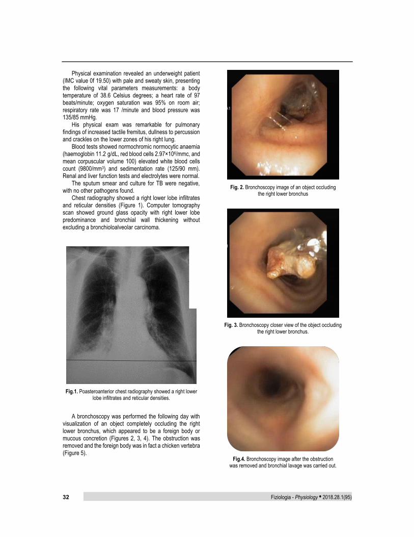
32 Fiziologia - Physiology • 2018.28.1(95)
Physical examination revealed an underweight patient (IMC value 0f 19.50) with pale and sweaty skin, presenting the following vital parameters measurements: a body temperature of 38.6 Celsius degrees; a heart rate of 97 beats/minute; oxygen saturation was 95% on room air; respiratory rate was 17 /minute and blood pressure was 135/85 mmHg.
His physical exam was remarkable for pulmonary findings of increased tactile fremitus, dullness to percussion and crackles on the lower zones of his right lung.
Blood tests showed normochromic normocytic anaemia (haemoglobin 11.2 g/dL, red blood cells 2.97×10⁶/mmc, and mean corpuscular volume 100) elevated white blood cells count (9800/mm3) and sedimentation rate (125/90 mm). Renal and liver function tests and electrolytes were normal.
The sputum smear and culture for TB were negative, with no other pathogens found.
Chest radiography showed a right lower lobe infiltrates and reticular densities (Figure 1). Computer tomography scan showed ground glass opacity with right lower lobe predominance and bronchial wall thickening without excluding a bronchioloalveolar carcinoma.
Fig.1. Poasteroanterior chest radiography showed a right lower
lobe infiltrates and reticular densities.
A bronchoscopy was performed the following day with visualization of an object completely occluding the right lower bronchus, which appeared to be a foreign body or mucous concretion (Figures 2, 3, 4). The obstruction was removed and the foreign body was in fact a chicken vertebra (Figure 5).
Fig. 2. Bronchoscopy image of an object occluding
the right lower bronchus
Fig. 3. Bronchoscopy closer view of the object occluding
the right lower bronchus.
Fig.4. Bronchoscopy image after the obstruction
was removed and bronchial lavage was carried out.
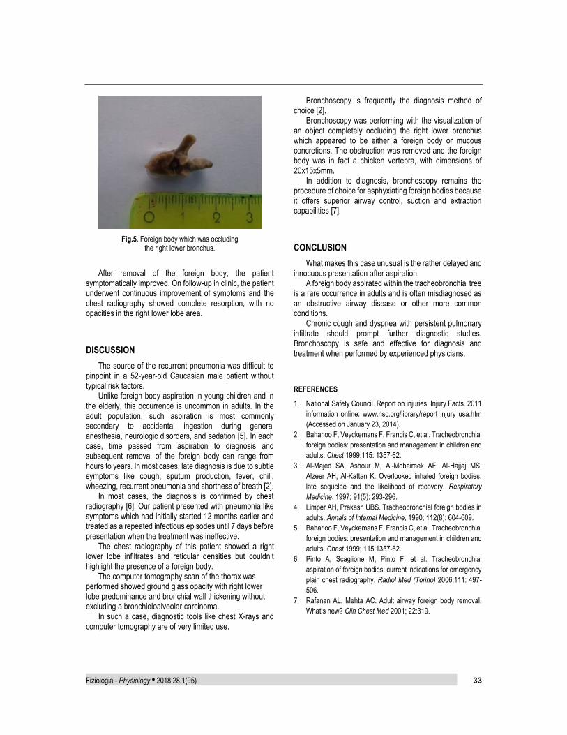
Fiziologia - Physiology • 2018.28.1(95) 33
Fig.5. Foreign body which was occluding
the right lower bronchus.
After removal of the foreign body, the patient symptomatically improved. On follow-up in clinic, the patient underwent continuous improvement of symptoms and the chest radiography showed complete resorption, with no opacities in the right lower lobe area.
DISCUSSION The source of the recurrent pneumonia was difficult to
pinpoint in a 52-year-old Caucasian male patient without typical risk factors.
Unlike foreign body aspiration in young children and in the elderly, this occurrence is uncommon in adults. In the adult population, such aspiration is most commonly secondary to accidental ingestion during general anesthesia, neurologic disorders, and sedation [5]. In each case, time passed from aspiration to diagnosis and subsequent removal of the foreign body can range from hours to years. In most cases, late diagnosis is due to subtle symptoms like cough, sputum production, fever, chill, wheezing, recurrent pneumonia and shortness of breath [2].
In most cases, the diagnosis is confirmed by chest radiography [6]. Our patient presented with pneumonia like symptoms which had initially started 12 months earlier and treated as a repeated infectious episodes until 7 days before presentation when the treatment was ineffective.
The chest radiography of this patient showed a right lower lobe infiltrates and reticular densities but couldn’t highlight the presence of a foreign body.
The computer tomography scan of the thorax was performed showed ground glass opacity with right lower lobe predominance and bronchial wall thickening without excluding a bronchioloalveolar carcinoma.
In such a case, diagnostic tools like chest X-rays and computer tomography are of very limited use.
Bronchoscopy is frequently the diagnosis method of choice [2].
Bronchoscopy was performing with the visualization of an object completely occluding the right lower bronchus which appeared to be either a foreign body or mucous concretions. The obstruction was removed and the foreign body was in fact a chicken vertebra, with dimensions of 20x15x5mm.
In addition to diagnosis, bronchoscopy remains the procedure of choice for asphyxiating foreign bodies because it offers superior airway control, suction and extraction capabilities [7].
CONCLUSION What makes this case unusual is the rather delayed and
innocuous presentation after aspiration. A foreign body aspirated within the tracheobronchial tree
is a rare occurrence in adults and is often misdiagnosed as an obstructive airway disease or other more common conditions.
Chronic cough and dyspnea with persistent pulmonary infiltrate should prompt further diagnostic studies. Bronchoscopy is safe and effective for diagnosis and treatment when performed by experienced physicians.
REFERENCES
1. National Safety Council. Report on injuries. Injury Facts. 2011 information online: www.nsc.org/library/report injury usa.htm (Accessed on January 23, 2014).
2. Baharloo F, Veyckemans F, Francis C, et al. Tracheobronchial foreign bodies: presentation and management in children and adults. Chest 1999;115: 1357-62.
3. Al-Majed SA, Ashour M, Al-Mobeireek AF, Al-Hajjaj MS, Alzeer AH, Al-Kattan K. Overlooked inhaled foreign bodies: late sequelae and the likelihood of recovery. Respiratory Medicine, 1997; 91(5): 293-296.
4. Limper AH, Prakash UBS. Tracheobronchial foreign bodies in adults. Annals of Internal Medicine, 1990; 112(8): 604-609.
5. Baharloo F, Veyckemans F, Francis C, et al. Tracheobronchial foreign bodies: presentation and management in children and adults. Chest 1999; 115:1357-62.
6. Pinto A, Scaglione M, Pinto F, et al. Tracheobronchial aspiration of foreign bodies: current indications for emergency plain chest radiography. Radiol Med (Torino) 2006;111: 497-506.
7. Rafanan AL, Mehta AC. Adult airway foreign body removal. What’s new? Clin Chest Med 2001; 22:319.

34 Fiziologia - Physiology • 2018.28.1(95)
DIAGNOSTIC GREȘIT AL UNUI CORP STRĂIN ASPIRAT BRONȘIC. RAPORT DE CAZ REZUMAT Aspirarea corpilor străini este de cele mai multe ori o condiție medicală severă, care necesită recunoaștere la timp și intervenție rapidă. Atunci când diagnosticul este întârziat, întârzierea consecutivă în aplicarea tratamentului este asociată cu complicații severe. Materiale și metode: Un pacient caucazian de sex masculin, care suferă de pneumonie recurentă de cauză necunoscută, în absența factorilor de risc tipici; este internat la Secție de Urgențe prezentând dispnee, tuse cronică productive, dureri toracice, febră moderată și scădere ponderală, care au debutat cu 12 luni înainte, dar au crescut ca intensitate în ultimele 7 zile, înainte de internare. Diagnosticul de corp strain de aspirație a fost stabilit după efectuarea bronhoscopiei, procedură efectuată datorită suspiciunii de carcinoma bronhoalveolar. Cuvinte cheie: corp străin, pneumonia la adult, bronhoscopie
Fiziologia - Physiology • 2018.28.1(95) 35
CLINICAL AND ETIOLOGICAL ASPECTS OF THE CONFUSIONAL SYNDROME
ALBU VALERIA CARMEN1*, SANDU RALUCA ELENA2*, PIRSCOVEANU DENISA1, BUMBEA ANA-MARIA3, PARVULESCU OCTAVIA CORINA4, BOGDAN CATALIN5, FLORESCU ANDREEA OANA6, BALSEANU TUDOR ADRIAN5, ENESCU AURELIA7, MIREA CECIL SORIN8, BARBULESCU ANDREEA LILI9
1Departament of Neurology, University of Medicine and Pharmacy of Craiova, Romania 2Departament of Biochemistry, University of Medicine and Pharmacy of Craiova, Romania 3Department of Medical Rehabilitation, Faculty of Medicine, University of Medicine and Pharmacy of Craiova, Romania 4Neuropsychiatry Hospital of Craiova, Romania 5Department of Functional Sciences, University of Medicine and Pharmacy of Craiova, Romania 6Departament of Radiology, University of Medicine and Pharmacy of Craiova, Romania 7Department of Medico-Surgical Emergencies, University of Medicine and Pharmacy of Craiova 8Department of Surgery, University of Medicine and Pharmacy of Craiova, Romania 9Departament of Pharmacology, University of Medicine and Pharmacy of Craiova, Romania
ABSTRACT Background: Acute confusional syndrome, also known as delirium, is one of the common reasons for hospital admission, especially among the elderly population. The aim of the study consisted in highlighting the demographic, clinical and laboratory characteristics of the patients admitted with the diagnosis of confusional syndrome. Methods: The observational retrospective study was conducted based on the analysis of the observation sheets of the 63 patients included in the study. Demographic, anamnestic laboratory data as well as, data obtained by clinical examination and information on the treatment and evolution of these patients were extracted. The analyzed paraclinic parameters included biological parameters (Complete blood count, erythrocyte sedimentation rate, C-reactive protein, lipidograma, glucose, total proteins, transaminase, urea, creatinine, urinalysis exam) and imagistics parameters, the latter being provided by the computer tomography exam. Results: The onset age of the confusional syndrome is between decade 6-7, reaching a peak in the range 70-80 years. The calculated average age of the study group is 71.5 ± 10 being slightly higher than the one reported by other studies (ID est 65 ± 10 years.). Gender distribution highlights the predominance of female patients: 57% compared to 43% for male gender. A third of the patients have fulfilled on the admission only the classical criteria of the confusional status (35%). As regards the comorbidities of patients included in this study, the majority presented one or more associated conditions. We identified the following in a descending order as frequency: hypertension, mainly of the second degree, found at more than half of the patients (59%), atrial fibrillation, with a prevalence of 27% and dyslipidemia, an important cardiovascular risk factor, found in 24% of cases. In terms of imaging changes, a significant proportion of 37% of patients did not show pathological changes, this result is similar to the results obtained in other studies. In what laboratory parameters are concerned, the most frequent change was hyperglycemia, recorded in 37 patients (58.7%), followed by anemic syndrome, found in 34 patients (53.9%) and hypercholesterolemia, situated on the third place, a change that corresponds to 26 patients. Conclusions: The spectrum of clinical symptoms presented by the patients with confusional syndrome is extremely variable and includes, in addition to classical symptoms, nonspecific symptoms such as: gait and balance disorders, dizziness, headache. From the imaging point of view, the main change observed in the patients included in the study was cortical atrophy, which is also a common neuroimaging characteristic found in elderly patients with confusional syndrome Keywords: Acute confusional state, delirium, risk factor, elderly patients, comorbidities
Received May 9th 2018. Accepted May 21st 2018. Address for correspondence: Cecil Sorin Mirea, Lecturer, MD, PhD, Department of Surgery, University of Medicine and Pharmacy of Craiova,2 Petru Rareş Street, 200349 Craiova, Romania; Phone +40351–443 500, e-mail: [email protected]

36 Fiziologia - Physiology • 2018.28.1(95)
INTRODUCTION
Acute confusional syndrome, also known as delirium, is one of the common reasons for hospital admission, especially among the elderly population. It is characterized by the alteration of the state of consciousness and by the degradation of cognition, centered on the inability to orientation, support and modular attention [4].As all cognitive processes are carried out based on the clarity and lucidity of the state of consciousness, they will be quantitatively and/or qualitatively affected in proportion to the degree of consciousness impairment. Hence it results the diversity of clinical presentation forms of this syndrome. Diagnosis is essentially clinical and it is based on detecting the following clinical elements:
‐ Hypoprosexia: deficit of targeting, concentration, maintenance and mobility attention to the most severe form of aprosexia.
‐ Deterioration of consciousness: obtundation, characterized by a state of apathy, drowsiness that increases the perceptive threshold, diminishes contact with the environment and causes disorientation in space and time.
‐ Perceptive disorders: distortions such as illusions or hallucinations most commonly visual type
‐ Sleep-wake cycle disorders such as partial or total insomnia, dyssomnia with fragmented sleep, daytime sleepiness or reversal of sleep-wake rhythm [13].
‐ Nevertheless speech disorders starting from mutism to increased verbal flow with incoherent speech, memory disturbances in particular recent memory, behavioral affective disorders with rapid and unpredictable fluctuations between anxious, depressed and euphoric states have to be taken into account.
This highly complex spectrum of clinical elements is determined by the interdependence of the cognitive functions so affecting any of them destabilizes the others. As generally accepted scientific instruments to support the diagnosis of acute confusional syndrome can be used: CAM (Assesment Method) or DSM 5 (Diagnostic and statistical Manual of Mental disorders) which contains the essential diagnostic criteria and the severity assessment can be performed by Delirium Rating Scale.
The defining elements are the sudden onset (hours, days), polymorphism and intermittent of clinical manifestations, acute evolution with daytime fluctuations and nocturnal global worsening [2, 13].
There are three clinical forms: - Hyperactive form (20% of cases): characterized by
psychomotor agitation predominates, psychotic symptoms.
- Hypoactive form (29%): characterized by apathy, lethargy, absence of cooperation.
- Mixed form (43%): having elements characteristic for hyper and hypoactive forms [13, 14].
From the pathophysiological point of view, acute confusional syndrome derives from metabolic disturbance and neurotransmission at diffuse brain level, under the action of intrinsic local factors or systemic extrinsic factors. The imbalance generated involves a variety of neurotransmitters, most commonly incriminated being acetylcholine and dopamine [9]. The etiology is extremely varied and can be classified as follows:
a) Metabolic causes: ‐ hypo/hyperglycemia, hypercapnia, hypoxia
(secondary to a pulmonary pathology, hypotension, congestive heart failure).
‐ electrolyte and acid-base balance disorders: hypo/hypernatremia, hypercalcemia, diabetic ketoacidosis.
‐ hepatic encephalopathy/uremic, porphyria, endocrinopathies (hypo-/hyperfunction of thyroid, adrenal glands, parathyroid glands), hypo/ hyperthermia.
‐ hypovitaminosis: B12, niacin (pellagra), thiamine (Wernicke encephalopathy).
b) Infectious causes with extra-cerebral onset: pneumonia, endocarditis, peritonitis, sepsis.
c) Alcoholic or drug intoxication: opiates, anticoagulants, barbiturates, sedatives, corticosteroids, anticonvulsants, serotonin antidepressants, dopamine agonists.
d) Alcohol, barbiturates, sedative-hypnotics withdrawal. e) Psychiatric causes: acute manic state, depression or
extreme anxiety, schizophrenia, hysterical fugue states. f) Toxic exposure: heavy metals (copper, mercury),
carbon monoxide, pesticides, cyanides, solvents, various industrial substances.
Neurological pathology (commonly associated with the clinical and focal signs):
a) Vascular disorders: ischemic stroke, intracranial/subdural hemorrhage, hypertensive encephalopathy, vasculitis (systemic erythematosus lupus, arteritis with gigantic cells, periarteria nodosa), gas embolism.
b) Tumors: primary cerebral tumors, cerebral metastases (which generate cerebral edema, hydrocephalus or compression of the brainstem) meningeal carcinomatosis, paraneoplastic syndromes.
c) Infections: meningitis and encephalitis of viral, fungal or parasitic origin, abscesses, progressive multifocal leukoencephalopathy.
d) Epilepsy: postictal state, temporal lobe epilepsy (partially complex epilepsy)- anamnesis and electroencephalogram can establish the diagnose [6, 7, 12]

Fiziologia - Physiology • 2018.28.1(95) 37
This study was conducted in the Neurology Clinic of the Neuropsychiatry Hospital of Craiova, on a group of 63 patients who were admitted to the hospital between 01.01.2017 and 31.12.2017 having the diagnosis of confusional syndrome
OBJECTIVES
The aim of the study consisted in highlighting the demographic, clinical and laboratory characteristics of the patients admitted with the diagnosis of confusional syndrome.
MATERIALS AND METHODS
The observational retrospective study was conducted based on the analysis of the observation sheets of the 63 patients included in the study. Demographic, anamnestic laboratory data as well as, data obtained by clinical examination and information on the treatment and evolution of these patients were extracted. The analyzed paraclinic parameters included biological parameters (Complete blood count, erythrocyte sedimentation rate, C-reactive protein, lipidograma, glucose, total proteins, transaminase, urea, creatinine, urinalysis exam) and imagistics parameters, the latter being provided by the computer tomography exam. The data was collected and processed by using Windows Microsoft excell 2013 program.
RESULTS
From a demographic point of view, the analyzed group presented the following characteristics:As stated in literature [8], the diagnosis of acute confusional syndrome is most commonly found in the elderly population. This feature can be found in our study, where the onset age of the confusional syndrome is between decade 6-7, reaching a peak in the range 70-80 years. The calculated average age of the study group is 71.5 ± 10 being slightly higher than the one reported by other studies (ID est 65 ± 10 years) [11]. The predominance of this diagnosis to the elderly population has as a pathophysiological hypothesis the association of multiple comorbidities specific to this age group [5,15,12]. Gender distribution highlights the predominance of female patients: 57% compared to 43% for male gender. Also,women have a tendency to lately develop this pathology, their average age is 72.3 ± 8.5 compared to 70.3 ± 11.9 in men (Figure 1).
Fig. 1. Graphic representation with age of patients
over decades of age
Another aspect that we submitted to the statistical analysis was represented by the reasons for hospital admission. So, also as stated in literature, we noticed that the spectrum of subjective symptoms of the patients with confusional syndrome can be multiple and varied. A third of the patients have fulfilled on the admission only the classical criteria of the confusional status (35%), the rest associated more or less specific symptoms of this syndrome, represented as frequency in the following image (Figure 2).
Fig. 2. Graphic representation -the reasons
for hospitalization of the patients in the studied group
As regards the comorbidities of patients included in this study, the majority presented one or more associated conditions. Thus, in the studied group, cardiovascular pathology occupied the first position as prevalence, being represented by: congestive heart failure, valvular insufficiency (mitral/aortic), myocardial infarction, chronic ischemic cardiopathy, hypertension, atrial fibrillation. We identified the following in a descending order as frequency: hypertension, mainly of the second degree, found at more than half of the patients (59%), atrial fibrillation, with a prevalence of 27% and dyslipidemia, an important

38 Fiziologia - Physiology • 2018.28.1(95)
cardiovascular risk factor, found in 24% of cases. All this contributes to primary or secondary damage, via cardiac dysfunction of the cerebral activity. There are studies that prove that cerebrovascular pathology represents a common precipitating factor in the confusion state (25% of the cases), being on the second position as frequency after infectious causes [10].
A significant percentage of the patients experienced glucose metabolism disorders, unbalanced glycemic status being one of the pathophysiological causes of brain activity disruption. Thus, 24% had type II diabetes and 16% had elevated blood glucose levels. Chronic kidney disease can also lead to neurological complications by failing to treat substances that may act as false neurotransmitters or interfere with ion pump activity. In the study group 7 patients with chronic kidney disease were identified. As we have shown in the introduction, liver pathology can generate metabolic imbalances with impact on cerebral homeostasis by favoring neuroinhibition. In the studied group we found hepatic dysfunction at different stages: hepatitis (HCV viral etiology in 5 cases or HBV in 2 cases), liver cirrhosis (3 cases) and decompensated stage with the installation of hepatic encephalopathy (2 cases). Also in the spectrum of metabolic disorders with cerebral, chronic alcoholism, found in patients of our study was included (7 patients).
Computer tomography is a reliable method of imaging exploration in terms of cost-benefit ratio. In terms of imaging changes, a significant proportion of 37% of patients did not show pathological changes, this result is similar to the results obtained in other studies. Both recent and older studies have shown that confusional syndrome has a positive and significant neuroimaging correlation with cortical atrophy (Figure 3).
Fig. 3. Graphic representation of CT performed in patients
in the studied group.
This clinical-imaging aspect was also found in the present study, in which, for cortical atrophy, we obtained the highest prevalence, the only imaging change in most cases (32%) or in combination with stroke sequelae (24 %).
In the category entitled other detected changes meningioma, cerebral meningoangiopathy, temporo-parietal tumor with cavern appearance, subarachnoid cyst and recent stroke are included.
In what laboratory parameters are concerned, the most frequent change was hyperglycemia, recorded in 37 patients (58.7%), followed by anemic syndrome, found in 34 patients (53.9%) and hypercholesterolemia, situated on the third place, a change that corresponds to 26 patients (Figure 4).
Fig. 4. Graphic representation of paraclinical characteristics.
CONCLUSIONS
1. From the demographic point of view, the studied group respects the characteristic of the confusional syndrome, that of being found among the elderly population
2. Etiology is in the majority of the cases multifactorial, being generated by the combination of imbalances caused by multiple and varied comorbidities of the elderly patients. In the studied group, cardiovascular pathology was predominant, followed by diabetes mellitus, dyslipidemia, renal, hepatic and cerebrovascular pathology (ishemic sechelar stroke).
3. The spectrum of clinical symptoms presented by the patients with confusional syndrome is extremely variable and includes, in addition to classical symptoms, nonspecific symptoms such as: gait and balance disorders, dizziness, headache
4. From the imaging point of view, the main change observed in the patients included in the study was cortical atrophy, which is also a common neuroimaging characteristic found in elderly patients with confusional syndrome
AUTHOR CONTRIBUTION Albu Valeria Carmen and Sandu Raluca Elena have equally contributed to this paper

Fiziologia - Physiology • 2018.28.1(95) 39
REFERENCES
1. Adamis D, Treloar A, McDonal AJ, et al. Concurrent validity of two instruments (the Confusion Asessment Method and Delirium Rating Scale) in the detection of delirium among older medical inpatients. Age Ageing, 2005, 34:72.
2. Akunne A, O’Mahony R. Guideline Development Group: Diagnosis, prevention and management of delirium: Summary of NICE guidance. BMJ, 2010; 341:c3704.
3. M D Cavallari M, PhD Hshieh TT, M D Guttmann C, et al. Brain atrophy and white matter hyperintensities are not significantly associated with incidence and severity of postoperative delirium in older persons without dementia. Neurobiol Aging. 2015; 36(6): 2122-2129.
4. Francis J, Aminoff M,Schmader EK. Delirium and acute confusional states, 2013 UpToDate
5. George J, Bleasdale S, Singleton SJ. Causes and prognosis of delirium in elderly patients admitted to a district general hospital. Age Ageing, 2006, 35:350-64.
6. Goldman-Rakic PS. Working memory and the mind, 1992, Sci Am, 267:110
7. Kopelman MD. Disorders of memory, 2002, Brain 125:2152
8. Lorenzl S, Fusgen I, Noachtar S. Acute Confusional States in the Elderly - Diagnosis and Treatment, Deutsches Arzteblatt, 2012 ,109(21): 391-400.
9. O Keeffe ST, Ni CA. Postoperative delirium in elderly. Anaesthesia Br J , 1994,73:673-87.
10. Rahkonen T, Makel H, Paanila S, et al. Delirium in elderly people without severe predisposing disordrs: etiology and one year prognosis after discharge. Psychogeriatric, 2000,12:473-81
11. Raj D, Garg RK, Malhotra HS, et al. Acute confusional state/delirium: An etiological and prognostic evaluation. Ann Indian Acad Neurol, 2014; 17(1): 30-34.
12. Ropper AH, Samuels MA, Klein JP. Adams and Victor s Principles of Neurology, tenth edition, 2014, section5, chapter 20.
13. Saxena S, Lawley D. Delirium in elderly: a clinical review. Postgrad Med J, 2009, 85: 405-413.
14. Singler B. Das postoperative Delir. Geriatrie Journal, 2010; 2:41-42.
15. Schorl JD, Levkoff SE, Lipsitz LA, et al. Risk factors for delirium in hospitalized elderly. JAMA, 1992, 267:827-31.
ASPECTE CLINICO-ETIOLOGICE ALE SINDROMULUI CONFUZIONAL REZUMAT Sindromul confuzional acut, cunoscut și ca delir în literatura de specialitate, este unul dintre motivele frecvente de internare în spital, mai ales în rândul populației vârstnice. Scopul studiului a constat în evidențierea caracteristicilor demografice, clinice și paraclinice ale pacienților internați cu diagnosticul de sindrom confuzional. Metode: Studiul de tip retrospectiv observațional a fost realizat pe baza analizei foilor de observație ale celor 63 de pacienți inclusi în studiu. Au fost extrase date de ordin demografic, anamnestic, de examen clinic și paraclinic, precum și informații cu privire la tratamentul și evoluția acestor pacienti. Elementele paraclinice analizate au fost de ordin biologic (Hemoleucograma, Viteza de sedimentare a hematiilor, Proteina C reactiva, lipidograma, glicemie, proteine totale, transaminaze, uree, creatinină, examen sumar de urină) și imagistic, acestea din urmă fiind furnizate de examenul computer tomograf. Rezultate: Vârsta de debut a sindromului confuzional se situează în decadele 6-7, atingând un vârf în intervalul 70-80 de ani. Vârsta medie calculată a lotului de studiu este de 71,5±10 fiind ușor mai ridicată față de cea raportată de alte studii (id est 65±10 ani.). Distribuția pe sexe pune în evidență predominanța pacienților de sex feminin: 57% față de 43% pentru sexul masculin. O treime dintre pacienti au indeplinit la internare doar criteriile clasice ale stării confuzionale (35%). Am identificat în ordine descrescătoare ca frecvență: hipertensiune arterială, preponderent de gradul II, regăsită la mai mult de jumătate dintre pacienți (59%), fibrilație atrială, cu o prevalență de 27% și dislipidemia, important factor de risc cardiovascular, regăsită în 24% din cazuri. Din punct de vedere al modificărilor imagistice, un procent considerabil de 37% dintre pacienți nu au prezentat modificări patologice, ponderea acestui grup fiind asemănătoare cu cea obținută în alte studii. Din punct de vedere al parametrilor de laborator, cea mai frecventă modificare decelată este hiperglicemia, înregistrată la un număr de 37 de pacienți (58,7%), urmată de sindromul anemic, regăsit la 34 de pacienți (53,9%) și pe locul trei de hipercolesterolemie, modificare ce corespunde unui număr de 26 pacienți. Concluzii: Spectrul acuzelor clinice ale pacienților internați cu diagnosticul de sindrom confuzional este extrem de variabil și cuprinde, pe lângă simptomele clasice, simptome nespecifice de tipul: tulburări de mers și echilibru, amețeală, cefalee. Din punct de vedere imagistic, principala modificare decelată la pacienții din studiu a fost atrofia corticală, aceasta fiind dealtfel o caracteristică neuroimagistică frecvent identificată la pacienții vârstnici cu sindrom confuzional Cuvinte cheie: sindrom confuzional acut, delir, factorii de risc, populatie vârstnică, comorbidități
40 Fiziologia - Physiology • 2018.28.1(95)
CLINICAL PATTERNS OF NEUROPSYCHIATRIC SYSTEMIC LUPUS ERYTHEMATOSUS SANDU RALUCA ELENA1*, BARBULESCU ANDREEA LILI2*, FLORESCU ANDREEA OANA3*, PIRSCOVEANU DENISA 4, BUMBEA ANA-MARIA5, PARVULESCU OCTAVIA CORINA6, TARTEA ANCA ELENA6, BOGDAN CATALIN7, BALSEANU TUDOR ADRIAN7, ENESCU AURELIA8, MIREA CECIL SORIN9, ALBU VALERIA CARMEN4, BURADA EMILIA7 1Departament of Biochemistry, University of Medicine and Pharmacy of Craiova, Romania 2Departament of Pharmacology, University of Medicine and Pharmacy of Craiova, Romania 3Departament of Radiology, University of Medicine and Pharmacy of Craiova, Romania 4Departament of Neurology, University of Medicine and Pharmacy of Craiova, Romania 5Department of Medical Rehabilitation, Faculty of Medicine, University of Medicine and Pharmacy of Craiova, Romania 6Neuropsychiatry Hospital of Craiova, Romania 7Department of Functional Sciences, University of Medicine and Pharmacy of Craiova, Romania 8Department of Medico-Surgical Emergencies, University of Medicine and Pharmacy of Craiova 9Department of Surgery, University of Medicine and Pharmacy of Craiova, Romania
ABSTRACT Systemic lupus erythematosus is an autoimmune, systemic, chronic disease with relapsing-remitting episodes whose etiology is unknown and with heterogeneous manifestations. It includes various manifestations, both in the neurological and psychiatric spheres. Neuropsychiatric manifestations may embrace various aspects such as headaches, seizures, psychosis, acute confusional state and cognitive impairment. These neuropsychiatric manifestations are called systemic neuropsychiatric lupus erythematosus (NPSLE). The risk factors consistently associated with neuropsychiatric manifestations of the systemic lupus erythematosus are (1) the general activity of the disease, in particular the damage caused by seizure disorders and severe cognitive impairment (2) previous events or other NPSLE intercurrent manifestations and (3) antiphospholipid antibodies (persistently moderate-to-high increased titre of anticardiolipin, anti-β2-glycoprotein IgG/IgM or lupus anticoagulant), especially for cerebrovascular disease (CVD), seizure disorder, moderate-to-severe cognitive impairement, myelopathy and movement disorder. This article provides a comprehensive and critical review of the literature on the pathophysiology, diagnosis, and management of NPSLE. Keywords: neuropsychiatric systemic lupus erythematosus (NPSLE); immunosuppressive treatment; antiphospholipid antibodies; treatment of neuropsychiatric systemic lupus erythematosus
CLINICAL PATTERNS
Systemic lupus erythematosus is an autoimmune, systemic, chronic disease with relapsing-remitting episodes whose etiology is unknown and with heterogeneous manifestations. It includes various manifestations, both in the neurological and psychiatric spheres [15]. Neuropsychiatric manifestations may embrace various
aspects such as headaches, seizures, psychosis, acute confusional state and cognitive impairment. Renal and neuropsychiatric involvements are associated with a higher mortality and morbidity rate [38].
These neuropsychiatric manifestations are called systemic neuropsychiatric lupus erythematosus (NPSLE) (Figure 1).
Received April 14th 2018. Accepted May 2nd 2018. Address for correspondence: Albu Valeria Carmen, Lecturer, MD, PhD, Department of Neurology, University of Medicine and Pharmacy of Craiova,2 Petru Rareş Street, 200349 Craiova, Romania; Phone +40351–443 500, e-mail: [email protected]
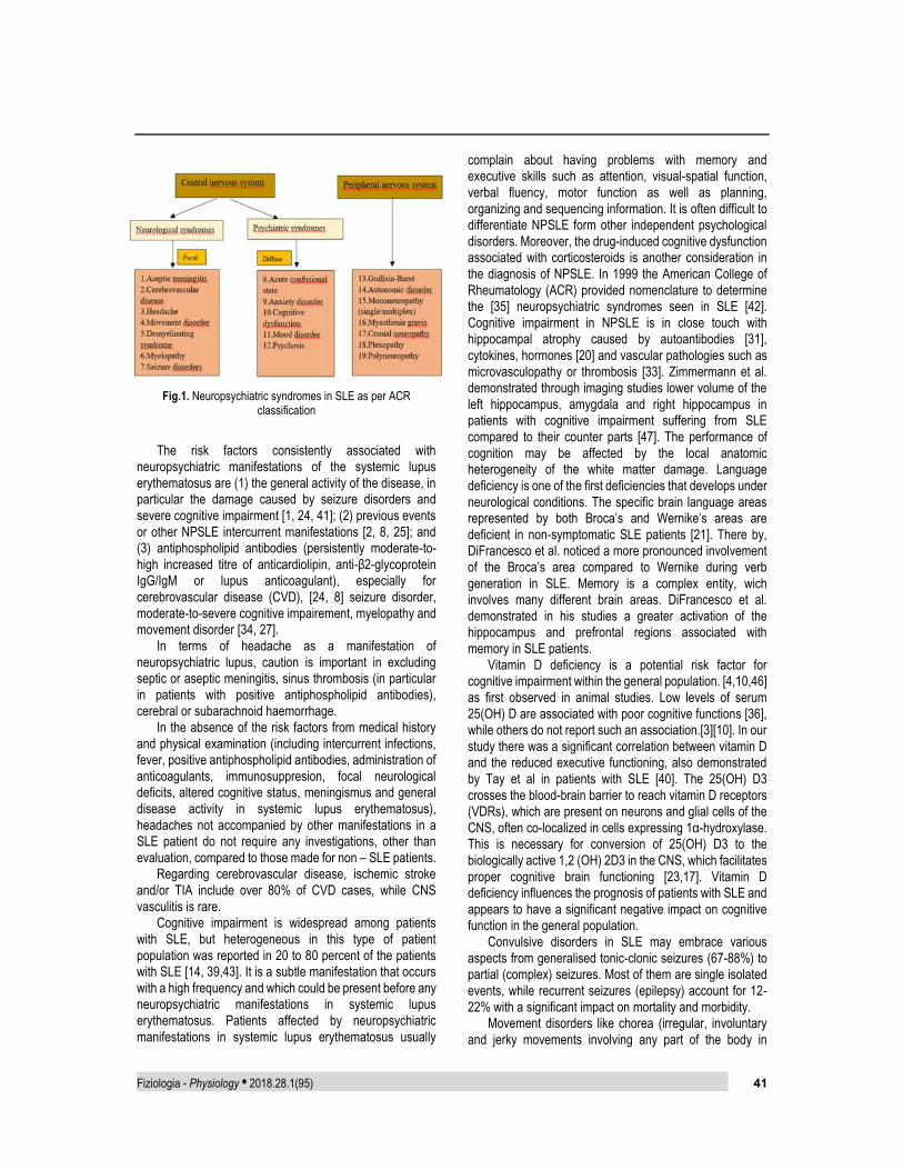
Fiziologia - Physiology • 2018.28.1(95) 41
Fig.1. Neuropsychiatric syndromes in SLE as per ACR
classification
The risk factors consistently associated with neuropsychiatric manifestations of the systemic lupus erythematosus are (1) the general activity of the disease, in particular the damage caused by seizure disorders and severe cognitive impairment [1, 24, 41]; (2) previous events or other NPSLE intercurrent manifestations [2, 8, 25]; and (3) antiphospholipid antibodies (persistently moderate-to-high increased titre of anticardiolipin, anti-β2-glycoprotein IgG/IgM or lupus anticoagulant), especially for cerebrovascular disease (CVD), [24, 8] seizure disorder, moderate-to-severe cognitive impairement, myelopathy and movement disorder [34, 27].
In terms of headache as a manifestation of neuropsychiatric lupus, caution is important in excluding septic or aseptic meningitis, sinus thrombosis (in particular in patients with positive antiphospholipid antibodies), cerebral or subarachnoid haemorrhage.
In the absence of the risk factors from medical history and physical examination (including intercurrent infections, fever, positive antiphospholipid antibodies, administration of anticoagulants, immunosuppresion, focal neurological deficits, altered cognitive status, meningismus and general disease activity in systemic lupus erythematosus), headaches not accompanied by other manifestations in a SLE patient do not require any investigations, other than evaluation, compared to those made for non – SLE patients.
Regarding cerebrovascular disease, ischemic stroke and/or TIA include over 80% of CVD cases, while CNS vasculitis is rare.
Cognitive impairment is widespread among patients with SLE, but heterogeneous in this type of patient population was reported in 20 to 80 percent of the patients with SLE [14, 39,43]. It is a subtle manifestation that occurs with a high frequency and which could be present before any neuropsychiatric manifestations in systemic lupus erythematosus. Patients affected by neuropsychiatric manifestations in systemic lupus erythematosus usually
complain about having problems with memory and executive skills such as attention, visual-spatial function, verbal fluency, motor function as well as planning, organizing and sequencing information. It is often difficult to differentiate NPSLE form other independent psychological disorders. Moreover, the drug-induced cognitive dysfunction associated with corticosteroids is another consideration in the diagnosis of NPSLE. In 1999 the American College of Rheumatology (ACR) provided nomenclature to determine the [35] neuropsychiatric syndromes seen in SLE [42]. Cognitive impairment in NPSLE is in close touch with hippocampal atrophy caused by autoantibodies [31], cytokines, hormones [20] and vascular pathologies such as microvasculopathy or thrombosis [33]. Zimmermann et al. demonstrated through imaging studies lower volume of the left hippocampus, amygdala and right hippocampus in patients with cognitive impairment suffering from SLE compared to their counter parts [47]. The performance of cognition may be affected by the local anatomic heterogeneity of the white matter damage. Language deficiency is one of the first deficiencies that develops under neurological conditions. The specific brain language areas represented by both Broca’s and Wernike’s areas are deficient in non-symptomatic SLE patients [21]. There by, DiFrancesco et al. noticed a more pronounced involvement of the Broca’s area compared to Wernike during verb generation in SLE. Memory is a complex entity, wich involves many different brain areas. DiFrancesco et al. demonstrated in his studies a greater activation of the hippocampus and prefrontal regions associated with memory in SLE patients.
Vitamin D deficiency is a potential risk factor for cognitive impairment within the general population. [4,10,46] as first observed in animal studies. Low levels of serum 25(OH) D are associated with poor cognitive functions [36], while others do not report such an association.[3][10]. In our study there was a significant correlation between vitamin D and the reduced executive functioning, also demonstrated by Tay et al in patients with SLE [40]. The 25(OH) D3 crosses the blood-brain barrier to reach vitamin D receptors (VDRs), which are present on neurons and glial cells of the CNS, often co-localized in cells expressing 1α-hydroxylase. This is necessary for conversion of 25(OH) D3 to the biologically active 1,2 (OH) 2D3 in the CNS, which facilitates proper cognitive brain functioning [23,17]. Vitamin D deficiency influences the prognosis of patients with SLE and appears to have a significant negative impact on cognitive function in the general population.
Convulsive disorders in SLE may embrace various aspects from generalised tonic-clonic seizures (67-88%) to partial (complex) seizures. Most of them are single isolated events, while recurrent seizures (epilepsy) account for 12-22% with a significant impact on mortality and morbidity.
Movement disorders like chorea (irregular, involuntary and jerky movements involving any part of the body in

42 Fiziologia - Physiology • 2018.28.1(95)
unnatural order) is the most common movement disorder in SLE, and it is also associated with antiphospholipid antibodies.
Acute confusional state is characterised by fluctuating level of consciousness with acut onset and decreased attention.
Lupic psychosis can dress the appearance of hallucinations (perceptions in the absence of external stimuli) or delusions (false beliefs refuted by objective evidence). Cortisone psychosis is common in 10% of patients treated with prednisone 1 mg/kg or more and it manifests primarily as a mood disorder (93%) more than psychosis [9]. In prospective studies anti-ribosomal-P antibodies have been associated with psychiatric SLE (sensitivity 25–27%, specificity 75–80%) [7,13].
Lupus myelopathy is manifested either as a transverse myelitis with rapid progressive development or as an ischemic/thrombotic myelopathy. Patients usually present with symptoms due to lesions of lower motor neuron such as flaccid paralysis, loss of muscle tone, hyporeflexia or upper motor neuron wich often presents with spastic paralysis, severe hipertonia and hyperreflexia. The latter can rather be associated with neuromyelitis optica (NMO) and antiphospholipid syndrome [6].
Most frequent cranial neuropathies involve the eighth, the oculomotor (third, fourth and sixth), and less commonly the fifth and seventh nerves. Optic neuropathy includes inflammatory optic neuritis and ischaemic/thrombotic optic neuropathy.
Peripheral nervous system disorders include polyneuropathy (2-3%) and less commonly mononeuropathy (single, multiplex), acute inflammatory demyelinating polyradiculopathy, myastenia gravis, plexopathy which are characterized by painful sensations, muscle weakness and atrophy. When focal neurological signs as gait disturbance, visual or urinary disorder, increased tendon reflexes and/or muscle tone are present, neuroimaging is necessary to exclude the involvement of the central nervous system.
Autoantibodies orientated against certain receptors found in the brain and against proteins found in the cellular membranes. Interferons and cytokines may also be involved. Some of the antibodies that may be involved are mentioned in Figure 2.
Fig. 2. Autoantibodies in neuropsychiatric SLE
Fig. 3. Autoantibodies associated with cognitive dysfunction,
psychosis and depression in SLE
Certain antibodies including antiphospholipid antibodies, anti-ribosomal P antibodies, anti-N-methyl-D-aspartate (NMDA) receptor antibodies and anti-endothelial cell antibodies can be related to central nervous system (CNS) symptoms such as seizures and stroke. NMDA receptors play a role in maintaining the synapse conduction and the plasticity of the central nervous system. These receptors being ligand-gated ion channels are important in memory, cognition and also in neural function.
Fig. 4. Pathopsysiology of neuropsychiatric SLE
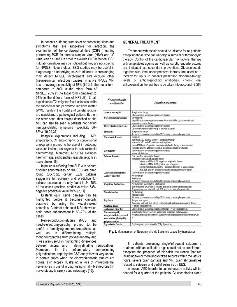
Fiziologia - Physiology • 2018.28.1(95) 43
In patients suffering from fever or presenting signs and symptoms that are suggestive for infection, the examination of the cerebrospinal fluid (CSF) (meaning performing PCR for herpes simplex virus (HSV) and JC virus) can be useful in order to exclude CNS infection. CSF mild abnormalities may be noticed but they are not specific for NPSLE. Nevertheless, EEG studies may be useful in diagnosing an underlying seizure disorder. Neuroimaging may detect NPSLE involvement and exclude other (neurosurgical, infectious) causes. In active NPSLE MRI has an average sensitivity of 57% (64% in the major form compared to 30% in the minor form of NPSLE, 76% in the focal form compared to 51% in the diffuse form of NPSLE). Small hyperintense T2-weighted focal lesions found in the subcortical and periventricular white matter (WM), mainly in the frontal and parietal regions are considered a pathological pattern. But, on the other hand, thes lesions described on the MRI can also be seen in patients not having neuropsychiatric symptoms (specificity 60–82%) [19,29,37].
Imagistic explorations including MRI angiography, CT angiography, or conventional angiography proved to be useful in detecting vascular lesions, aneurysms in subarachnoid haemorrhage. Moreover, MRI/DWI excludes haemorrhage, and identifies vascular regions in acute stroke [16].
In patients suffering from SLE with seizure disorder abnormalities on the EEG are often found (60-70%), certain EEG patterns suggestive for epilepsy and predictive for seizure recurrence are only found in 24–50% of the cases (positive predictive value 73%, negative predictive value 79%) [2,11].
Bilateral optic nerve damage can be highlighted before it becomes clinically observed by using the visual-evoked potentials. Contrast-enhanced MRI shows an optic nerve enhancement in 60–70% of the cases.
Nerve-conduction-studies (NCS) and needle-electromyography proved to be useful in identifying mononeuropathies, as well as in differentiating multiple mononeuropathies from polyneuropathy and it was also useful in highlighting differences between axonal and demyelinating neuropathies. Moreover, in the inflammatory demyelinating polyradiculoneuropathy the CSF analysis was very useful. In certain cases when the electrodiagnostic studies are normal skin biopsy illustrating a loss of intraepidermal nerve fibres is useful in diagnosing small-fibre neuropathy, nerve biopsy is rarely used nowadays [45].
GENERAL TREATMENT
Treatment with aspirin should be initiated for all patients excepting those who can undergo a surgical or thrombolytic therapy. Control of the cardiovascular risk factors, therapy with antiplatelet agents as well as carotid endarterectomy are indicated as secondary prevention. Glucocorticoids together with immunosuppressive therapy are used as a therapy for lupus. In patients presenting moderate-to-high levels of antiphospholipid antibodies, chronic oral anticoagulation therapy has to be taken into account [18,28].
In patients presenting single/infrequent seizures a treatment with antiepileptic drugs should not be considered, excepting the presence of high-risk recurrence features including two or more unprovoked seizures within the last 24 hours, severe brain damage and MRI brain abnormalities related to seizures and partial seizures on EEG.
A second AED in order to control seizure activity will be needed for a quarter of the patients. Glucocorticoids alone
Fig. 5. Management of Neuropsychiatric Systemic Lupus Erythematosus

44 Fiziologia - Physiology • 2018.28.1(95)
or combined with immunosuppressive drugs may be needed if seizures are a reflection of an acute inflammatory event or lupus flare is present. Refractory seizures responded well to the treatment combining intravenous methylprednisolone with cyclophosphamide [5].
For controlling NPSLE activity dopamine antagonists are used as a symptomatic therapy as well as immunosuppressive agents (azathioprine, cyclophosphamide) in combination with glucocorticoids for movement disorders.
Antidepressive and antipsychotic agents are included in the management of psychiatric disorders, as well as biofeedback-assisted cognitive behavioral treatment, that proved useful in treating depressive symptoms [12]. Glucocorticoids and immunosuppressive drugs proved significant results (60–80% response) in treating psychiatric disorders. Relapses were also stated (up to 50%) [32,22,26,30]. Rituximab showed a significant improvement in psychiatric refractory manifestations [44].
Only 20% of SLE patients develop in time chronic psychotic disorder.
25 SLE-related optic neuritis accompanied by weak visual outcome, only a percentage of 30% of the patients have a visual acuity greater than 20/25. Treatment for this type of neuropathy consists of a combination between methylprednisolone and cyclophosphamide, both with intravenous administration. In antiphospholipid-positive patients that do not respond to immunosuppressive drugs anticoagulants may be a solution.
In treating peripheral nervous system disorders glucocorticoids alone or in combination with immunosuppressive drugs had good results (60–75% response rate). In severe cases plasma exchange, rituximab and immunoglobulin with intravenous administration proved good results. AUTHOR CONTRIBUTION Sandu Raluca Elena, Barbulescu Andreea Lili and Florescu Andreea Oana have equally contributed to this paper
REFERENCES
1. Andrade RM, Alarcón GS, González LA et al: Seizures in patients with systemic lupus erythematosus. LUMINA, a multiethnic cohort (LUMINA LIV). Ann Rheum Dis 2008; 67: 829–34.
2. Appenzeller S, Cendes F, Costallat LT: Epileptic seizures in systemic lupus erythematosus. Neurology 2004; 63: 1808-12 .
3. Aung K, Burnett J, Smith SM et al: Vitamin D deficiency associated with self-neglect in the elderly. J Elder Abuse Negl. 2006;18(4):63–78.
4. Balion C, Griffith LE, Strifler L et al: Vitamin D, cognition, and dementia: A systematic review and meta-analysis. Neurology. 2012;79(13): 1397–1405.
5. Barile-Fabris L, Ariza-Andraca R, Olguín-Ortega L et al: Controlled clinical trial of IV cyclophosphamide versus IV methylprednisolone in severe neurological manifestations in
systemic lupus erythematosus. Ann Rheum Dis 2005; 64: 620–5.
6. Birnbaum J, Petri M, Thompson R et al: Distinct subtypes of myelitis in systemic lupus erythematosus. Arthritis Rheum 2009; 60 : 3378 – 87.
7. Briani C, Lucchetta M, Ghirardello A et al: Neurolupus is associated with anti-ribosomal P protein antibodies: an inception cohort study. J Autoimmun 2009; 32 : 79 – 84.
8. Buján S, Ordi-Ros J, Paredes J et al: Contribution of the initial features of systemic lupus erythematosus to the clinical evolution and survival of a cohort of Mediterranean patients. Ann Rheum Dis 2003; 62 : 859–65.
9. Chau SY, Mok CC. Factors predictive of corticosteroid psychosis in patients with systemic lupus erythematosus. Neurology 2003; 61: 104-7.
10. Etgen T, Sander D, Bickel H et al: Vitamin D deficiency, cognitive impairment and dementia: a systematic review and meta-analysis. Dement Geriatr Cogn Disord. 2012;33(5):297–305.
11. González-Duarte A, Cantú-Brito CG, Ruano-Calderón L et al: Clinical description of seizures in patients with systemic lupus erythematosus. Eur Neurol 2008; 59: 320–3.
12. Greco CM, Rudy TE, Manzi S: Effects of a stress-reduction program on psychological function, pain and physical function of systemic lupus erythematosus patients: a randomized controlled trial. Arthritis Rheum 2004; 51: 625–34.
13. Hanly JG, Urowitz MB, Siannis F et al: Autoantibodies and neuropsychiatric events at the time of systemic lupus erythematosus diagnosis: results from an international inception cohort study. Arthritis Rheum 2008; 58: 843 – 53.
14. Harrison MJ, Ravdin LD: Cognitive dysfunction in neuropsychiatric systemic lupus erythematosus. Curr Opin Rheumatol. 2002;14(5):510–514.
15. Hoda AH MD, Lamiaa ID MD, Nermeen AF MD et al. Does Vitamin D Deficiency Contribute to Cognitive Dysfunction in Patients with Systemic Lupus Erythematosus? Innov Clin Neurosci 2018:15(9–10):25–29.
16. Ioannidis JPA, Aringer M, Bollen E et al: EULAR recommendations for the management of systemic lupus erythematosus with neuropsychiatric manifestations: report of a task force of the EULAR standing committee for clinical affairs. Dis 2010;69:2074–2082. doi:10.1136/ard.2010.130476.
17. Johansson P, Almqvist EG, Johansson J-O et al: Cerebrospinal fluid (CSF) 25-hydroxyvitamin D concentration and CSF acetylcholinesterase activity are reduced in patients with Alzheimer’s disease. PLoS One. 2013;8(11):e81989.
18. Khamashta MA, Cuadrado MJ, Mujic Fet al:The management of thrombosis in theantiphospholipid-antibody syndrome. N Engl J Med 1995; 332 : 993 –7.
19. Kozora E, West SG, Kotzin BL et al Magnetic resonance imaging abnormalities and cognitive deficits in systemic lupus erythematosus patients without overt central nervous system disease. Arthritis Rheum 1998; 41: 41–7.
20. Kozora E, Laudenslager M, Lemieux A et al: Inflammatory and hormonal measures predict neuropsychological functioning in systemic lupus erythematosus and rheumatoid arthritis patients. J Int Neuropsychol Soc. 2001;7(6):745-54.
21. Maeshima E, Yamada Y, Yukawa S: Higher cortical dysfunction, antiphospholipid antibodies and neuroradiological examinations in systemic lupus erythematosus. Intern Med. 1992;31(10):1169-74.
Fiziologia - Physiology • 2018.28.1(95) 45
22. Martin-Suarez I, D’Cruz D, Mansoor Met al:Immunosuppressive treatmentin severe connective tissue diseases: effects of low dose intravenouscyclophosphamide. Ann Rheum Dis 1997; 56 481–7.
23. McGrath J, Feron F, Eyles D et al: Vitamin D: the neglected neurosteroid? Trends Neurosci. 2001;24(10):570–572.
24. Mikdashi J, Handwerger B: Predictors of neuropsychiatric damage in systemic lupus erythematosus. Maryland lupus cohort. Rheumatology (Oxford) 2004; 43: 1555–60 .
25. Mikdashi J, Krumholz A, Handwerger B: Factors at diagnosis predict subsequent occurrence of seizures in systemic lupus erythematosus. Neurology 2005; 64: 2102–7.
26. Mok CC, Lau CS, Wong RW: Treatment of lupus psychosis with oralcyclophosphamide followed by azathioprine maintenance: an open-label study. Am J Med 2003; 115: 59–62.
27. Mok MY, Chan EY, Fong DY et al: Antiphospholipid antibody profi les and their clinical associations in Chinese patients with systemic lupus erythematosus. J Rheumatol 2005; 32: 622–8.
28. Muñoz-Rodriguez FJ, Font J, Cervera R et al: Clinical study and follow-up of 100 patients with the antiphospholipid syndrome. Semin Arthritis Rheum 1999; 29: 182–90.
29. Nomura K, Yamano S, Ikeda Y et al: Asymptomatic cerebrovascular lesions detected by magnetic resonance imaging in patients with systemic lupus erythematosus lacking a history of neuropsychiatric events. Intern Med 1999; 38: 785–95.
30. Pego-Reigosa JM, Isenberg DA: Psychosis due to systemic lupus erythematosus:characteristics and long-term outcome of this rare manifestation of the disease. Rheumatology (Oxford) 2008; 47: 1498–502.
31. Peretti CS, Peretti CR, Kozora E et al: Cognitive impairment in systemic lupus erythematosus women with elevated autoantibodies and normal single photon emission computerized tomography. Psychother Psychosom. 2012;81(5):276-85.
32. Rahman P, Humphrey-Murto S, Gladman DD et al: Cytotoxic therapy in systemic lupus erythematosus. Experience from a single center. Medicine (Baltimore)1997; 76: 432–7.
33. Sankowski R, Mader S, Valdes-Ferrer SI: Systemic inflammation and the brain: novel roles of genetic, molecular, and environmental cues as drivers of neurodegeneration. Front Cell Neurosci. 2015;9:28.
34. Sanna G, Bertolaccini ML, Cuadrado MJ et al: Neuropsychiatric manifestations in systemic lupus erythematosus: prevalence and association with antiphospholipid antibodies. J Rheumatol 2003; 30: 985– 92.
35. Sarbu N, Alobeidi F, Toledano P et al: Brain abnormalities in newly diagnosed neuropsychiatric lupus: systematic MRI approach and correlation with clinical and laboratory data in a large multicenter cohort. Autoimmun Rev. 2015;14(2):153-9.
36. Sato Y, Honda Y, Hayashida N et al: Vitamin K deficiency and osteopenia in elderly women with Alzheimer’s disease. Arch Phys Med Rehabil. 2005;86(3):576–581.
37. Sibbitt WL Jr, Sibbitt RR, Griffey RH et al: Magnetic resonance and computed tomographic imaging in the evaluation of acute neuropsychiatric disease in systemic lupus erythematosus. Ann Rheum Dis 1989; 48: 1014–22.
38. Tamirou F, Arnaud LTR, et al: Systemic lupus erythematosus: state of the art on clinical practice guidelines. RMD Open 2018: 4: e000793. doi:10.1136/ rmdopen-2018-000793.
39. Tay SH, Mak A: Anti-NR2A/B antibodies and other major molecular mechanisms in the pathogenesis of cognitive dysfunction in systemic lupus erythematosus. Int J Mol Sci. 2015;16(5): 10281–10300.
40. Tay SH, Ho CS, Ho RC-M et al: 25-Hydroxyvitamin D3 deficiency independently predicts cognitive impairment in patients with systemic lupus erythematosus. PLoS One. 2015;10(12):e0144149.
41. Tomietto P, Annese V, D’Agostini S et al: General and specific factors associated with severity of cognitive impairment in systemic lupus erythematosus. Arthritis Rheum 2007; 57: 1461–72 .
42. The American College of Rheumatology nomenclature and case definitions for neuropsychiatric lupus syndromes. Arthritis Rheum. 1999;42(4):599-608.
43. The American College of Rheumatology nomenclature and case definitions for neuropsychiatric lupus syndromes. Arthritis Rheum. 1999;42:599–608.
44. Tokunaga M, Saito K, Kawabata D et al: Efficacy of rituximab (anti-CD20) for refractory systemic lupus erythematosus involving the central nervous system. Ann Rheum Dis 2007; 66: 470–5.
45. Tseng MT, Hsieh SC, Shun CT et al: Skin denervation and cutaneous vasculitis in systemic lupus erythematosus. Brain 2006; 129: 977–85.
46. Yap KS, Morand EF: Vitamin D and systemic lupus erythematosus: continued evolution. Int J Rheum Dis. 2015;18(2):242–249.
47. Zimmermann N, Corrêa DG, Kubo TA et al: Global cognitive impairment in systemic lupus erythematosus patients: a structural MRI study. Clin Neuroradiol. 2017;27(1):23-9.
46 Fiziologia - Physiology • 2018.28.1(95)
AFECTAREA NEUROPSIHIATRICA IN LUPUS ERITEMATOS SISTEMIC - ASPECTE CLINICE REZUMAT Lupusul eritematos sistemic este o boală sistemică,autoimună, cronică, cu episoade recurent-remisive, a cărei etiologie este necunoscută și cu manifestări heterogene. Această boală include diverse simptome, atât în sfera neurologică, cât și în cea psihiatrică. Manifestările neuropsihiatrice pot cuprinde diverse aspecte, cum ar fi dureri de cap, convulsii, psihoze, stare de confuzie acută și tulburări cognitive. Aceste manifestări neuropsihiatrice fac parte din lupusul eritematos sistemic neuropsihiatric (NPSLE). Factorii de risc frecvent asociați cu manifestările neuropsihiatrice ale lupusului eritematos sistemic sunt (1) activitatea generală a bolii, în special leziunile provocate de convulsii și de tulburările cognitive severe (2) evenimentele anterioare sau alte manifestări intercurente ale NPSLE și (3) anticorpiantifosfolipidici (titruri anticardiolipin, IgG / IgM anti-β2-glicoprotein anti-β2 sau anticoagulant lupus), în special pentru boala cerebrovasculară (CVD), tulburări convulsive, tulburări cognitive moderate până la severe, mielopatie și tulburări de mișcare. Ne propunem o trecere în revistă a principalelor aspecte referitoare la fiziopatologia, diagnosticul și managementul manifestarilor neuropsihiatrice din lupusului eritematos sistemic. Cuvinte cheie: manifestări neuropsihiatrice în Lupus eritematos sistemic; tratament imunospresiv, anticorpi antifosfolipidici; tratamentul lupusului eritematos sistemic cu manifestări neuropsihiatrice