STUDIUL FENOTIPIC ȘI GENOTIPIC AL TULPINILOR DE ... · Stafilococii pătrund în organism la...
44
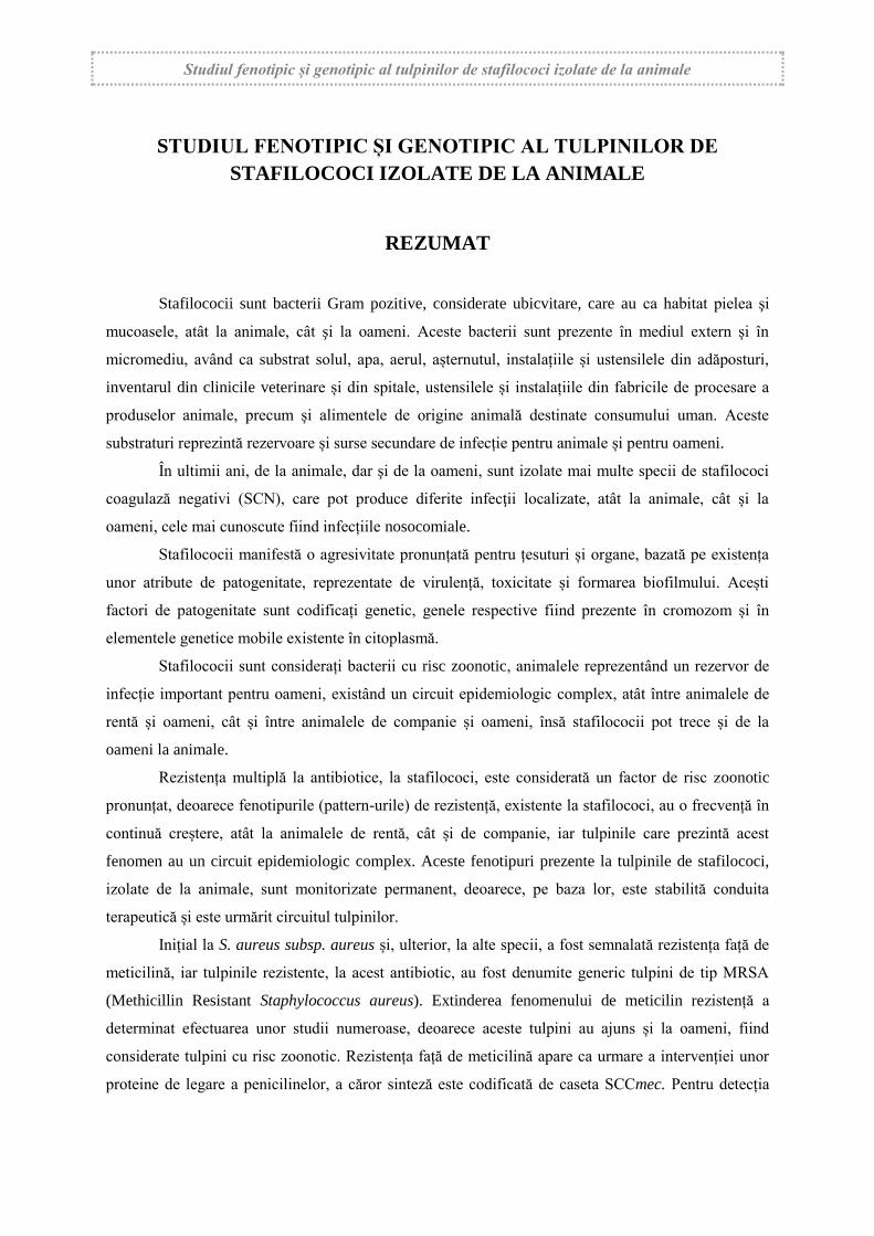
Studiul fenotipic și genotipic al tulpinilor de stafilococi izolate de la animale STUDIUL FENOTIPIC ȘI GENOTIPIC AL TULPINILOR DE STAFILOCOCI IZOLATE DE LA ANIMALE REZUMAT Stafilococii sunt bacterii Gram pozitive, considerate ubicvitare, care au ca habitat pielea şi mucoasele, atât la animale, cât şi la oameni. Aceste bacterii sunt prezente în mediul extern și în micromediu, având ca substrat solul, apa, aerul, așternutul, instalațiile și ustensilele din adăposturi, inventarul din clinicile veterinare și din spitale, ustensilele și instalațiile din fabricile de procesare a produselor animale, precum și alimentele de origine animală destinate consumului uman. Aceste substraturi reprezintă rezervoare și surse secundare de infecție pentru animale și pentru oameni. În ultimii ani, de la animale, dar şi de la oameni, sunt izolate mai multe specii de stafilococi coagulază negativi (SCN), care pot produce diferite infecţii localizate, atât la animale, cât și la oameni, cele mai cunoscute fiind infecțiile nosocomiale. Stafilococii manifestă o agresivitate pronunțată pentru țesuturi și organe, bazată pe existența unor atribute de patogenitate, reprezentate de virulență, toxicitate și formarea biofilmului. Acești factori de patogenitate sunt codificați genetic, genele respective fiind prezente în cromozom și în elementele genetice mobile existente în citoplasmă. Stafilococii sunt considerați bacterii cu risc zoonotic, animalele reprezentând un rezervor de infecție important pentru oameni, existând un circuit epidemiologic complex, atât între animalele de rentă și oameni, cât și între animalele de companie și oameni, însă stafilococii pot trece și de la oameni la animale. Rezistența multiplă la antibiotice, la stafilococi, este considerată un factor de risc zoonotic pronunțat, deoarece fenotipurile (pattern-urile) de rezistență, existente la stafilococi, au o frecvență în continuă creștere, atât la animalele de rentă, cât și de companie, iar tulpinile care prezintă acest fenomen au un circuit epidemiologic complex. Aceste fenotipuri prezente la tulpinile de stafilococi, izolate de la animale, sunt monitorizate permanent, deoarece, pe baza lor, este stabilită conduita terapeutică și este urmărit circuitul tulpinilor. Inițial la S. aureus subsp. aureus și, ulterior, la alte specii, a fost semnalată rezistența față de meticilină, iar tulpinile rezistente, la acest antibiotic, au fost denumite generic tulpini de tip MRSA (Methicillin Resistant Staphylococcus aureus). Extinderea fenomenului de meticilin rezistență a determinat efectuarea unor studii numeroase, deoarece aceste tulpini au ajuns și la oameni, fiind considerate tulpini cu risc zoonotic. Rezistența față de meticilină apare ca urmare a intervenției unor proteine de legare a penicilinelor, a căror sinteză este codificată de caseta SCCmec. Pentru detecția
Transcript of STUDIUL FENOTIPIC ȘI GENOTIPIC AL TULPINILOR DE ... · Stafilococii pătrund în organism la...
Studiul fenotipic și genotipic al tulpinilor de stafilococi izolate de la animale
STUDIUL FENOTIPIC ȘI GENOTIPIC AL TULPINILOR DE
STAFILOCOCI IZOLATE DE LA ANIMALE
REZUMAT
Stafilococii sunt bacterii Gram pozitive, considerate ubicvitare, care au ca habitat pielea şi
mucoasele, atât la animale, cât şi la oameni. Aceste bacterii sunt prezente în mediul extern și în
micromediu, având ca substrat solul, apa, aerul, așternutul, instalațiile și ustensilele din adăposturi,
inventarul din clinicile veterinare și din spitale, ustensilele și instalațiile din fabricile de procesare a
produselor animale, precum și alimentele de origine animală destinate consumului uman. Aceste
substraturi reprezintă rezervoare și surse secundare de infecție pentru animale și pentru oameni.
În ultimii ani, de la animale, dar şi de la oameni, sunt izolate mai multe specii de stafilococi
coagulază negativi (SCN), care pot produce diferite infecţii localizate, atât la animale, cât și la
oameni, cele mai cunoscute fiind infecțiile nosocomiale.
Stafilococii manifestă o agresivitate pronunțată pentru țesuturi și organe, bazată pe existența
unor atribute de patogenitate, reprezentate de virulență, toxicitate și formarea biofilmului. Acești
factori de patogenitate sunt codificați genetic, genele respective fiind prezente în cromozom și în
elementele genetice mobile existente în citoplasmă.
Stafilococii sunt considerați bacterii cu risc zoonotic, animalele reprezentând un rezervor de
infecție important pentru oameni, existând un circuit epidemiologic complex, atât între animalele de
rentă și oameni, cât și între animalele de companie și oameni, însă stafilococii pot trece și de la
oameni la animale.
Rezistența multiplă la antibiotice, la stafilococi, este considerată un factor de risc zoonotic
pronunțat, deoarece fenotipurile (pattern-urile) de rezistență, existente la stafilococi, au o frecvență în
continuă creștere, atât la animalele de rentă, cât și de companie, iar tulpinile care prezintă acest
fenomen au un circuit epidemiologic complex. Aceste fenotipuri prezente la tulpinile de stafilococi,
izolate de la animale, sunt monitorizate permanent, deoarece, pe baza lor, este stabilită conduita
terapeutică și este urmărit circuitul tulpinilor.
Inițial la S. aureus subsp. aureus și, ulterior, la alte specii, a fost semnalată rezistența față de
meticilină, iar tulpinile rezistente, la acest antibiotic, au fost denumite generic tulpini de tip MRSA
(Methicillin Resistant Staphylococcus aureus). Extinderea fenomenului de meticilin rezistență a
determinat efectuarea unor studii numeroase, deoarece aceste tulpini au ajuns și la oameni, fiind
considerate tulpini cu risc zoonotic. Rezistența față de meticilină apare ca urmare a intervenției unor
proteine de legare a penicilinelor, a căror sinteză este codificată de caseta SCCmec. Pentru detecția
Studiul fenotipic și genotipic al tulpinilor de stafilococi izolate de la animale
II
rezistenței la meticilină a fost propusă, din anul 2004, oxacilina, iar din anul 2005, cefoxitinul,
deoarece rezultatele obținute, cu aceste două beta-lactamine, sunt mult mai fiabile.
Teza de doctorat cuprinde 284 pagini, 38 tabele și 87 figuri, în care sunt incluse 52 de imagini
originale și 35 de grafice. Suportul științific al tezei este reprezentat de 300 titluri bibliografice, care
includ lucrări științifice, tratate, teze de doctorat și pagini web.
Teza de doctorat este structurată în două părți, respectiv “Cercetările bibliografice”, incluse în
partea a I-a și “Cercetările proprii”, incluse în partea a II-a.
Partea a I-a
CERCETĂRI BIBLIOGRAFICE
Prima parte a tezei, reprezintă un studiu bibliografic, structurat în două capitole, fiind extinsă
pe un număr de 74 pagini (26,06%). În această parte se regăsesc 3 tabele și 3 figuri reprezentative
alături de date actuale privind genul Staphylococcus.
CAPITOLUL 1. FAMILIA STAPHYLOCOCCACEAE. GENUL
STAPHYLOCOCCUS
Primul capitol reprezintă o sinteză a datelor din literatura de specialitate, riguros selectate cu
privire la sistematica genului Staphylococcus. Este prezentată taxonomia acestui gen, preluată din
www.bacterio.net/-allnamessz.html, care include List of Prokaryotic names with Standing in
Nomenclature, coordonat de Prof. Dr. EUSZÉBY J. P.
În cadrul capitolului, sunt prezentate sintetic ultimile date privind ecologia, morfologia,
structura antigenică și factorii de patogentitate. De asemenea, capitolul include și date actuale privind
genetica acestor bacterii, antibiorezistența multiplă și fenomenul de meticilin rezistență. Sunt
prezentate și speciile de stafilococi patogene pentru animalele de rentă și de companie, precum și
entitățile morbide produse.
CAPITOLUL 2. DIAGNOSTICUL DE LABORATOR ÎN INFECȚIILE
CU STAFILOCOCI LA ANIMALE
În acest capitol este prezentată o amplă sinteză bibliografică, privind diagnosticul infecțiilor
stafilococice la animale. Sunt redate tehnicile de recoltare a probelor, materialele patologice, precum
și transportul acestora. O atenție deosebită este acordată examenului bacteriologic, privind atât
identificarea primară, cât și identificarea definitivă, precum și mediile de cultură, testele curente și
Studiul fenotipic și genotipic al tulpinilor de stafilococi izolate de la animale
III
sistemele comerciale. De asemenea, este prezentată identificarea clonală, detecția toxinei și testele de
biologie moleculară, utilizate în ultimii ani.
Partea a II-a
CERCETĂRI PROPRII
Partea a II-a include cercetările proprii, fiind structurată în 6 capitole (3-8), extinse pe un
număr de 210 pagini (73,94%). Această parte a tezei este ilustrată de 35 tabele și 84 figuri, în care
sunt incluse 49 imagini originale și 35 de grafice.
CAPITOLUL 3. SCOPUL, MOTIVAȚIA ȘI OBIECTIVELE
CERCETĂRII
Stafilococii sunt bacterii patogene, condiționat patogene și oportuniste, în funcție de specie și
în funcție de prezența unor factori favorizanți. Au tropism marcat pentru piele și pentru mucoase,
producând infecții localizate supurative, septicemii și entități infecțioase, bine conturate, care
evoluează la animale și la oameni, denumite generic stafilococii. În unele situații, pot interveni şi ca
agenţi patogeni secundari, în principal după unele viroze .
La animale evoluează stafilococii, bine conturate, ca entităţi infecţioase, distincte
epidemiologic şi anatomoclinic, cu evoluţie septicemică şi/sau localizată, distinngându-se ca
frecvență, importanță economică și sanitară mastitele infecțioase ale vacilor de lapte.
În funcție de patogenitatea speciei de stafilococ și de evoluția clinică a entităților produse,
majoritatea cercetătorilor fac distincție între S. aureus subsp. aureus, specia tip a genului, considerată
specia cea mai patogenă și alte specii de stafilococi coagulază pozitivi și coagulază negativi, incluse
într-o grupă denumită generic stafilococi “non-S. aureus”.
Mecanismele de patogenitate se desfășoară într-o succesiune complexă, în care intervin
enzimele extracelulare și toxinele, care acționează ca și inele într-o reacție biochimică ramnificată și
mai puțin sub formă individualizată. Stafilococii pătrund în organism la nivelul pielii și mucoaselor
prin glandele sebacee, sudoripare, foliculii piloși, microleziuni și leziuni de dimensiuni mai mari.
Procesul infecțios incipient (local) este consecința numărului, virulenței și toxicității tulpinii și a
capacității mecanismelor de apărare locală, a organismelor gazdă, care intervin prin fagocitoză,
respectiv prin granulocitele neutrofile mobilizate și potențate prin opsonizare și prin liză de către
sistemul complement. Infecțiile stafilococice repetate, la nivelul pielii, pot declanșa o
hipersensibilizare sau alergie față de unele antigene stafilococice.
Studiile efectuate au demonstrat că antibiorezistența este determinată genetic, având ca suport
foarte multe gene de rezistență situate în cromozomul bacterian, plasmidele R, intergoni și
Studiul fenotipic și genotipic al tulpinilor de stafilococi izolate de la animale
IV
transpozoni, care reprezintă elementele genetice mobile. Prin intermediul acestora, genele care
codifică rezistența la antibiotice pot fi transferate între tulpinile aceleiași specii bacteriene (transmitere
intraspecifică), precum și între tulpini care aparțin altor specii bacteriene (transmitere interspecifică).
De asemenea, cercetările au mai demonstrat că plasmidele R reprezintă principalii factori ai rezistenței
extracromozomale la antibiotice, mai ales cele de tip conjugativ care conțin 3 grupe de gene, respectiv
gene care controlează transferul, gene care codifică autoreplicarea și gene care codifică rezistența față
de antibiotice. Aceste gene au fost detectate și studiate cu ajutorul unor tehnici de biologie
moleculară, cea mai utilizată tehnica PCR, în mai multe variante.
Numeroase colective de cercetare efectuează studii ample de screening, pentru a monitoriza
circuitul epidemiologic al tulpinilor meticilin rezistente, în acest scop, fiind utilizate atât tehnicile
clasice fenotipice, cât și metodele de biologie moleculară, care sunt mai rapide și care permit detecția
SCCmec, care codifică rezistența față de acest antibiotic.
Având în vedere aspectele prezentate anterior, în cadrul cercetărilor care fac obiectul tezei de
doctorat, au fost urmărite mai multe obiective:
studiul cultural, morfologic și tinctorial al tulpinilor de stafilococi izolate;
studiul profilului biochimic, cu ajutorul unui sistem multitest și pe baza activității
glucidolitice;
utilizarea unor medii cromogene pentru discriminarea tulpinilor de S. aureus subsp. aureus
și a tulpinilor de tip MRSA;
stabilirea frecvenței unor specii de stafilococi izolate de la animale;
studiul unor factori de patogenitate existenți la tulpinile izolate;
simplificarea schemei de izolare și de tipizare primară a tulpinilor izolate;
cercetarea prezenței coagulazei libere ca factor de patogenitate și de diferențiere a tulpinilor
în cele două grupe;
studierea fenotipurilor (pattern-urilor) de rezistență la tulpinile izolate;
studierea fenomenului de meticilin rezistență la tulpinile izolate;
detecția genei mec la tulpinile de stafilococi meticilin rezistente, cu ajutorul tehnicii PCR;
diferențierea tulpinilor de S. intermedius față de tulpinile de S. pseudintermedius, cu ajutorul
tehnicii PCR;
identificarea clonală a tulpinilor de S. intermedius/S. pseudintermedius izolate de la câini și
pisici.
Studiul fenotipic și genotipic al tulpinilor de stafilococi izolate de la animale
V
CAPITOLUL 4. IZOLAREA ȘI CARACTERIZAREA FENOTIPICĂ A
TULPINILOR DE STAFILOCOCI
La animale și la oameni stafilococii produc diferite infecții localizate sau boli infecțioase care
sunt bine conturate, prezentând importanță economică și sanitară. În ultimii ani, a crescut rolul
stafilococilor coagulază negativi (SCN) în etiologia unor infecții localizate, dintre care se disting ca
frecvență și importanță mastitele subclinice la vacile de lapte sau diferite infecții cutanate sau cu
localizare în unele organe la suine și la animalele de companie. Aceste bacterii au tropism pentru
epitelii (piele și mucoase), însă datorită echipamentului agresiv, de care dispun, reprezentat de
enzime, toxine și alți factori de patogenitate, pot invada orice țesut sau organ. Infecțiile naturale, la
oameni și animale, sunt influențate de mai mulți factori legați de: organismele gazdă, existența unor
factori favorizanți și de speciile de stafilococi.
Cercetările care fac obiectul acestui capitol au avut ca obiectiv principal caracterizarea
fenotipică a tulpinilor de stafilococi izolate de la animale de rentă și de companie, sănătoase sau cu
diferite infecții localizate sau generalizate.
4.1. MATERIALE ȘI METODE
Probele de material patologic au fost prelevate de la mai multe specii de animale, urmând a fi
supuse examenului bacteriologic, efectuat în conformitate cu metodologia de izolare și tipizare a
stafilococilor.
4.1.1. Recoltarea, transportul și însâmânțarea primară a probelor
Probele cu material patologic au fost prelevate de la animale din mai multe specii și categorii
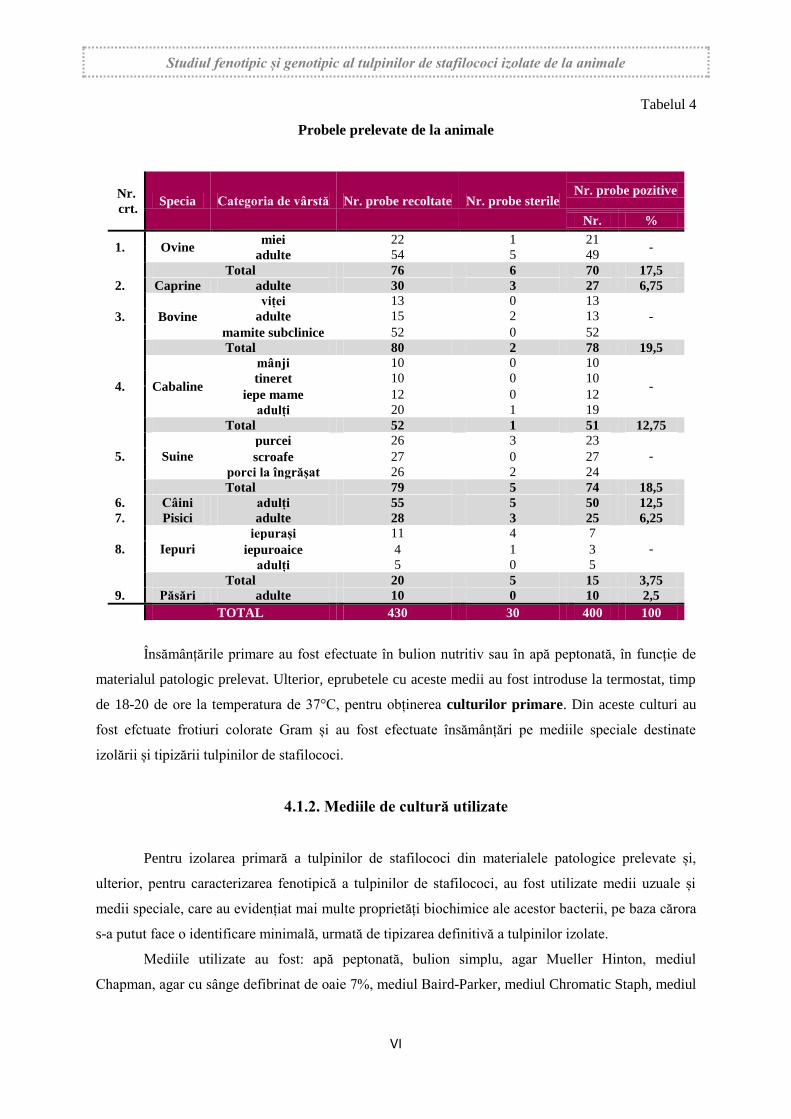
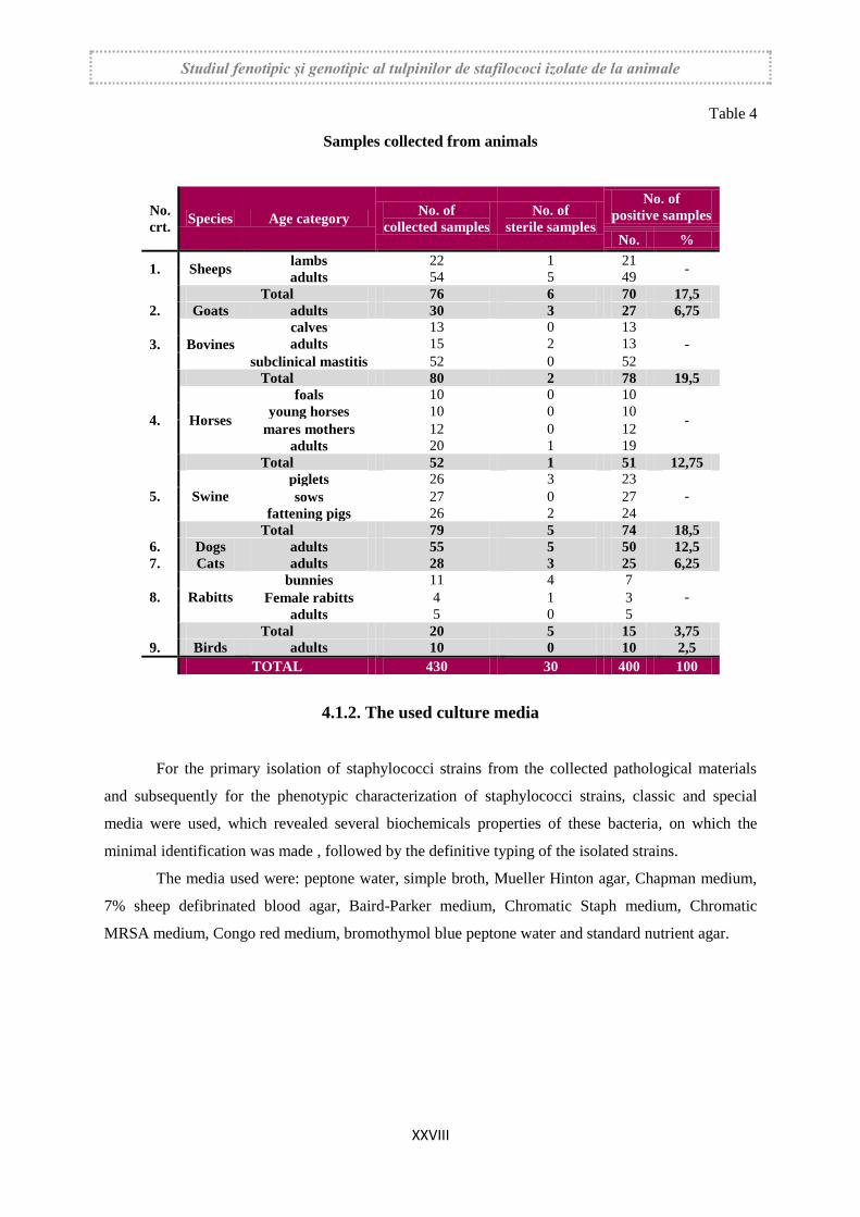
de vârstă, cu diferite afecțiuni, sau clinic sănătoase, de rentă și de companie (tabelul 4).
Pentru prelevarea probelor, de pe piele, au fost utilizate tampoanele sterile, secrețiile nazale
au fost prelevate tot cu tampoane sterile, iar secrețiile uterovaginale au fost prelevate cu tampoane de
vată sterile fixate pe o tijă de plastic mai lungă.
Probele de lapte mastitic au fost recoltate, steril, de la un număr de 22 de vaci primipare, la
care mastitele subclinice au debutat și evoluat la 45-60 zile de la fătare, fiind afectate unul, două sau
chiar trei sferturi mamare, iar de la oi și capre cu mamită gangrenoasă probele de secreții patologice
mamare au fost recoltate, în flacoane sterile.
Studiul fenotipic și genotipic al tulpinilor de stafilococi izolate de la animale
VI
Tabelul 4
Probele prelevate de la animale
Nr.
crt. Specia Categoria de vârstă Nr. probe recoltate Nr. probe sterile
Nr. probe pozitive
Nr. %
1. Ovine miei 22 1 21
- adulte 54 5 49
Total 76 6 70 17,5
2. Caprine adulte 30 3 27 6,75
3. Bovine
viței 13 0 13
- adulte 15 2 13
mamite subclinice 52 0 52
Total 80 2 78 19,5
4. Cabaline
mânji 10 0 10
- tineret 10 0 10
iepe mame 12 0 12
adulți 20 1 19
Total 52 1 51 12,75
5. Suine
purcei 26 3 23
- scroafe 27 0 27
porci la îngrășat 26 2 24
Total 79 5 74 18,5
6. Câini adulți 55 5 50 12,5
7. Pisici adulte 28 3 25 6,25
8. Iepuri
iepurași 11 4 7
- iepuroaice 4 1 3
adulți 5 0 5
Total 20 5 15 3,75
9. Păsări adulte 10 0 10 2,5
TOTAL 430 30 400 100
Însămânțările primare au fost efectuate în bulion nutritiv sau în apă peptonată, în funcție de
materialul patologic prelevat. Ulterior, eprubetele cu aceste medii au fost introduse la termostat, timp
de 18-20 de ore la temperatura de 37°C, pentru obținerea culturilor primare. Din aceste culturi au
fost efctuate frotiuri colorate Gram și au fost efectuate însămânțări pe mediile speciale destinate
izolării și tipizării tulpinilor de stafilococi.
4.1.2. Mediile de cultură utilizate
Pentru izolarea primară a tulpinilor de stafilococi din materialele patologice prelevate și,
ulterior, pentru caracterizarea fenotipică a tulpinilor de stafilococi, au fost utilizate medii uzuale și
medii speciale, care au evidențiat mai multe proprietăți biochimice ale acestor bacterii, pe baza cărora
s-a putut face o identificare minimală, urmată de tipizarea definitivă a tulpinilor izolate.
Mediile utilizate au fost: apă peptonată, bulion simplu, agar Mueller Hinton, mediul
Chapman, agar cu sânge defibrinat de oaie 7%, mediul Baird-Parker, mediul Chromatic Staph, mediul
Studiul fenotipic și genotipic al tulpinilor de stafilococi izolate de la animale
VII
Chromatic MRSA, mediul cu roșu de Congo, apa peptonată cu albastru de bromtimol și agarul nutritiv
standard.
4.1.3. Sistemul API Staph
Identificarea, la nivel de specie, a tulpinilor de stafilococi (64 tulpini), izolate din probele de
lapte mastitic, recoltate numai de la vaci primipare, cu mastite subclinice, a fost realizată prin
utilizarea microtestului API Staph şi a programului software Apiweb de interpretare.
4.1.4. Testul evidențierii catalazei
Pentru evidențierea prezenței catalazei a fost utilizat peroxidul de hidrogen, soluție 3%, care a
fost adăugat în eprubetele cu culturile de stafilococi, în cantitate de 1 ml, culturile fiind examinate
timp 5-10 minute pentru evidențierea bulelor de gaz.
4.1.5. Testele pentru evidențierea coagulazei libere și legate
Coagulaza liberă, difuzibilă, a fost evidențiată prin testul în tuburi cu plasma de iepure
liofilizată și cu ajutorul mediului Baird-Parker, iar coagulaza legată, denumită și “clumping factor” a
fost evidenţiată cu ajutorul kiturilor Staphylo Rapid Test și Staph Latex kit.
4.1.6. Examenul bacterioscopic și cultural
Examenul cultural. Culturile obținute, în urma însămânțărilor primare și în urma
însămânțărilor pe mediile amintite anterior, au fost examinate cu ochiul liber și cu lupa stereoscopică,
fiind apreciate următoarele caractere: turbiditatea pe mediile lichide, forma, mărimea,
pigmentogeneza și virarea culorii pe mediile solide utilizate, prezentate anterior.
Examenul bacterioscopic. Au fost efectuate frotiuri, colorate Gram, din colonii izolate,
caracteristice, cu scopul de a evidenția caracterelor morfologice și tinctoriale.
4.1.7. Testarea rezistenței la furazolidon și novobiocin
Testul de sensibilitate la novobiocin a fost efectuat pentru a diferenţia unele specii de
stafilococi coagulază negativi faţă de speciile de stafilococi coagulază pozitivi.
Studiul fenotipic și genotipic al tulpinilor de stafilococi izolate de la animale
VIII
Testul de sensibilitate la furazolidon a fost folosit pentru a diferenţia tulpinile de stafilococi
față de tulpinile de micrococi. Principiul acestui test constă în faptul că stafilococii sunt sensibili la
compuşii bacteriostatici, din clasa furanilor, în timp ce micrococii sunt rezistenţi.
4.2. REZULTATELE OBȚINUTE
4.2.1. Rezultatele obținute privind identificarea preliminară
4.2.1.1. Rezultatele examenului cultural și bacterioscopic
În apa peptonată, culturile primare au produs o turbiditate variabilă, unele culturi producând
și un depozit necaracteristic, ușor omogenizabil, iar în bulion, culturile primare au produs o
turbiditate intensă cu un depozit necaracteristic, ușor omogenizabil, iar unele tulpini au format un inel
discret la suprafață.
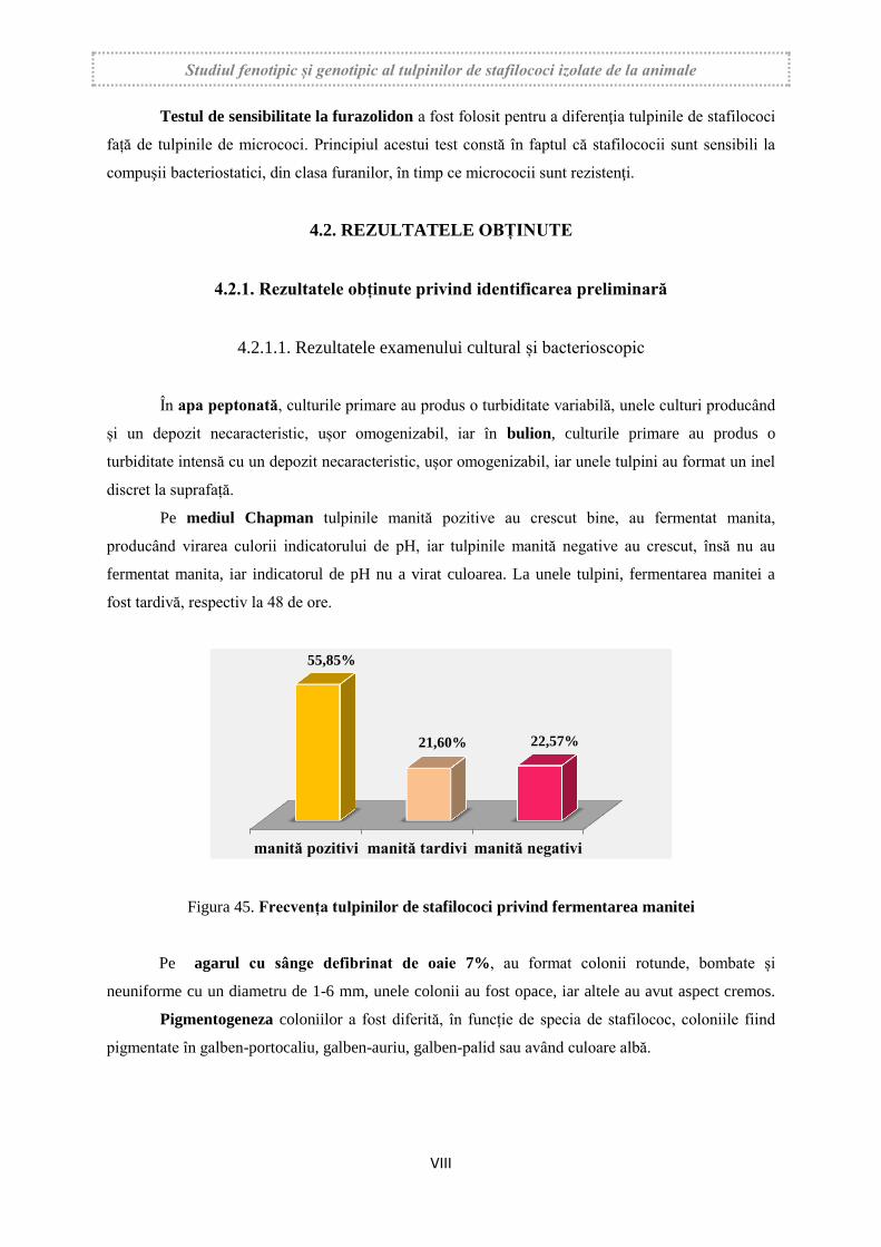
Pe mediul Chapman tulpinile manită pozitive au crescut bine, au fermentat manita,
producând virarea culorii indicatorului de pH, iar tulpinile manită negative au crescut, însă nu au
fermentat manita, iar indicatorul de pH nu a virat culoarea. La unele tulpini, fermentarea manitei a
fost tardivă, respectiv la 48 de ore.
Figura 45. Frecvența tulpinilor de stafilococi privind fermentarea manitei
Pe agarul cu sânge defibrinat de oaie 7%, au format colonii rotunde, bombate și
neuniforme cu un diametru de 1-6 mm, unele colonii au fost opace, iar altele au avut aspect cremos.
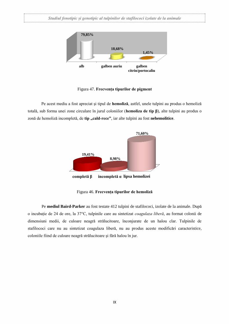
Pigmentogeneza coloniilor a fost diferită, în funcție de specia de stafilococ, coloniile fiind
pigmentate în galben-portocaliu, galben-auriu, galben-palid sau având culoare albă.
manită pozitivi manită tardivi manită negativi
55,85%
21,60% 22,57%
Studiul fenotipic și genotipic al tulpinilor de stafilococi izolate de la animale
IX
Figura 47. Frecvența tipurilor de pigment
Pe acest mediu a fost apreciat și tipul de hemoliză, astfel, unele tulpini au produs o hemoliză
totală, sub forma unei zone circulare în jurul coloniilor (hemoliza de tip β), alte tulpini au produs o
zonă de hemoliză incompletă, de tip „cald-rece”, iar alte tulpini au fost nehemolitice.
Figura 46. Frecvența tipurilor de hemoliză
Pe mediul Baird-Parker au fost testate 412 tulpini de stafilococi, izolate de la animale. După
o incubație de 24 de ore, la 37°C, tulpinile care au sintetizat coagulaza liberă, au format colonii de
dimensiuni medii, de culoare neagră strălucitoare, înconjurate de un halou clar. Tulpinile de
stafilococi care nu au sintetizat coagulaza liberă, nu au produs aceste modificări caracteristice,
coloniile fiind de culoare neagră strălucitoare și fără halou în jur.
alb galben auriu galben
citrin/portocaliu
79,85%
18,68% 1,45%
completă β incompletă α lipsa hemolizei
19,41% 8,98%
71,60%
Studiul fenotipic și genotipic al tulpinilor de stafilococi izolate de la animale
X
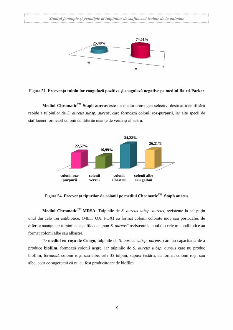
Figura 51. Frecvența tulpinilor coagulază pozitive și coagulază negative pe mediul Baird-Parker
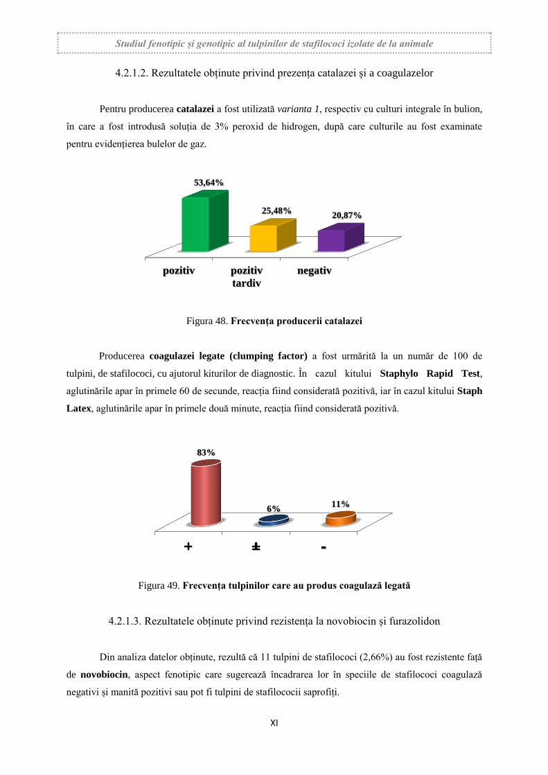
Mediul ChromaticTM
Staph aureus este un mediu cromogen selectiv, destinat identificării
rapide a tulpinilor de S. aureus subsp. aureus, care formează colonii roz-purpurii, iar alte specii de
stafilococi formează colonii cu diferite nuanțe de verde și albastru.
Figura 54. Frecvența tipurilor de colonii pe mediul ChromaticTM
Staph aureus
Mediul ChromaticTM
MRSA. Tulpinile de S. aureus subsp. aureus, rezistente la cel puțin
unul din cele trei antibiotice, (MET, OX, FOX) au format colonii colorate mov sau portocaliu, de
diferite nuanțe, iar tulpinile de stafilococi „non-S. aureus” rezistente la unul din cele trei antibiotice au
format colonii albe sau albastre.
Pe mediul cu roșu de Congo, tulpinile de S. aureus subsp. aureus, care au capacitatea de a
produce biofilm, formează colonii negre, iar tulpinile de S. aureus subsp. aureus care nu produc
biofilm, formează colonii roșii sau albe, cele 35 tulpini, supuse testării, au format colonii roșii sau
albe, ceea ce sugerează că nu au fost producătoare de biofilm.
25,48% 74,51%
colonii roz-
purpurii
colonii
verzui
colonii
albăstrui
colonii albe
sau gălbui
22,57% 16,99%
34,22%
26,21%
Studiul fenotipic și genotipic al tulpinilor de stafilococi izolate de la animale
XI
4.2.1.2. Rezultatele obținute privind prezența catalazei și a coagulazelor
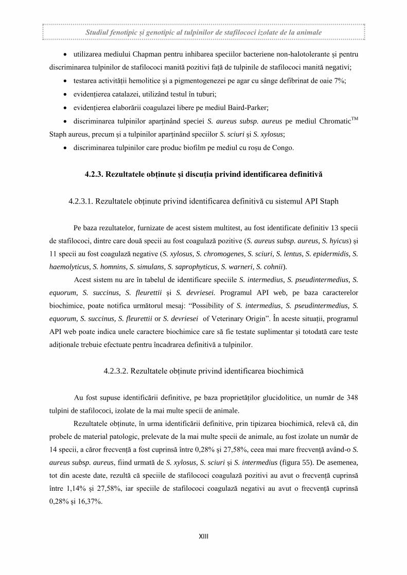
Pentru producerea catalazei a fost utilizată varianta 1, respectiv cu culturi integrale în bulion,
în care a fost introdusă soluția de 3% peroxid de hidrogen, după care culturile au fost examinate
pentru evidențierea bulelor de gaz.
Figura 48. Frecvența producerii catalazei
Producerea coagulazei legate (clumping factor) a fost urmărită la un număr de 100 de
tulpini, de stafilococi, cu ajutorul kiturilor de diagnostic. În cazul kitului Staphylo Rapid Test,
aglutinările apar în primele 60 de secunde, reacția fiind considerată pozitivă, iar în cazul kitului Staph
Latex, aglutinările apar în primele două minute, reacția fiind considerată pozitivă.
Figura 49. Frecvența tulpinilor care au produs coagulază legată
4.2.1.3. Rezultatele obținute privind rezistența la novobiocin și furazolidon
Din analiza datelor obținute, rezultă că 11 tulpini de stafilococi (2,66%) au fost rezistente față
de novobiocin, aspect fenotipic care sugerează încadrarea lor în speciile de stafilococi coagulază
negativi și manită pozitivi sau pot fi tulpini de stafilococii saprofiți.
pozitiv pozitiv
tardiv
negativ
53,64%
25,48% 20,87%
83%
6% 11%

Studiul fenotipic și genotipic al tulpinilor de stafilococi izolate de la animale
XII
Figura 52. Frecvența tulpinilor sensibile față de novobiocin
Din analiza rezultatelor obținute, rezultă că 27 de tulpini (6,55%), din tulpinile supuse testării,
au fost rezistente față de furazolidon, probabil ca urmare a apariției unor mutante rezistente la acest
chimioterapic, care au apărut fie spontan, fie ca urmare a unor tratamente efectuate cu substanțe
antimicrobiene din aceeași grupă.
Figura 53. Frecvența tulpinilor sensibile față de furazolidon
4.2.2. Discuția rezultatelor obținute privind identificarea preliminară
Pe baza rezultatelor obținute, în cadrul cercetărilor proprii, putem recomanda o schemă de
identificare preliminară privind infecțiile stafilococice la animale, care permite:
încadrarea tulpinilor izolate în cadrul genului Staphylococcus;
încadrarea tulpinilor izolate în cadrul celor două grupe, respectiv stafilococi coagulază
pozitivi și stafilococi coagulază negativi;
discriminarea speciei S. aureus subsp. aureus față de alte specii de stafilococi;
discriminarea tulpinilor care produc biofilm.
Aceste obiective pot fi realizate folosind următoarele etape:
obținerea culturilor primare în apă peptonată;
Sensibil Intermediar Rezistent
75,00%
22,33%
2,66%
Sensibil Intermediar Rezistent
72,33%
21,11%
6,55%
Studiul fenotipic și genotipic al tulpinilor de stafilococi izolate de la animale
XIII
utilizarea mediului Chapman pentru inhibarea speciilor bacteriene non-halotolerante și pentru
discriminarea tulpinilor de stafilococi manită pozitivi față de tulpinile de stafilococi manită negativi;
testarea activității hemolitice și a pigmentogenezei pe agar cu sânge defibrinat de oaie 7%;
evidențierea catalazei, utilizând testul în tuburi;
evidențierea elaborării coagulazei libere pe mediul Baird-Parker;
discriminarea tulpinilor aparținând speciei S. aureus subsp. aureus pe mediul ChromaticTM
Staph aureus, precum și a tulpinilor aparținând speciilor S. sciuri și S. xylosus;
discriminarea tulpinilor care produc biofilm pe mediul cu roșu de Congo.
4.2.3. Rezultatele obținute și discuția privind identificarea definitivă
4.2.3.1. Rezultatele obținute privind identificarea definitivă cu sistemul API Staph
Pe baza rezultatelor, furnizate de acest sistem multitest, au fost identificate definitiv 13 specii
de stafilococi, dintre care două specii au fost coagulază pozitive (S. aureus subsp. aureus, S. hyicus) și
11 specii au fost coagulază negative (S. xylosus, S. chromogenes, S. sciuri, S. lentus, S. epidermidis, S.
haemolyticus, S. homnins, S. simulans, S. saprophyticus, S. warneri, S. cohnii).
Acest sistem nu are în tabelul de identificare speciile S. intermedius, S. pseudintermedius, S.
equorum, S. succinus, S. fleurettii și S. devriesei. Programul API web, pe baza caracterelor
biochimice, poate notifica următorul mesaj: “Possibility of S. intermedius, S. pseudintermedius, S.
equorum, S. succinus, S. fleurettii or S. devriesei of Veterinary Origin”. În aceste situații, programul
API web poate indica unele caractere biochimice care să fie testate suplimentar și totodată care teste
adiționale trebuie efectuate pentru încadrarea definitivă a tulpinilor.
4.2.3.2. Rezultatele obținute privind identificarea biochimică
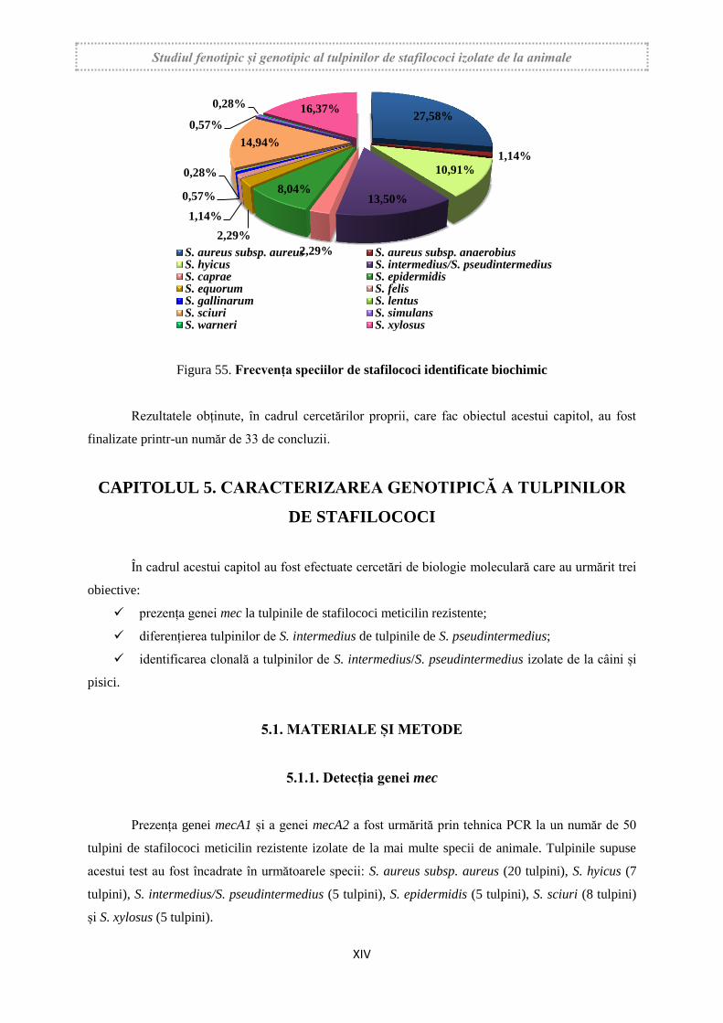
Au fost supuse identificării definitive, pe baza proprietăților glucidolitice, un număr de 348
tulpini de stafilococi, izolate de la mai multe specii de animale.
Rezultatele obținute, în urma identificării definitive, prin tipizarea biochimică, relevă că, din
probele de material patologic, prelevate de la mai multe specii de animale, au fost izolate un număr de
14 specii, a căror frecvență a fost cuprinsă între 0,28% și 27,58%, ceea mai mare frecvență având-o S.
aureus subsp. aureus, fiind urmată de S. xylosus, S. sciuri și S. intermedius (figura 55). De asemenea,
tot din aceste date, rezultă că speciile de stafilococi coagulază pozitivi au avut o frecvență cuprinsă
între 1,14% și 27,58%, iar speciile de stafilococi coagulază negativi au avut o frecvență cuprinsă
0,28% și 16,37%.
Studiul fenotipic și genotipic al tulpinilor de stafilococi izolate de la animale
XIV
Figura 55. Frecvența speciilor de stafilococi identificate biochimic
Rezultatele obținute, în cadrul cercetărilor proprii, care fac obiectul acestui capitol, au fost
finalizate printr-un număr de 33 de concluzii.
CAPITOLUL 5. CARACTERIZAREA GENOTIPICĂ A TULPINILOR
DE STAFILOCOCI
În cadrul acestui capitol au fost efectuate cercetări de biologie moleculară care au urmărit trei
obiective:
prezența genei mec la tulpinile de stafilococi meticilin rezistente;
diferențierea tulpinilor de S. intermedius de tulpinile de S. pseudintermedius;
identificarea clonală a tulpinilor de S. intermedius/S. pseudintermedius izolate de la câini și
pisici.
5.1. MATERIALE ȘI METODE
5.1.1. Detecția genei mec
Prezența genei mecA1 și a genei mecA2 a fost urmărită prin tehnica PCR la un număr de 50
tulpini de stafilococi meticilin rezistente izolate de la mai multe specii de animale. Tulpinile supuse
acestui test au fost încadrate în următoarele specii: S. aureus subsp. aureus (20 tulpini), S. hyicus (7
tulpini), S. intermedius/S. pseudintermedius (5 tulpini), S. epidermidis (5 tulpini), S. sciuri (8 tulpini)
și S. xylosus (5 tulpini).
27,58%
1,14%
10,91%
13,50%
2,29%
8,04%
2,29%
1,14%
0,57%
0,28%
14,94%
0,57%
0,28% 16,37%
S. aureus subsp. aureus S. aureus subsp. anaerobiusS. hyicus S. intermedius/S. pseudintermediusS. caprae S. epidermidisS. equorum S. felisS. gallinarum S. lentusS. sciuri S. simulansS. warneri S. xylosus
Studiul fenotipic și genotipic al tulpinilor de stafilococi izolate de la animale
XV
5.1.2. Diferențierea speciei S. intermedius de S. pseudintermedius
Au fost supuse tehnicii PCR, pentru diferențiere, între aceste două specii, un număr de 25 de
tulpini izolate de la câini și pisici care, pe baza caracterelor fenotipice, au fost încadrate în specia S.
intermedius. Având în vedere faptul că, în ultimii ani, este dominantă specia S. pseudintermedius și că
aceste două specii nu pot fi diferențiate fenotipic, în cadrul acestor cercetări, discriminarea, între
aceste specii, a fost făcută prin tehnica de biologie moleculară amintită.
5.1.3. Amprentarea genetică prin amplificare arbitrară - RAPD
Pentru identificarea clonală, a tulpinilor de S. intermedius/S. pseudintermedius (34 tulpini),
izolate de la câini şi pisici, a fost utilizat testul de biologie moleculară denumit Amprentarea genetică
prin amplificare arbitrară.
5.2. REZULTATELE OBȚINUTE
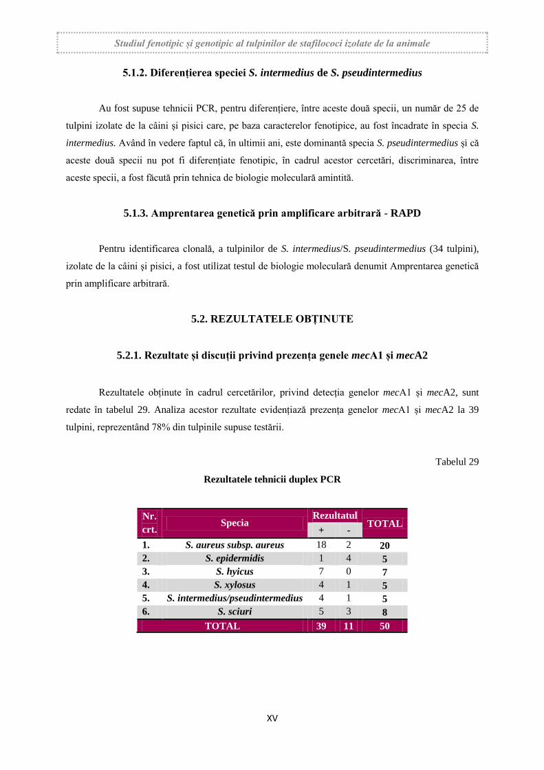
5.2.1. Rezultate și discuții privind prezența genele mecA1 și mecA2
Rezultatele obținute în cadrul cercetărilor, privind detecția genelor mecA1 și mecA2, sunt
redate în tabelul 29. Analiza acestor rezultate evidențiază prezența genelor mecA1 și mecA2 la 39
tulpini, reprezentând 78% din tulpinile supuse testării.
Tabelul 29
Rezultatele tehnicii duplex PCR
Nr.
crt. Specia
Rezultatul TOTAL
+ -
1. S. aureus subsp. aureus 18 2 20
2. S. epidermidis 1 4 5
3. S. hyicus 7 0 7
4. S. xylosus 4 1 5
5. S. intermedius/pseudintermedius 4 1 5
6. S. sciuri 5 3 8
TOTAL 39 11 50
Studiul fenotipic și genotipic al tulpinilor de stafilococi izolate de la animale
XVI
5.2.2. Rezultate și discuții privind diferențierea speciilor S. intermedius și S.
pseudintermedius
Pentru diferențierea genotipică au fost supuse testării 25 tulpini de stafilococi, din care 22
tulpini au fost izolate de la câini și 3 tulpini au fost izolate de la pisici.
Cu ajutorul acestei tehnici de biologie moleculară și prin utilizarea a doi primeri specifici, 9
tulpini, respectiv 36%, au fost incluse în specia S. intermedius, 6 tulpini fiind izolate de la câini și 3
tulpini fiind izolate de la pisici, iar 16 tulpini, respectiv 64%, au fost incluse în specia S.
pseudintermedius, toate fiind izolate numai de la câini.
5.2.3. Rezultate și discuții privind amprentarea genetică prin amplificare arbitrară –
RAPD
Prin această tehnică, de biologie moleculară, au fost testate 34 tulpini de S.intermedius/S.
pseudintermedius, respectiv 22 tulpini de origine canină și 12 tulpini de origine felină. Amprentarea
genetică, prin această tehnică, a fost realizată cu amorsele P1 și P2, pe molecule de ADN genomic
extrase din cele 34 tulpini.
Tulpinile de S.intermedius/S. pseudintermedius, izolate de la câini (22 tulpini) sunt
îndepărtate filogenetic faţă de tulpinile de origine felină (12 tulpini), având fiecare propriul cluster,
respectiv origini diferite.
Tulpinile de S.intermedius/S. pseudintermedius, izolate de la pisici, au amprente genetice
similare având aceeaşi origine, ceea ce demonstrează înrudirea lor filogenetică.
Rezultatele obținute, în cadrul cercetărilor proprii, care fac obiectul acestui capitol, au fost
finalizate printr-un număr de 8 de concluzii.
CAPITOLUL 6. CERCETĂRI PRIVIND FRECVENȚA
FENOTIPURILOR DE REZISTENȚĂ LA ANTIBIOTICE LA
TULPINILE DE STAFILOCOCI
Antibiorezistența, la bacterii, reprezintă o problemă de mare actualitate în medicina veterinară
și umană, deoarece este considerată un fenomen cu risc zoonotic pronunțat. Fenotipurile de rezistență
la bacterii patogene pentru animale, inclusiv la stafilococi, au o frecvență în continuă creștere datorită
utilizării produselor medicinale veterinare, pe bază de antibiotice, în special la animalele crescute în
sistem intensiv.
Studiul fenotipic și genotipic al tulpinilor de stafilococi izolate de la animale
XVII
Cercetările efectuate, în acest capitol, au urmărit stabilirea fenotipică a antibiorezistenței,
respectiv stabilirea frecvenței fenotipurilor de rezistență la tulpinile de stafilcoci izolate de la animale.
6.1. MATERIALE ȘI METODE
Profilul de rezistență la antibiotice a fost determinat prin metoda disc-difuzimetrică (metoda
KIRBY-BAUER), folosind, în acest scop, mediul MUELLER-HINTON, biodiscuri cu 20 antibiotice
din mai multe grupe și 412 tulpini de stafilococi, care au fost testate, încadrate în 22 de specii.
6.2. REZULTATE OBȚINUTE ȘI DISCUȚIA REZULTATELOR
Rezultatele obținute, în forma primară, au fost prelucrate, centralizate și redate în mai multe
tabele și sub formă grafică, în funcție de: antiobiotic, specia de animal și speciile de stafilococi
izolate.
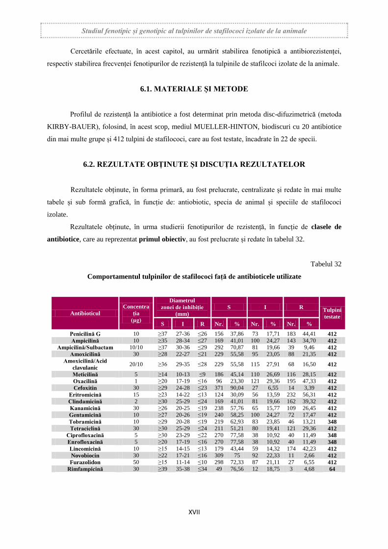
Rezultatele obținute, în urma studierii fenotipurilor de rezistență, în funcție de clasele de
antibiotice, care au reprezentat primul obiectiv, au fost prelucrate și redate în tabelul 32.
Tabelul 32
Comportamentul tulpinilor de stafilococi față de antibioticele utilizate
Antibioticul
Concentra
ția
(µg)
Diametrul
zonei de inhibiție
(mm)
S I R Tulpini
testate
S I R Nr. % Nr. % Nr. %
Penicilină G 10 ≥37 27-36 ≤26 156 37,86 73 17,71 183 44,41 412
Ampicilină 10 ≥35 28-34 ≤27 169 41,01 100 24,27 143 34,70 412
Ampicilină/Sulbactam 10/10 ≥37 30-36 ≤29 292 70,87 81 19,66 39 9,46 412
Amoxicilină 30 ≥28 22-27 ≤21 229 55,58 95 23,05 88 21,35 412
Amoxicilină/Acid
clavulanic 20/10 ≥36 29-35 ≤28 229 55,58 115 27,91 68 16,50 412
Meticilină 5 ≥14 10-13 ≤9 186 45,14 110 26,69 116 28,15 412
Oxacilină 1 ≥20 17-19 ≤16 96 23,30 121 29,36 195 47,33 412
Cefoxitin 30 ≥29 24-28 ≤23 371 90,04 27 6,55 14 3,39 412
Eritromicină 15 ≥23 14-22 ≤13 124 30,09 56 13,59 232 56,31 412
Clindamicină 2 ≥30 25-29 ≤24 169 41,01 81 19,66 162 39,32 412
Kanamicină 30 ≥26 20-25 ≤19 238 57,76 65 15,77 109 26,45 412
Gentamicină 10 ≥27 20-26 ≤19 240 58,25 100 24,27 72 17,47 412
Tobramicină 10 ≥29 20-28 ≤19 219 62,93 83 23,85 46 13,21 348
Tetraciclină 30 ≥30 25-29 ≤24 211 51,21 80 19,41 121 29,36 412
Ciprofloxacină 5 ≥30 23-29 ≤22 270 77,58 38 10,92 40 11,49 348
Enrofloxacină 5 ≥20 17-19 ≤16 270 77,58 38 10,92 40 11,49 348
Lincomicină 10 ≥15 14-15 ≤13 179 43,44 59 14,32 174 42,23 412
Novobiocin 30 ≥22 17-21 ≤16 309 75 92 22,33 11 2,66 412
Furazolidon 50 ≥15 11-14 ≤10 298 72,33 87 21,11 27 6,55 412
Rimfampicină 30 ≥39 35-38 ≤34 49 76,56 12 18,75 3 4,68 64
Studiul fenotipic și genotipic al tulpinilor de stafilococi izolate de la animale
XVIII
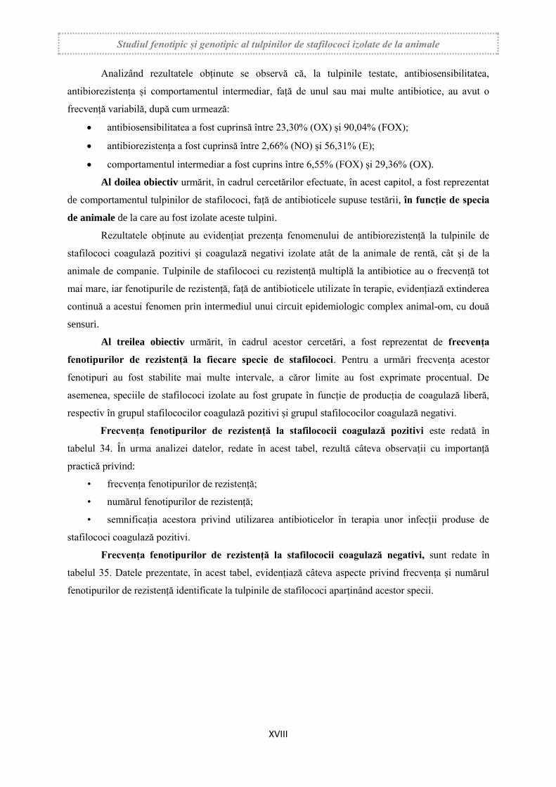
Analizând rezultatele obținute se observă că, la tulpinile testate, antibiosensibilitatea,
antibiorezistența și comportamentul intermediar, față de unul sau mai multe antibiotice, au avut o
frecvență variabilă, după cum urmează:
antibiosensibilitatea a fost cuprinsă între 23,30% (OX) și 90,04% (FOX);
antibiorezistența a fost cuprinsă între 2,66% (NO) și 56,31% (E);
comportamentul intermediar a fost cuprins între 6,55% (FOX) și 29,36% (OX).
Al doilea obiectiv urmărit, în cadrul cercetărilor efectuate, în acest capitol, a fost reprezentat
de comportamentul tulpinilor de stafilococi, față de antibioticele supuse testării, în funcție de specia
de animale de la care au fost izolate aceste tulpini.
Rezultatele obținute au evidențiat prezența fenomenului de antibiorezistență la tulpinile de
stafilococi coagulază pozitivi și coagulază negativi izolate atât de la animale de rentă, cât și de la
animale de companie. Tulpinile de stafilococi cu rezistență multiplă la antibiotice au o frecvență tot
mai mare, iar fenotipurile de rezistență, față de antibioticele utilizate în terapie, evidențiază extinderea
continuă a acestui fenomen prin intermediul unui circuit epidemiologic complex animal-om, cu două
sensuri.
Al treilea obiectiv urmărit, în cadrul acestor cercetări, a fost reprezentat de frecvența
fenotipurilor de rezistență la fiecare specie de stafilococi. Pentru a urmări frecvența acestor
fenotipuri au fost stabilite mai multe intervale, a căror limite au fost exprimate procentual. De
asemenea, speciile de stafilococi izolate au fost grupate în funcție de producția de coagulază liberă,
respectiv în grupul stafilococilor coagulază pozitivi și grupul stafilococilor coagulază negativi.
Frecvența fenotipurilor de rezistență la stafilococii coagulază pozitivi este redată în
tabelul 34. În urma analizei datelor, redate în acest tabel, rezultă câteva observații cu importanță
practică privind:
• frecvența fenotipurilor de rezistență;
• numărul fenotipurilor de rezistență;
• semnificația acestora privind utilizarea antibioticelor în terapia unor infecții produse de
stafilococi coagulază pozitivi.
Frecvența fenotipurilor de rezistență la stafilococii coagulază negativi, sunt redate în
tabelul 35. Datele prezentate, în acest tabel, evidențiază câteva aspecte privind frecvența și numărul
fenotipurilor de rezistență identificate la tulpinile de stafilococi aparținând acestor specii.
Studiul fenotipic și genotipic al tulpinilor de stafilococi izolate de la animale
XIX
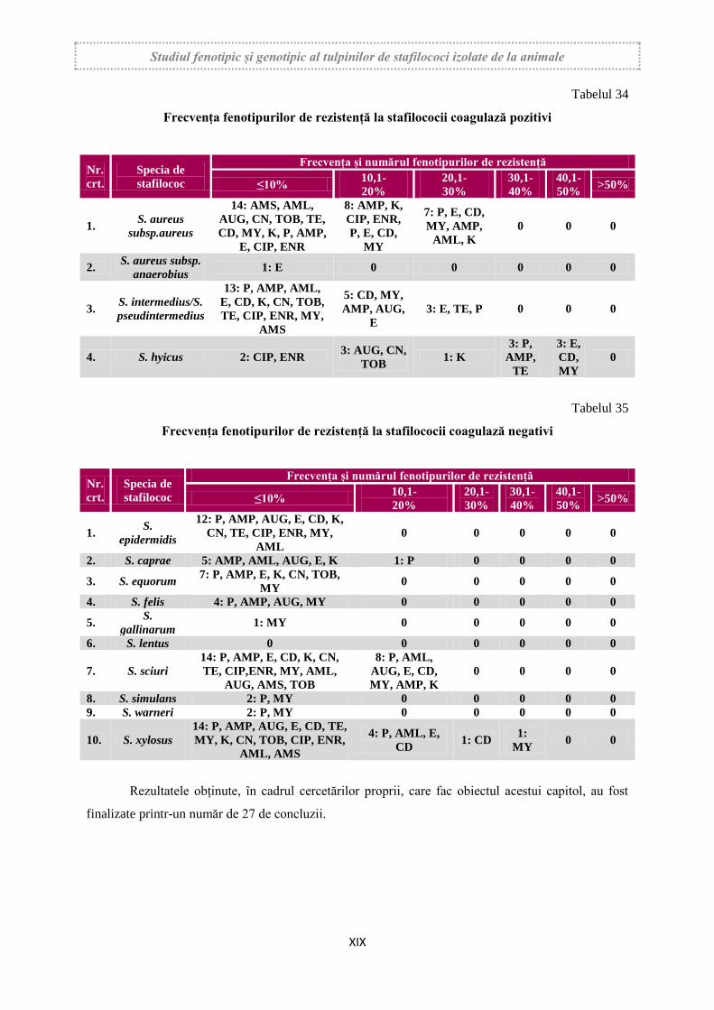
Tabelul 34
Frecvența fenotipurilor de rezistență la stafilococii coagulază pozitivi
Nr.
crt.
Specia de
stafilococ
Frecvența și numărul fenotipurilor de rezistență
≤10% 10,1-
20%
20,1-
30%
30,1-
40%
40,1-
50% >50%
1. S. aureus
subsp.aureus
14: AMS, AML,
AUG, CN, TOB, TE,
CD, MY, K, P, AMP,
E, CIP, ENR
8: AMP, K,
CIP, ENR,
P, E, CD,
MY
7: P, E, CD,
MY, AMP,
AML, K
0 0 0
2. S. aureus subsp.
anaerobius 1: E 0 0 0 0 0
3. S. intermedius/S.
pseudintermedius
13: P, AMP, AML,
E, CD, K, CN, TOB,
TE, CIP, ENR, MY,
AMS
5: CD, MY,
AMP, AUG,
E
3: E, TE, P 0 0 0
4. S. hyicus 2: CIP, ENR 3: AUG, CN,
TOB 1: K
3: P,
AMP,
TE
3: E,
CD,
MY
0
Tabelul 35
Frecvența fenotipurilor de rezistență la stafilococii coagulază negativi
Nr.
crt.
Specia de
stafilococ
Frecvența și numărul fenotipurilor de rezistență
≤10% 10,1-
20%
20,1-
30%
30,1-
40%
40,1-
50% >50%
1. S.
epidermidis
12: P, AMP, AUG, E, CD, K,
CN, TE, CIP, ENR, MY,
AML
0 0 0 0 0
2. S. caprae 5: AMP, AML, AUG, E, K 1: P 0 0 0 0
3. S. equorum 7: P, AMP, E, K, CN, TOB,
MY 0 0 0 0 0
4. S. felis 4: P, AMP, AUG, MY 0 0 0 0 0
5. S.
gallinarum 1: MY 0 0 0 0 0
6. S. lentus 0 0 0 0 0 0
7. S. sciuri
14: P, AMP, E, CD, K, CN,
TE, CIP,ENR, MY, AML,
AUG, AMS, TOB
8: P, AML,
AUG, E, CD,
MY, AMP, K
0 0 0 0
8. S. simulans 2: P, MY 0 0 0 0 0
9. S. warneri 2: P, MY 0 0 0 0 0
10. S. xylosus
14: P, AMP, AUG, E, CD, TE,
MY, K, CN, TOB, CIP, ENR,
AML, AMS
4: P, AML, E,
CD 1: CD
1:
MY 0 0
Rezultatele obținute, în cadrul cercetărilor proprii, care fac obiectul acestui capitol, au fost
finalizate printr-un număr de 27 de concluzii.

Studiul fenotipic și genotipic al tulpinilor de stafilococi izolate de la animale
XX
CAPITOLUL 7. CERCETĂRI PRIVIND FRECVENȚA TULPINILOR
METICILIN REZISTENTE
O atenție deosebită este acordată, în ultimii ani, tulpinilor de stafilococi meticilin rezistente,
cunoscute generic sub denumirea de stafilococi MRSA (Methicillin Resistant Staphylococcus aureus).
Aceste tulpini au un risc zoonotic pronunțat și un circuit epidemiologic complex, fiind întâlnite
frecvent și la oameni. De asemenea, rezistența la meticilină este asociată cu rezistența multiplă la
antibiotice, mai ales cu rezistența față de peniciline, cefalosporine și aztreonam.
Datele existente în literatură demonstrează că meticilin-rezistența este prezentă atât la
stafilococii coagulază pozitivi, cât și la stafilococii coagulază negativi. Extinderea meticilin-
rezistenței, de la tulpinile de S. aureus subsp. aureus la alte specii de stafilococi, este generată de
mecanismele de transmitere ale casetei cromozomiale stafilococice mec (Staphylococcal Chromosome
Cassete mec- SCCmec), existentă în elementele genetice mobile din citolpasma stafilococilor.
7.1. MATERIALE ȘI METODE
Frecvența tulpinilor meticilin rezistente a fost urmărită la un număr de 412 tulpini de
stafilococi, încadrate în 22 de specii. Frecvența tulpinilor meticilin rezistente a fost stabilită fenotipic
prin două teste și confirmată la un număr de 50 tulpini prin reacția polimerazică în lanț.
Frecvența tulpinilor meticilin rezistente a fost stabilită fenotipic prin metoda disc-
difuzimetrică (Kirby-Bauer), utilizând biodiscuri cu meticilină (5 µg), oxacilină (1 µg) și cefoxitin
(30 µg).
Un număr de 210 tulpini de stafilococi coagulază pozitivi și coagulază negativi au fost
selecționate, pentru a fi pasate pe mediul Chromatic MRSA, după următoarele criterii:
au manifestat rezistență fenotipică față de cel puțin unul din cele trei antibiotice amintite;
au avut comportament intermediar la cel puțin două din cele trei antibiotice.
7.2. REZULTATE OBȚINUTE
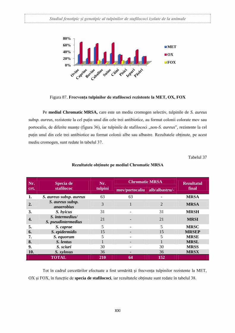
Rezultatele obținute, prezentate figura 87, redau frecvența tulpinilor meticilin rezistente,
detectate prin metoda disc-difuzimetrică, din totalul tulpinilor de stafilococi supuse testării și în
funcție de specia de animale.
Studiul fenotipic și genotipic al tulpinilor de stafilococi izolate de la animale
XXI
Figura 87. Frecvența tulpinilor de stafilococi rezistente la MET, OX, FOX
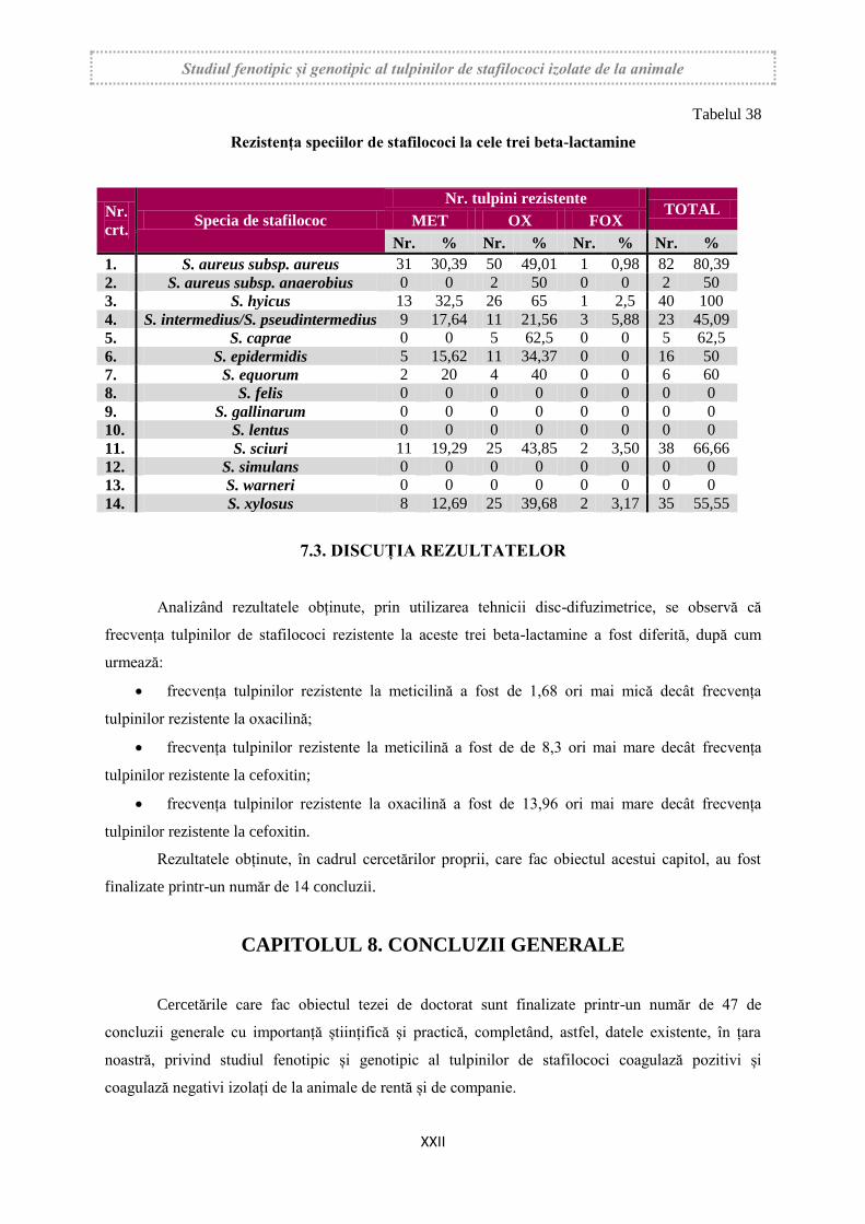
Pe mediul Chromatic MRSA, care este un mediu cromogen selectiv, tulpinile de S. aureus
subsp. aureus, rezistente la cel puțin unul din cele trei antibiotice, au format colonii colorate mov sau
portocaliu, de diferite nuanțe (figura 36), iar tulpinile de stafilococi „non-S. aureus”, rezistente la cel
puțin unul din cele trei antibiotice au format colonii albe sau albastre. Rezultatele obținute, pe acest
mediu cromogen, sunt redate în tabelul 37.
Tabelul 37
Rezultatele obținute pe mediul Chromatic MRSA
Nr.
crt.
Specia de
stafilococ
Nr.
tulpini
Chromatic MRSA Rezultatul
final mov/portocaliu alb/albastru/-
1. S. aureus subsp. aureus 63 63 - MRSA
2. S. aureus subsp.
anaerobius 3 1 2 MRSA
3. S. hyicus 31 - 31 MRSH
4. S. intermedius/
S. pseudintermedius 21 - 21 MRSI
5. S. caprae 5 - 5 MRSC
6. S. epidermidis 15 - 15 MRSEP
7. S. equorum 5 - 5 MRSE
8. S. lentus 1 - 1 MRSL
9. S. sciuri 30 - 30 MRSS
10. S. xylosus 36 - 36 MRSX
TOTAL 210 64 152
Tot în cadrul cercetărilor efectuate a fost urmărită și frecvența tulpinilor rezistente la MET,
OX și FOX, în funcțtie de specia de stafilococi, iar rezultatele obținute sunt redate în tabelul 38.
0%
20%
40%
60%
80%
MET
OX
FOX
Studiul fenotipic și genotipic al tulpinilor de stafilococi izolate de la animale
XXII
Tabelul 38
Rezistența speciilor de stafilococi la cele trei beta-lactamine
Nr.
crt. Specia de stafilococ
Nr. tulpini rezistente TOTAL
MET OX FOX
Nr. % Nr. % Nr. % Nr. %
1. S. aureus subsp. aureus 31 30,39 50 49,01 1 0,98 82 80,39
2. S. aureus subsp. anaerobius 0 0 2 50 0 0 2 50
3. S. hyicus 13 32,5 26 65 1 2,5 40 100
4. S. intermedius/S. pseudintermedius 9 17,64 11 21,56 3 5,88 23 45,09
5. S. caprae 0 0 5 62,5 0 0 5 62,5
6. S. epidermidis 5 15,62 11 34,37 0 0 16 50
7. S. equorum 2 20 4 40 0 0 6 60
8. S. felis 0 0 0 0 0 0 0 0
9. S. gallinarum 0 0 0 0 0 0 0 0
10. S. lentus 0 0 0 0 0 0 0 0
11. S. sciuri 11 19,29 25 43,85 2 3,50 38 66,66
12. S. simulans 0 0 0 0 0 0 0 0
13. S. warneri 0 0 0 0 0 0 0 0
14. S. xylosus 8 12,69 25 39,68 2 3,17 35 55,55
7.3. DISCUȚIA REZULTATELOR
Analizând rezultatele obținute, prin utilizarea tehnicii disc-difuzimetrice, se observă că
frecvența tulpinilor de stafilococi rezistente la aceste trei beta-lactamine a fost diferită, după cum
urmează:
frecvența tulpinilor rezistente la meticilină a fost de 1,68 ori mai mică decât frecvența
tulpinilor rezistente la oxacilină;
frecvența tulpinilor rezistente la meticilină a fost de de 8,3 ori mai mare decât frecvența
tulpinilor rezistente la cefoxitin;
frecvența tulpinilor rezistente la oxacilină a fost de 13,96 ori mai mare decât frecvența
tulpinilor rezistente la cefoxitin.
Rezultatele obținute, în cadrul cercetărilor proprii, care fac obiectul acestui capitol, au fost
finalizate printr-un număr de 14 concluzii.
CAPITOLUL 8. CONCLUZII GENERALE
Cercetările care fac obiectul tezei de doctorat sunt finalizate printr-un număr de 47 de
concluzii generale cu importanță științifică și practică, completând, astfel, datele existente, în țara
noastră, privind studiul fenotipic și genotipic al tulpinilor de stafilococi coagulază pozitivi și
coagulază negativi izolați de la animale de rentă și de companie.
Studiul fenotipic și genotipic al tulpinilor de stafilococi izolate de la animale
XXIII
PHENOTYPIC AND GENOTYPIC STUDY OF STAPHYLOCOCCI
STRAINS ISOLATED FROM ANIMALS
ABSTRACT
Staphylococci are Gram positive bacteria, considered as ubiquitous, which have skin and
mucous habitat in both animals and humans. These bacteria are present in the external environment
and in the micromedium, having, as a substrate, the soil, water, air, bedding, facilities and utensils in
the shelters, inventory in veterinary and hospital clinics, utensils and installations in plants processing
animal products, and also food of animal origin intended for human consumption. These substrates
represent reservoirs and secondary sources of infection for animals and humans.
In recent years, several species of negative coagulase staphylococci (SCN) have been isolated
from animals and humans, which can produce localized infections in both animals and humans, most
notably nosocomial infections.
Staphylococci manifests a pronounced aggression for tissues and organs, based on the
existence of pathogenicity attributes, represented by virulence, toxicity and biofilm formation. These
pathogenicity factors are genetically encoded, having the respective genes present in the chromosome
and in the mobile genetic elements existing in the cytoplasm.
Staphylococci are considered to be zoonotic-risk bacteria, animals being an important
infection reservoir for humans, with a complex epidemiological circuit, both between livestock and
humans, and between pets and humans, however staphylococci can also move from people to animals.
Multiple resistance to antibiotics, in staphylococci, is considered a pronounced zoonotic risk
factor, because staphylococcal resistance phenotypes have a steadily increasing rate in both livestock
and pets and the strains presenting this phenomenon have a complex epidemiological circuit. These
phenotypes present in staphylococcus strains, isolated from animals, are permanently monitored
because, based on them, therapeutic conduct is established and the strain circuit is followed.
Resistance to methicillin was reported, initially, to S. aureus subsp. aureus and then to other
species and the resistant strainsto this antibiotic were called MRSA strains (Methicillin Resistant
Staphylococcus aureus). Extending the methicillin resistance phenomenon led to numerous studies, as
these strains have also reached to humans, therefore considered strains with zoonotic risk. Resistance
to methicillin occurs as a result of penicillin binding proteins intervention, whose synthesis is encoded
by the SCCmec cassette. For the detection of methicillin resistance, oxacillin has been proposed since
2004, and cefoxitin since 2005, because the results obtained with these two beta-lactamases are much
more reliable.

Studiul fenotipic și genotipic al tulpinilor de stafilococi izolate de la animale
XXIV
The Ph D thesis contains 284 pages, 38 tables and 87 figures, which includes 52 original
images and 35 graphics. The scientific support of the thesis is represented by 300 bibliographic titles,
including scientific papers, papers, doctoral thesis and web pages.
The doctoral thesis is structured in two parts, namely "Bibliographic research", included in
Part I and "Own research", included in Part II.
Part I
BIBLIOGRAPHICAL RESEARCH
The first part of the thesis is a bibliographic study, structured in two chapters and extended
to 74 pages (26,06%). In this part, there are 3 tables and 3 representative figures along with current
data on the Staphylococcus genus.
CHAPTER 1. THE STAPHYLOCOCCACEAE FAMILY. THE
STAPHYLOCOCCUS GENUS
The first chapter is a synthesis of data from the literature, rigorously selected on systematics
of Staphylococcus genus. The taxonomy of this genus, taken from www.bacterio.net/-
allnamessz.html, which includes the List of Prokaryotic names with Standing in Nomenclature, is
coordinated by Prof. Dr. EUSZÉBY J. P.
Within the chapter, the latest data on ecology, morphology, antigenic structure and pathogenic
factors are presented. Also, the chapter includes current data on the genetics of these bacteria, multiple
antibiotic resistance and the phenomenon of methicillin resistance. It is also presented the pathogenic
staphylococci species to livestock and pets and the morbid entities products.
CHAPTER 2. LABORATORY DIAGNOSIS OF STAPHYLOCOCCAL
INFECTIONS IN ANIMALS
This chapter presents an extensive bibliographic synthesis on the diagnosis of staphylococcal
infections in animals. Techniques of sampling, pathological materials, as well as their transport are
presented. Particular attention is given to the bacteriological examination, regarding both primary and
definitive identification, as well as culture media, current tests and commercial systems. It is also
presented the clonal identification, toxin detection and molecular biology tests used in recent years.
Studiul fenotipic și genotipic al tulpinilor de stafilococi izolate de la animale
XXV
Part II
OWN RESEARCH
Part II includes its own research and it’s structured in 6 chapters (3-8), extended to 210 pages
(73.94%). This part of the thesis is illustrated by 35 tables and 84 figures, including 49 original
images and 35 graphs.
CAPITOLUL 3. GOAL, MOTIVATION AND RESEARCH OBJECTIVES
Staphylococci are pathogenic, conditioned pathogenic and opportunistic pathogenic bacteria
depending on the species and the presence of favorable factors. They have tropism for skin and
mucous membranes, producing localized suppurative infections, septicemia and infectious entity,
well-contoured, that evolve in animals and humans, generically called staphylococci. In some
situations, they may also act as secondary pathogens, mainly after some viroses.
Staphylococci evolve in animals as distinct epidemiological and anatomoclinic, with septic
and/or localized evolution. Of these, the infectious mastitis of dairy cows is distinguished as the
frequency, economic and health importance.
Depending on the pathogenicity of the staphylococci species and the clinical evolution of the
produced entities, most researchers distinguish between S. aureus subsp. aureus, the type species of
the genus, considered the most pathogenic species and other species of coagulase positive and
negative staphylococci, included in a group generically called "non-S. aureus " staphylococci.
Pathogenicity mechanisms are carried out in a complex sequence, involving extracellular
enzymes and toxins, which act as rings in a biochemical reaction, and less as individualized form.
Staphylococci penetrate the body into the skin and mucous membranes through sebaceous glands,
sweat glands, hair follicles, microlesions and larger lesions. Incipient (locally) infectious process is
the consequence of strain number, virulence and toxicity, and the ability of local defense mechanisms,
of host organisms, that interact through phagocytosis, mainly through the neutrophil granulocytes
mobilized and potentiated by opsonization and through lysis by the complement system. Repeated
staphylococcal infections in the skin may trigger a hypersensitivity or allergy to some staphylococcal
antigens.
Studies showed that antibiotic resistance is genetically determined, with the support of many
resistance genes located in the bacterial chromosome, plasmids R, intergongs and transposons, which
are mobile genetic elements. Through these, genes encoding the antibiotic resistance can be
transferred between strains of the same bacterial species (intraspecific transmission) as well as
between strains belonging to other bacterial species (interspecific transmission). Also, the research
showed that R-plasmids are the main factors of extracromosomal resistance to antibiotics, especially
Studiul fenotipic și genotipic al tulpinilor de stafilococi izolate de la animale
XXVI
those of the conjugate type that contain 3 groups of genes, namely transfer control genes, genes
encoding autoreplication, and genes encoding resistance to antibiotics. These genes have been
detected and studied using molecular biology techniques, the most widely used PCR technique, in
several variants.
Numerous research teams undertake extensive screening studies to monitor the
epidemiological circuit of resistant methicillin strains, using both classical phenotypical techniques
and molecular biology methods that are faster and allow the detection of SCCmec, which encodes
resistance to this antibiotic.
Taking into consideration the above mentioned aspects, the following objectives were pursued
in the research underlying the PhD thesis:
cultural, morphological and tinctorial study of staphylococci isolates;
the study of the biochemical profile, using a multitest system and based on glucidolytic
activity;
the use of chromogenic media to discriminate strains of S. aureus subsp. aureus and MRSA
strains;
establishing the frequency of some staphylococci species isolated from animals;
the study of existing pathogenicity factors in the isolated strains;
simplifying the isolation and primary typing scheme of isolated strains;
investigating the presence of free coagulation as a pathogenic factor and differentiation of
the strains in the two groups;
studying the resistance phenotypes (patterns) of the isolated strains;
studying the phenomenon of methicillin resistance at the isolated strains;
the detection of mec gene in methicillin-resistant staphylococci strains, using the PCR
technique;
the differentiation of S. intermedius strains from S. pseudintermedius strains using PCR
technique;
clonal identification of S. intermedius/S. pseudintermedius strains isolated from dogs and
cats.
CHAPTER 4. ISOLATION AND PHENOTYPIC CHARACTERIZATION
OF STAPHYLOCOCCI STRAINS
In animals and humans, staphylococci produce various localized infections or infectious
diseases, that are well-rounded, with economic and health importance. In recent years, the role of
negative coagulase staphylococci (NCS) in the etiology of localized infections has increased, of which
stand out the subclinical mastitis in dairy cows or various cutaneous infections or with localization in
Studiul fenotipic și genotipic al tulpinilor de stafilococi izolate de la animale
XXVII
some organs in swine and pets. These bacteria have tropism for epithelial (skin and mucous), but due
to the aggressive equipment they possess, reprezented by the enzymes, toxins and other pathogenic
factors, they can invade any tissue or organ. Natural infections in humans and animals are influenced
by several factors related to host organisms, the existence of favorable factors and staphylococci
species.
The research underlying this chapter has as main objective the phenotypic characterization of
of staphylococci strains isolated from healthy and with different localized or generalized infections
animals from livestock and pets.
4.1. MATERIALS AND METHODS
Samples of pathological material were taken from several animal species that will be
subjected to the bacteriological examination, performed according to the staphylococcal isolation and
typing methodology.
4.1.1. Sampling, transporting and insemination of samples
Pathological samples were collected from livestock and companion animals of several species
and age categories, with different conditions, or clinically healthy (Table 4).
Sterile swabs were used to collect samples from the skin, nasal secretions were also taken
with sterile swabs, and uterovaginal secretions were taken with sterile cotton wipes fixed on a longer
plastic rod.
Mastic milk samples were sterile collected, from a number of 22 primiparous cows, where
subclinical mastitis begun and evolved 45-60 days after calving, affecting one, two or even three
quarters of the mammary gland, while from sheep and goats with gangrenous mastitis, the mastitic
pathological samples were collected in sterile vials.
Primary sowings were performed in nutrient broth or peptone water, depending on the
pathological material that was taken. Subsequently, the tubes with these media were placed on the
thermostat for 18-20 hours at 37°C to obtain the primary cultures. Gram stained smears were made
from these cultures and inseminations were made on the special media intended for the isolation and
typing of staphylococcus strains.
Studiul fenotipic și genotipic al tulpinilor de stafilococi izolate de la animale
XXVIII
Table 4
Samples collected from animals
No.
crt. Species Age category
No. of
collected samples
No. of
sterile samples
No. of
positive samples
No. %
1. Sheeps lambs 22 1 21
- adults 54 5 49
Total 76 6 70 17,5
2. Goats adults 30 3 27 6,75
3. Bovines
calves 13 0 13
- adults 15 2 13
subclinical mastitis 52 0 52
Total 80 2 78 19,5
4. Horses
foals 10 0 10
- young horses 10 0 10
mares mothers 12 0 12
adults 20 1 19
Total 52 1 51 12,75
5. Swine
piglets 26 3 23
- sows 27 0 27
fattening pigs 26 2 24
Total 79 5 74 18,5
6. Dogs adults 55 5 50 12,5
7. Cats adults 28 3 25 6,25
8. Rabitts
bunnies 11 4 7
- Female rabitts 4 1 3
adults 5 0 5
Total 20 5 15 3,75
9. Birds adults 10 0 10 2,5
TOTAL 430 30 400 100
4.1.2. The used culture media
For the primary isolation of staphylococci strains from the collected pathological materials
and subsequently for the phenotypic characterization of staphylococci strains, classic and special
media were used, which revealed several biochemicals properties of these bacteria, on which the
minimal identification was made , followed by the definitive typing of the isolated strains.
The media used were: peptone water, simple broth, Mueller Hinton agar, Chapman medium,
7% sheep defibrinated blood agar, Baird-Parker medium, Chromatic Staph medium, Chromatic
MRSA medium, Congo red medium, bromothymol blue peptone water and standard nutrient agar.
Studiul fenotipic și genotipic al tulpinilor de stafilococi izolate de la animale
XXIX
4.1.3. API Staph system
Identification to species level of staphylococci strains (64 strains), isolated from mastitic milk
samples, collected only from primiparous with subclinical mastitis was made by using the API Staph
microtest and the Apiweb interpretation software.
4.1.4. Catalase test
Hydrogen peroxide, a 3% solution, was used to demonstrate the presence of catalase, which
was added in staphylococci cultures tubes in an amount of 1 ml and after that the cultures were
examined for 5-10 minutes for gas bubbles.
4.1.5. Free and bound coagulase tests
Diffusible free coagulase was highlighted by lyophilised rabbit plasma tubes test and Baird-
Parker medium, and bound coagulase, also called clumping factor, was highlighted with the Staphylo
Rapid Test and Staph Latex kit.
4.1.6. Bacterioscopic and cultural examination
Cultural examination. Cultures obtained after primary inseminations and after inseminations
on the mentioned media were examined with the naked eye and the stereoscopic glass, appreciating
the following characteristics: turbidity on liquid media, shape, size, pigmentation and coloring on the
solid used media, presented above.
Bacterioscopic examination. Gram stained smears were performed from isolated,
characteristic colonies in order to emphasize morphological and tinctorial characters.
4.1.7. Furazolidone and novobiocin resistance testing
Novobiocin susceptibility test was performed to differentiate some negative coagulase
staphylococci species from positive coagulase staphylococci species.
The furazolidone susceptibility test was used to differentiate staphylococci strains from
micrococci strains. The principle of this test is that staphylococci are susceptible to bacteriostatic
compounds from the furans class, while the micrococci are resistant.
Studiul fenotipic și genotipic al tulpinilor de stafilococi izolate de la animale
XXX
4.2. THE OBTAINED RESULTS
4.2.1. The results regarding the preliminary identification
4.2.1.1. Cultural and bacterioscopic examination results
In peptone water, primary cultures produced variable turbidity, some cultures also producing
a non-characteristic, easy homogenisable deposit, while in the broth, the primary cultures produced
intense turbidity with a non-characteristic deposit, easily homogenizable, and some strains formed a
discrete ring on the surface.
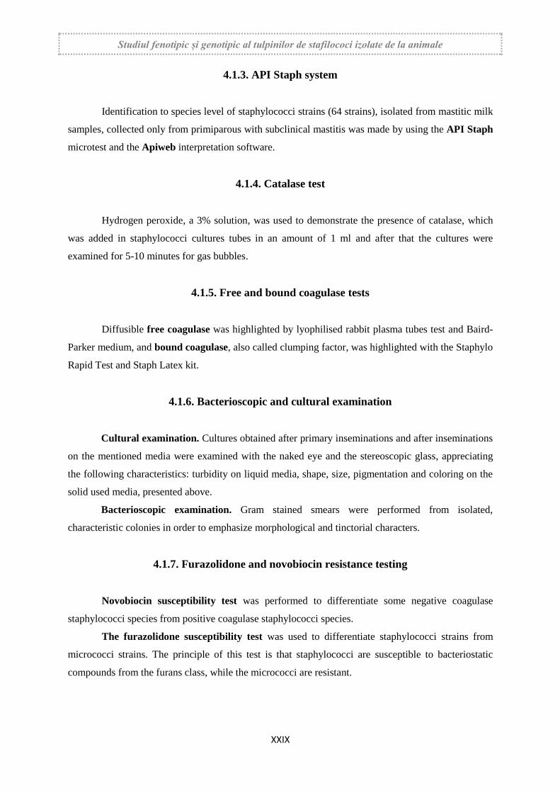
On the Chapman medium, the positive mannitol strains grew well, fermented the mannite,
producing the pH indicator coloration, and the negative mannitol strains grew but did not fermented
the mannite and the pH indicator did not turn the color. In some strains, the mannitol fermentation
was delayed, respectively at 48 hours.
Figure 45. Frequency of staphylococci strains regarding mannitol fermentation
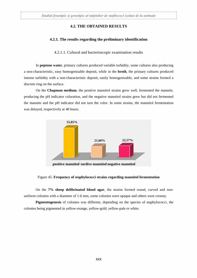
On the 7% sheep defibrinated blood agar, the strains formed round, curved and non-
uniform colonies with a diameter of 1-6 mm, some colonies were opaque and others were creamy.
Pigmentogenesis of colonies was different, depending on the species of staphylococci, the
colonies being pigmented in yellow-orange, yellow-gold, yellow-pale or white.
positive mannitol tardive mannitol negative mannitol
55,85%
21,60% 22,57%
Studiul fenotipic și genotipic al tulpinilor de stafilococi izolate de la animale
XXXI
Figure 47. Frequency of pigment types
On this medium has also been determined the type of hemolysis, thus, some strains produced
total haemolysis in the form of a circular zone around the colonies (type β hemolysis), while other
strains produced a zone of incomplete haemolysis, type "hot -cold" and other strains were non-
hemolytic.
Figure 46. Frequency of hemolysis types
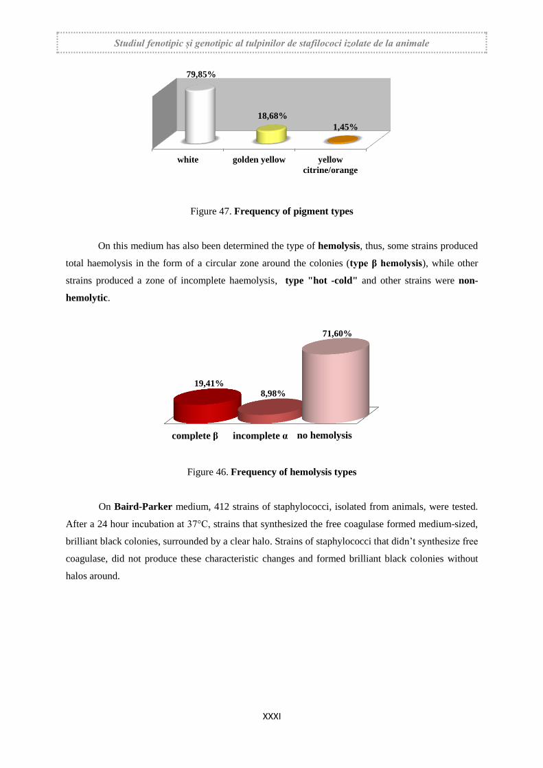
On Baird-Parker medium, 412 strains of staphylococci, isolated from animals, were tested.
After a 24 hour incubation at 37°C, strains that synthesized the free coagulase formed medium-sized,
brilliant black colonies, surrounded by a clear halo. Strains of staphylococci that didn’t synthesize free
coagulase, did not produce these characteristic changes and formed brilliant black colonies without
halos around.
white golden yellow yellow
citrine/orange
79,85%
18,68%
1,45%
complete β incomplete α no hemolysis
19,41% 8,98%
71,60%
Studiul fenotipic și genotipic al tulpinilor de stafilococi izolate de la animale
XXXII
Figure 51. Frequency of coagulase positive and negative strains on Baird-Parker medium
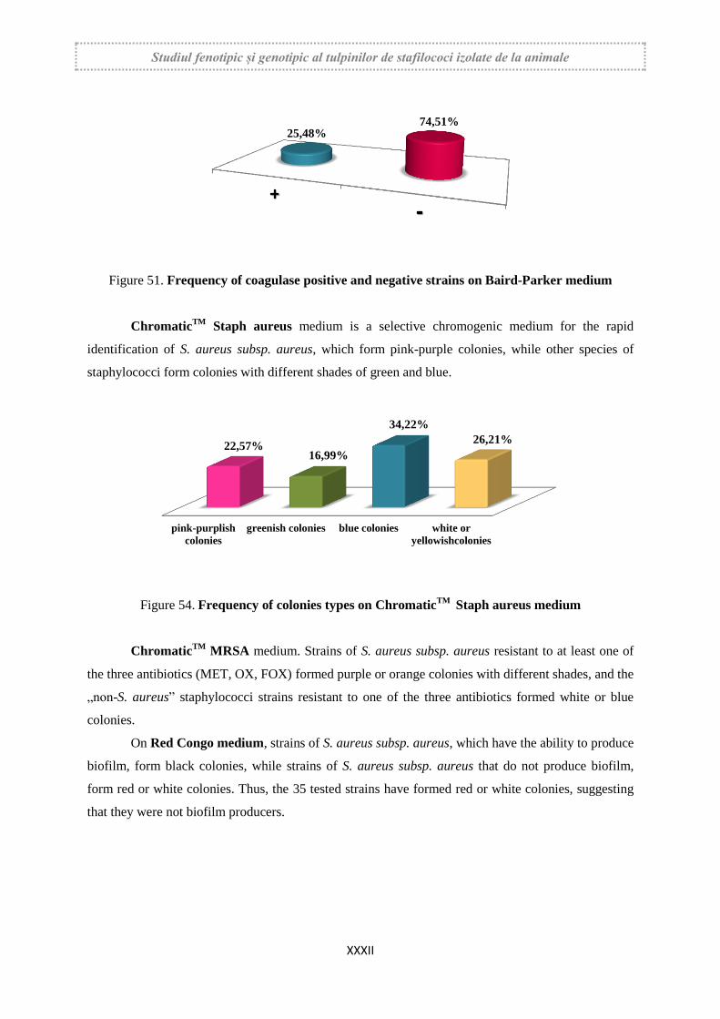
ChromaticTM
Staph aureus medium is a selective chromogenic medium for the rapid
identification of S. aureus subsp. aureus, which form pink-purple colonies, while other species of
staphylococci form colonies with different shades of green and blue.
Figure 54. Frequency of colonies types on ChromaticTM
Staph aureus medium
ChromaticTM
MRSA medium. Strains of S. aureus subsp. aureus resistant to at least one of
the three antibiotics (MET, OX, FOX) formed purple or orange colonies with different shades, and the
„non-S. aureus” staphylococci strains resistant to one of the three antibiotics formed white or blue
colonies.
On Red Congo medium, strains of S. aureus subsp. aureus, which have the ability to produce
biofilm, form black colonies, while strains of S. aureus subsp. aureus that do not produce biofilm,
form red or white colonies. Thus, the 35 tested strains have formed red or white colonies, suggesting
that they were not biofilm producers.
25,48% 74,51%
pink-purplish
colonies
greenish colonies blue colonies white or
yellowishcolonies
22,57% 16,99%
34,22%
26,21%
Studiul fenotipic și genotipic al tulpinilor de stafilococi izolate de la animale
XXXIII
4.2.1.2. The results obtained regarding the catalase and coagulase presence
For catalase production, variant 1, respectively whole broth cultures was used, in which the
3% hydrogen peroxide solution was introduced and after that the cultures were examined for evidence
of gas bubbles.
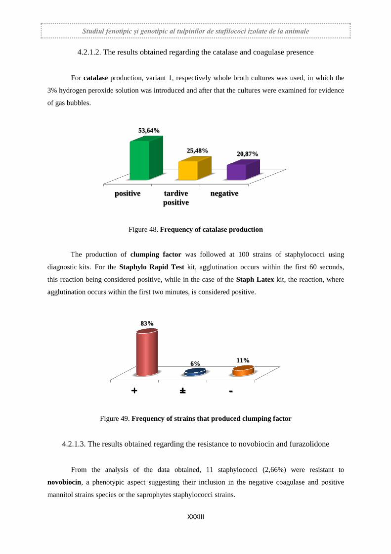
Figure 48. Frequency of catalase production
The production of clumping factor was followed at 100 strains of staphylococci using
diagnostic kits. For the Staphylo Rapid Test kit, agglutination occurs within the first 60 seconds,
this reaction being considered positive, while in the case of the Staph Latex kit, the reaction, where
agglutination occurs within the first two minutes, is considered positive.
Figure 49. Frequency of strains that produced clumping factor
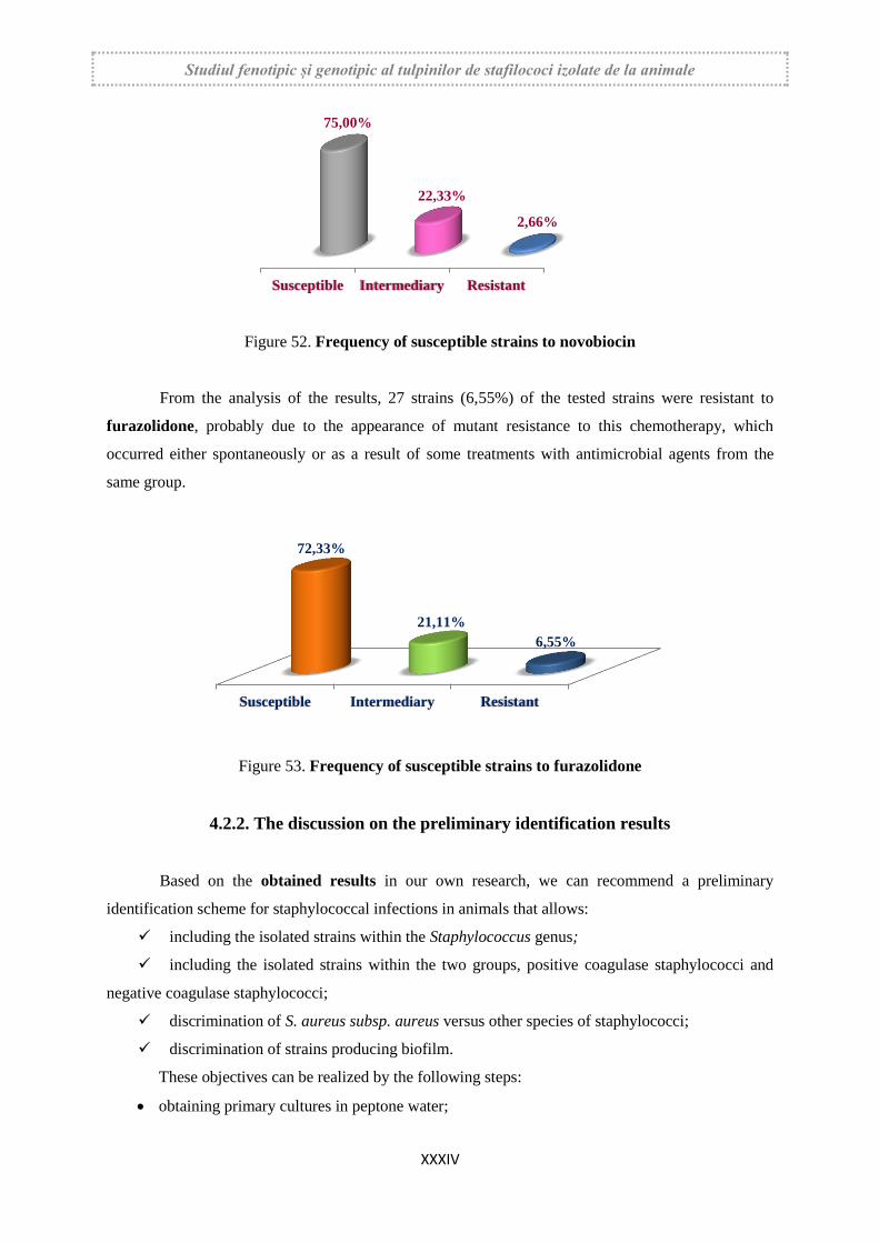
4.2.1.3. The results obtained regarding the resistance to novobiocin and furazolidone
From the analysis of the data obtained, 11 staphylococci (2,66%) were resistant to
novobiocin, a phenotypic aspect suggesting their inclusion in the negative coagulase and positive
mannitol strains species or the saprophytes staphylococci strains.
positive tardive
positive
negative
53,64%
25,48% 20,87%
83%
6% 11%
Studiul fenotipic și genotipic al tulpinilor de stafilococi izolate de la animale
XXXIV
Figure 52. Frequency of susceptible strains to novobiocin
From the analysis of the results, 27 strains (6,55%) of the tested strains were resistant to
furazolidone, probably due to the appearance of mutant resistance to this chemotherapy, which
occurred either spontaneously or as a result of some treatments with antimicrobial agents from the
same group.
Figure 53. Frequency of susceptible strains to furazolidone
4.2.2. The discussion on the preliminary identification results
Based on the obtained results in our own research, we can recommend a preliminary
identification scheme for staphylococcal infections in animals that allows:
including the isolated strains within the Staphylococcus genus;
including the isolated strains within the two groups, positive coagulase staphylococci and
negative coagulase staphylococci;
discrimination of S. aureus subsp. aureus versus other species of staphylococci;
discrimination of strains producing biofilm.
These objectives can be realized by the following steps:
obtaining primary cultures in peptone water;
Susceptible Intermediary Resistant
75,00%
22,33%
2,66%
Susceptible Intermediary Resistant
72,33%
21,11%
6,55%
Studiul fenotipic și genotipic al tulpinilor de stafilococi izolate de la animale
XXXV
use of Chapman medium for the inhibition of non- halotolerant bacterial species and for the
discrimination of positive mannitol staphylococci strains against negative mannitol staphylococci
strains;
testing of hemolytic activity and pigmentogenesis on 7% sheep defibrinated blood agar;
highlighting the catalase using the tube test;
highlighting the elaboration;
discrimination of strains included in S. aureus subsp. aureus species on ChromaticTM
Staph
aureus medium, as well as strains included in S. sciuri and S. xylosus species;
discrimination of strains that produce biofilm on the Red Congo medium.
4.2.3. The results and discussion regarding the final identification
4.2.3.1. The results regarding the final identification with API Staph system
Based on the results provided by this multitest system, 13 species of staphylococci were
definitively identified, of which two were positive coagulase species (S. aureus subsp. aureus, S.
hyicus) and 11 species were negative coagulase (S. xylosus, S. chromogenes, S. sciuri, S. lentus, S.
epidermidis, S. haemolyticus, S. homnins, S. simulans, S. saprophyticus, S. warneri, S. cohnii).
This system does not have S. intermedius, S. pseudintermedius, S. equorum, S. succinus, S.
fleurettii and S. devriesei species in the identification table. The API web program, based on
biochemical characters, can notify the following message: “Possibility of S. intermedius, S.
pseudintermedius, S. equorum, S. succinus, S. fleurettii or S. devriesei of Veterinary Origin”. In these
situations, the API web program may indicate some biochemical characters to be tested further, and
also which additional tests should be performed for the definitive classification of the strains.
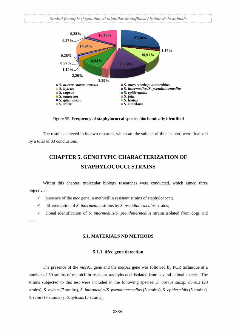
4.2.3.2. The results regarding the biochemical identification
Based on their glucidolytic properties, 348 strains of staphylococci isolated from several
animal species were definitive identified.
The results obtained after definitive identification by biochemical typing show that from the
samples of pathological material taken from several animal species, 14 species were isolated, with the
frequency between 0,28% and 27,58%, from which the highest was from S. aureus subsp. aureus,
followed by S. xylosus, S. sciuri and S. intermedius (figure 55). Also, all of these data show that
positive coagulase staphylococci species had a frequency between 1,14% and 27,58% and coagulase
negative species had a frequency between 0,28% and 16,37 %.
Studiul fenotipic și genotipic al tulpinilor de stafilococi izolate de la animale
XXXVI
Figure 55. Frequency of staphylococcal species biochemically identified
The results achieved in its own research, which are the subject of this chapter, were finalized
by a total of 33 conclusions.
CHAPTER 5. GENOTYPIC CHARACTERIZATION OF
STAPHYLOCOCCI STRAINS
Within this chapter, molecular biology researches were conducted, which aimed three
objectives:
presence of the mec gene in methicillin resistant strains of staphylococci;
differentiation of S. intermedius strains by S. pseudintermedius strains;
clonal identification of S. intermedius/S. pseudintermedius strains.isolated from dogs and
cats.
5.1. MATERIALS ND METHODS
5.1.1. Mec gene detection
The presence of the mecA1 gene and the mecA2 gene was followed by PCR technique at a
number of 50 strains of methicillin resistant staphylococci isolated from several animal species. The
strains subjected to this test were included in the following species: S. aureus subsp. aureus (20
strains), S. hyicus (7 strains), S. intermedius/S. pseudintermedius (5 strains), S. epidermidis (5 strains),
S. sciuri (8 strains) și S. xylosus (5 strains).
27,58%
1,14%
10,91%
13,50%
2,29%
8,04%
2,29%
1,14%
0,57%
0,28%
14,94%
0,57%
0,28% 16,37%
S. aureus subsp. aureus S. aureus subsp. anaerobiusS. hyicus S. intermedius/S. pseudintermediusS. caprae S. epidermidisS. equorum S. felisS. gallinarum S. lentusS. sciuri S. simulans
Studiul fenotipic și genotipic al tulpinilor de stafilococi izolate de la animale
XXXVII
5.1.2. Differentiating S. intermedius from S. pseudintermedius species
25 isolates from dogs and cats, that based on phenotypic characters were included in the S.
intermedius strain were subjected to the PCR technique for differentiation between these two species.
Considering the fact that S. pseudintermedius is dominant in the last years and that these two species
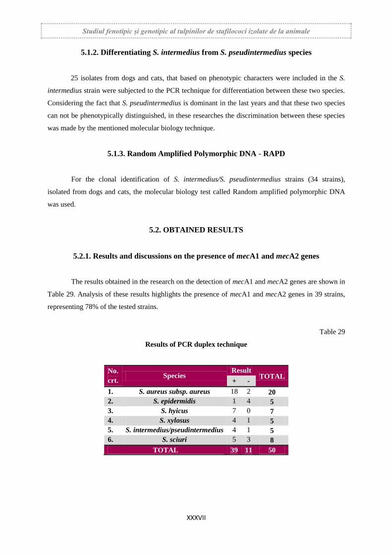
can not be phenotypically distinguished, in these researches the discrimination between these species
was made by the mentioned molecular biology technique.
5.1.3. Random Amplified Polymorphic DNA - RAPD
For the clonal identification of S. intermedius/S. pseudintermedius strains (34 strains),
isolated from dogs and cats, the molecular biology test called Random amplified polymorphic DNA
was used.
5.2. OBTAINED RESULTS
5.2.1. Results and discussions on the presence of mecA1 and mecA2 genes
The results obtained in the research on the detection of mecA1 and mecA2 genes are shown in
Table 29. Analysis of these results highlights the presence of mecA1 and mecA2 genes in 39 strains,
representing 78% of the tested strains.
Table 29
Results of PCR duplex technique
No.
crt. Species
Result TOTAL
+ -
1. S. aureus subsp. aureus 18 2 20
2. S. epidermidis 1 4 5
3. S. hyicus 7 0 7
4. S. xylosus 4 1 5
5. S. intermedius/pseudintermedius 4 1 5
6. S. sciuri 5 3 8
TOTAL 39 11 50
Studiul fenotipic și genotipic al tulpinilor de stafilococi izolate de la animale
XXXVIII
5.2.2. Results and discussions on the differentiation of S. intermedius and S.
pseudintermedius species
For the genotypic differentiation 25 strains of staphylococci were tested, of which 22 strains
were isolated from dogs and 3 strains were isolated from cats.
Using this technique of molecular biology and by using two specific primers, 9 strains (36%)
were included in S. intermedius species, 6 strains isolated from dogs and 3 strains isolated from cats,
while 16 strains, respectively 64%, were included in S. pseudintermedius species, all of them being
isolated only from dogs.
5.2.3. Results and discussions on Random Amplified Polymorphic DNA – RAPD
34 strains of S. intermedius/S. pseudintermedius were tested through this technique of
molecular biology, respectively 22 strains of canine origin and 12 strains of feline origin. Genetic
imprinting by this technique was performed with P1 and P2 primers on genomic DNA molecules
extracted from the 34 strains.
S. intermedius/S. pseudintermedius strains, isolated from dogs (22 strains) are
phylogenetically distant from the feline strains (12 strains), each having its own cluster, respectively
different origins.
S. intermedius/S. pseudintermedius strains, isolated from cats, have similar genetic
imprintings of the same origin, demonstrating their phylogenetic affinity.
The results achieved, in its own research, covered in this chapter were completed by a total of
8 conclusions.
CHAPTER 6. RESEARCH ON THE FREQUENCY OF ANTIBIOTIC
RESISTANT PHENOTYPES OF STAPHYLOCOCI STRAINS
Antibiotic resistance to bacteria is a major issue in veterinary and human medicine, because it
is considered a pronounced zoonotic risk phenomenon. The resistance phenotypes to pathogenic
bacteria for animals, including staphylococci, have a growing frequency due to the use of veterinary
medicinal products, based on antibiotics, especially in animals raised in intensive system.
The researches carried out in this chapter aimed to establish the phenotypic antibiotic
resistance, namely the establishment of resistance phenotypes frequency in staphylococci strains
isolated animals.
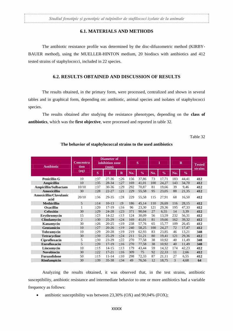
Studiul fenotipic și genotipic al tulpinilor de stafilococi izolate de la animale
XXXIX
6.1. MATERIALS AND METHODS
The antibiotic resistance profile was determined by the disc-difuzometric method (KIRBY-
BAUER method), using the MUELLER-HINTON medium, 20 biodiscs with antibiotics and 412
tested strains of staphylococci, included in 22 species.
6.2. RESULTS OBTAINED AND DISCUSSION OF RESULTS
The results obtained, in the primary form, were processed, centralized and shown in several
tables and in graphical form, depending on: antibiotic, animal species and isolates of staphylococci
species.
The results obtained after studying the resistance phenotypes, depending on the class of
antibiotics, which was the first objective, were processed and reported in table 32.
Table 32
The behavior of staphylococcal strains to the used antibiotics
Antibiotic
Concentra
tion
(µg)
Diameter of
inhibition zone
(mm)
S I R Tested
strains
S I R No. % No. % No. %
Penicillin G 10 ≥37 27-36 ≤26 156 37,86 73 17,71 183 44,41 412
Ampicillin 10 ≥35 28-34 ≤27 169 41,01 100 24,27 143 34,70 412
Ampicillin/Sulbactam 10/10 ≥37 30-36 ≤29 292 70,87 81 19,66 39 9,46 412
Amoxicillin 30 ≥28 22-27 ≤21 229 55,58 95 23,05 88 21,35 412
Amoxicillin/Clavulanic
acid 20/10 ≥36 29-35 ≤28 229 55,58 115 27,91 68 16,50 412
Methicillin 5 ≥14 10-13 ≤9 186 45,14 110 26,69 116 28,15 412
Oxacillin 1 ≥20 17-19 ≤16 96 23,30 121 29,36 195 47,33 412
Cefoxitin 30 ≥29 24-28 ≤23 371 90,04 27 6,55 14 3,39 412
Erythromycin 15 ≥23 14-22 ≤13 124 30,09 56 13,59 232 56,31 412
Clindamycin 2 ≥30 25-29 ≤24 169 41,01 81 19,66 162 39,32 412
Kanamycin 30 ≥26 20-25 ≤19 238 57,76 65 15,77 109 26,45 412
Gentamicin 10 ≥27 20-26 ≤19 240 58,25 100 24,27 72 17,47 412
Tobramycin 10 ≥29 20-28 ≤19 219 62,93 83 23,85 46 13,21 348
Tetracycline 30 ≥30 25-29 ≤24 211 51,21 80 19,41 121 29,36 412
Ciprofloxacin 5 ≥30 23-29 ≤22 270 77,58 38 10,92 40 11,49 348
Enrofloxacin 5 ≥20 17-19 ≤16 270 77,58 38 10,92 40 11,49 348
Lincomycin 10 ≥15 14-15 ≤13 179 43,44 59 14,32 174 42,23 412
Novobiocin 30 ≥22 17-21 ≤16 309 75 92 22,33 11 2,66 412
Furazolidone 50 ≥15 11-14 ≤10 298 72,33 87 21,11 27 6,55 412
Rimfampicin 30 ≥39 35-38 ≤34 49 76,56 12 18,75 3 4,68 64
Analyzing the results obtained, it was observed that, in the test strains, antibiotic
susceptibility, antibiotic resistance and intermediate behavior to one or more antibiotics had a variable
frequency as follows:
antibiotic susceptibility was between 23,30% (OX) and 90,04% (FOX);
Studiul fenotipic și genotipic al tulpinilor de stafilococi izolate de la animale
XL
antibiotic resistance was between 2,66% (NO) and 56,31% (E);
intermediate behavior was between 6,55% (FOX) and 29,36% (OX).
The second objective pursued in these researches was the behavior of staphylococcal strains
and the tested antibiotics, depending on the species of animals from which these strains were isolated.
The results showed the presence of the antibiotic resistance phenomenon in the coagulase-
positive and coagulase negative staphylococci strains isolated from both livestock animals and pets.
Staphylococci strains with multiple antibiotic resistance have a growing frequency and the resistance
phenotypes to antibiotics used in therapy show the continued spread of this phenomenon through a
two-way animal-human epidemiological circuit.
The third objective pursued in these researches was represented the frequency of resistance
phenotypes for each species of staphylococci. To monitor the frequency of these phenotypes,
several intervals were established, limits of which were expressed in percentage. Also, the isolates of
staphylococci were grouped according to the production of free coagulation, respectively in the group
of positive coagulase staphylococci and the group of negative coagulase staphylococci.
The frequency of resistance phenotypes from positive coagulase staphylococci is shown
in table 34Following the analysis of the data presented in this table, there are some practical
observations on:
• the frequency of resistance phenotypes;
• the number of resistance phenotypes;
• their significance for the use of antibiotics in the treatment of infections produced by
positive coagulase staphylococci.
The frequency of resistance phenotypes from negative coagulase staphylococci are shown
in table 35. The data presented in this table outline some aspects of the frequency and number of
resistance phenotypes identified in the strains of staphylococci belonging to these species.
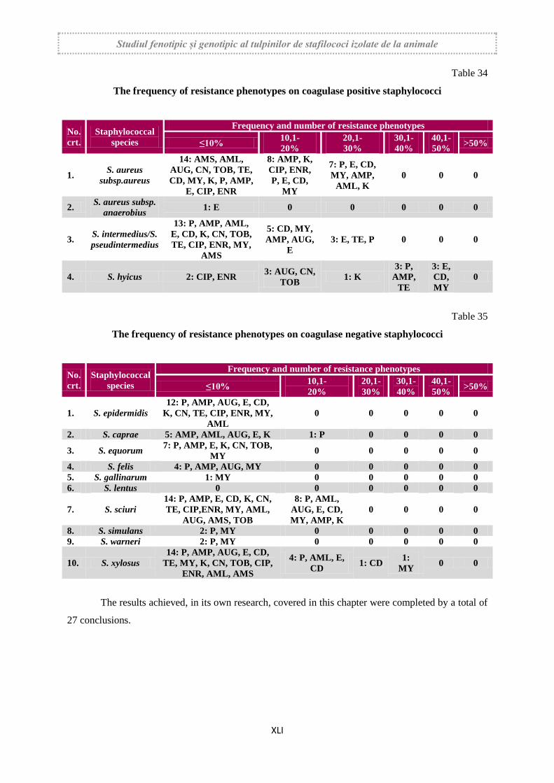
Studiul fenotipic și genotipic al tulpinilor de stafilococi izolate de la animale
XLI
Table 34
The frequency of resistance phenotypes on coagulase positive staphylococci
No.
crt.
Staphylococcal
species
Frequency and number of resistance phenotypes
≤10% 10,1-
20%
20,1-
30%
30,1-
40%
40,1-
50% >50%
1. S. aureus
subsp.aureus
14: AMS, AML,
AUG, CN, TOB, TE,
CD, MY, K, P, AMP,
E, CIP, ENR
8: AMP, K,
CIP, ENR,
P, E, CD,
MY
7: P, E, CD,
MY, AMP,
AML, K
0 0 0
2. S. aureus subsp.
anaerobius 1: E 0 0 0 0 0
3. S. intermedius/S.
pseudintermedius
13: P, AMP, AML,
E, CD, K, CN, TOB,
TE, CIP, ENR, MY,
AMS
5: CD, MY,
AMP, AUG,
E
3: E, TE, P 0 0 0
4. S. hyicus 2: CIP, ENR 3: AUG, CN,
TOB 1: K
3: P,
AMP,
TE
3: E,
CD,
MY
0
Table 35
The frequency of resistance phenotypes on coagulase negative staphylococci
No.
crt.
Staphylococcal
species
Frequency and number of resistance phenotypes
≤10% 10,1-
20%
20,1-
30%
30,1-
40%
40,1-
50% >50%
1. S. epidermidis
12: P, AMP, AUG, E, CD,
K, CN, TE, CIP, ENR, MY,
AML
0 0 0 0 0
2. S. caprae 5: AMP, AML, AUG, E, K 1: P 0 0 0 0
3. S. equorum 7: P, AMP, E, K, CN, TOB,
MY 0 0 0 0 0
4. S. felis 4: P, AMP, AUG, MY 0 0 0 0 0
5. S. gallinarum 1: MY 0 0 0 0 0
6. S. lentus 0 0 0 0 0 0
7. S. sciuri
14: P, AMP, E, CD, K, CN,
TE, CIP,ENR, MY, AML,
AUG, AMS, TOB
8: P, AML,
AUG, E, CD,
MY, AMP, K
0 0 0 0
8. S. simulans 2: P, MY 0 0 0 0 0
9. S. warneri 2: P, MY 0 0 0 0 0
10. S. xylosus
14: P, AMP, AUG, E, CD,
TE, MY, K, CN, TOB, CIP,
ENR, AML, AMS
4: P, AML, E,
CD 1: CD
1:
MY 0 0
The results achieved, in its own research, covered in this chapter were completed by a total of
27 conclusions.
Studiul fenotipic și genotipic al tulpinilor de stafilococi izolate de la animale
XLII
CHAPTER 7. RESEARCH REGARDING THE FREQUENCY OF
METHICILLIN RESISTANT STRAINS
Particular attention is paid, in recent years, to methicillin-resistant staphylococci strains,
generically known as MRSA staphylococci (Methicillin Resistant Staphylococcus aureus). These
strains have a pronounced zoonotic risk and a complex epidemiological circuit, being common in
humans as well. Also, resistance to methicillin is associated with multiple antibiotic resistance,
especially resistance to penicillins, cephalosporins and aztreonam
Existing data in the literature demonstrate that methicillin resistance is present in both
coagulase positive and negative coagulase staphylococci. Expansion of methicillin resistance, from
strains of S. aureus subsp. aureus to other staphylococci species, is generated by the Staphylococcal
Chromosome Cassete mec- SCCmec transmission mechanisms, present in the mobile genetic elements
of the staphylococcal cytoplasm.
7.1. MATERIALS AND METHODS
The frequency of methicillin resistant strains was pursued at 412 strains of staphylococci,
included in 22 species. The frequency of methicillin resistant strains was phenotypically established
by two tests and confirmed at 50 strains by the polymerase chain reaction.
The frequency of resistant methicillin strains was determined phenotypically by the disc-
difuzimetric method (Kirby-Bauer) using methicillin (5 μg), oxacillin (1 μg) and cefoxitin (30 μg).
A number of 210 positive and negative coagulase strains were selected to be passed on the
Chromatic MRSA medium according to the following criteria:
presented phenotypic resistance to at least one of the three antibiotics mentioned;
had intermediate behavior in at least two of those three antibiotics.
7.2. OBTAINED RESULTS
The results obtained, shown in figure 87, show the frequency of resistant methicillin strains
detected by the disc-diffusometric method, from the total strains of tested staphylococci and
depending on the species of animals.
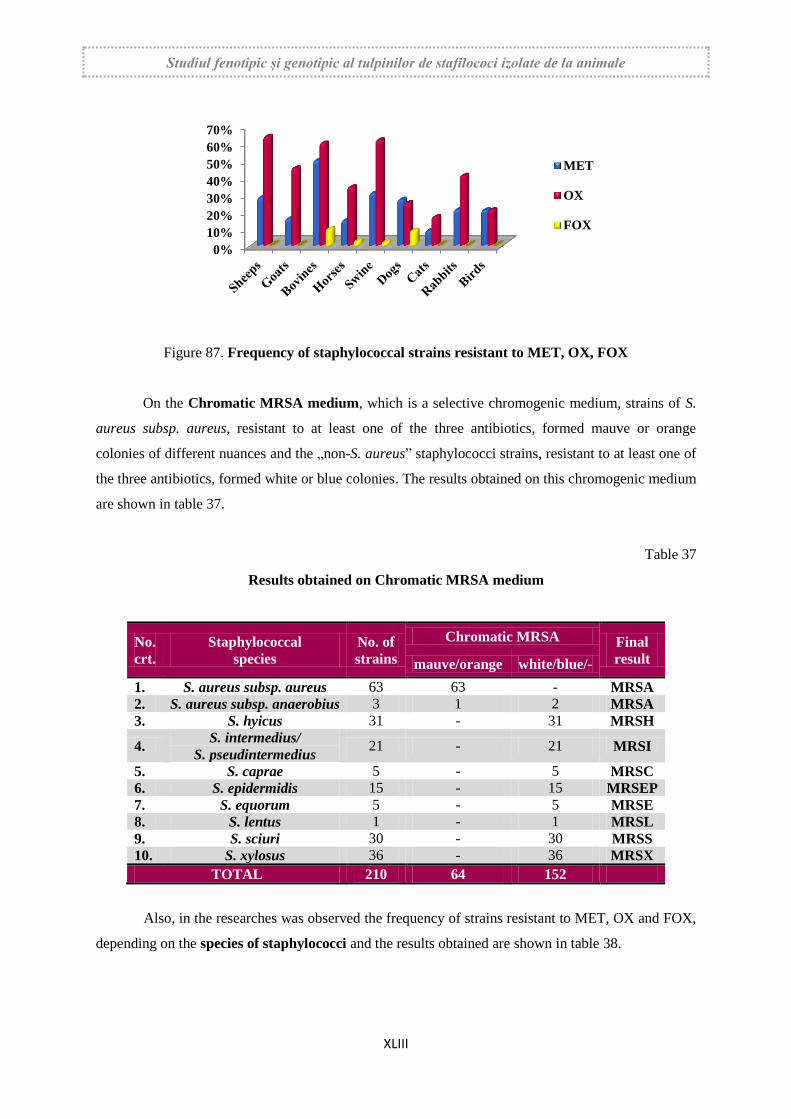
Studiul fenotipic și genotipic al tulpinilor de stafilococi izolate de la animale
XLIII
Figure 87. Frequency of staphylococcal strains resistant to MET, OX, FOX
On the Chromatic MRSA medium, which is a selective chromogenic medium, strains of S.
aureus subsp. aureus, resistant to at least one of the three antibiotics, formed mauve or orange
colonies of different nuances and the „non-S. aureus” staphylococci strains, resistant to at least one of
the three antibiotics, formed white or blue colonies. The results obtained on this chromogenic medium
are shown in table 37.
Table 37
Results obtained on Chromatic MRSA medium
No.
crt.
Staphylococcal
species
No. of
strains
Chromatic MRSA Final
result mauve/orange white/blue/-
1. S. aureus subsp. aureus 63 63 - MRSA
2. S. aureus subsp. anaerobius 3 1 2 MRSA
3. S. hyicus 31 - 31 MRSH
4. S. intermedius/
S. pseudintermedius 21 - 21 MRSI
5. S. caprae 5 - 5 MRSC
6. S. epidermidis 15 - 15 MRSEP
7. S. equorum 5 - 5 MRSE
8. S. lentus 1 - 1 MRSL
9. S. sciuri 30 - 30 MRSS
10. S. xylosus 36 - 36 MRSX
TOTAL 210 64 152
Also, in the researches was observed the frequency of strains resistant to MET, OX and FOX,
depending on the species of staphylococci and the results obtained are shown in table 38.
0%
10%
20%
30%
40%
50%
60%
70%
MET
OX
FOX
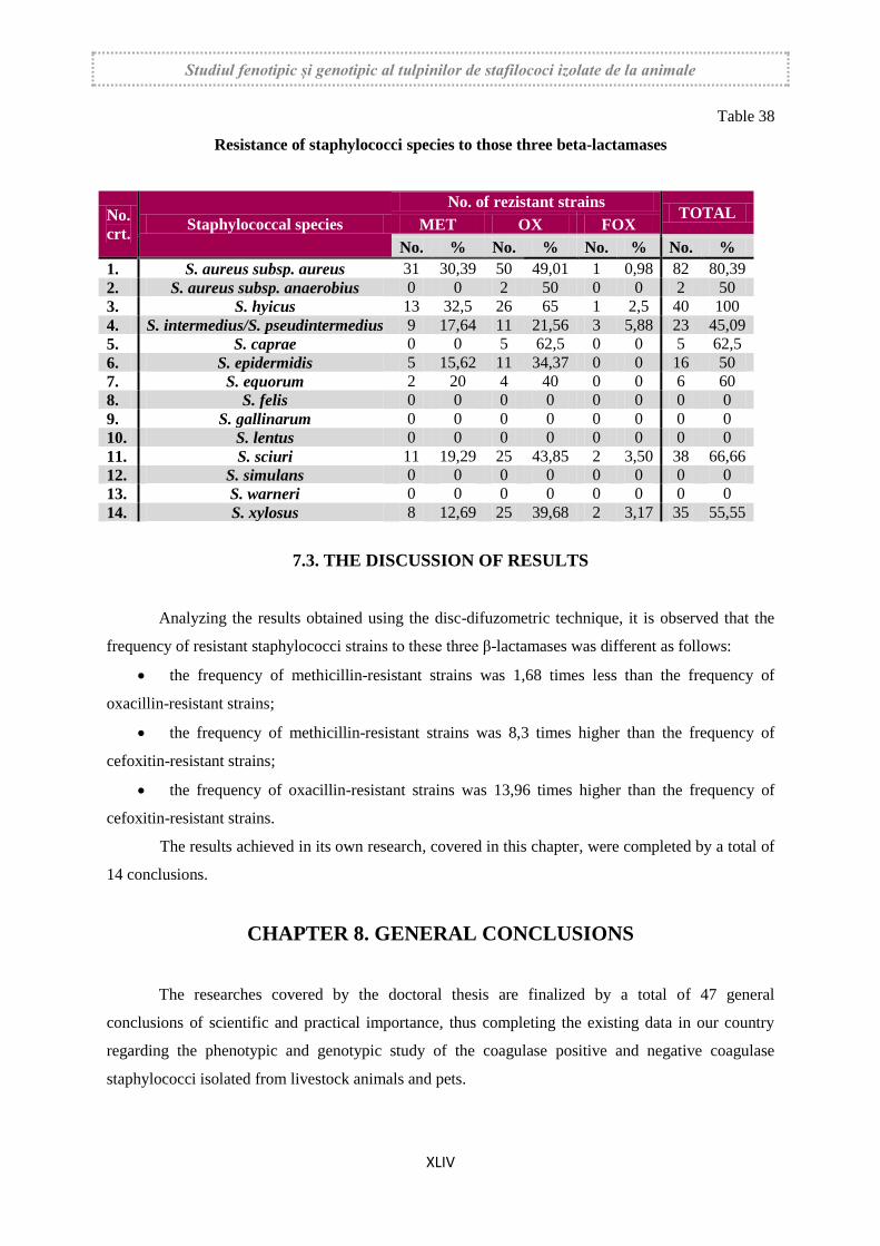
Studiul fenotipic și genotipic al tulpinilor de stafilococi izolate de la animale
XLIV
Table 38
Resistance of staphylococci species to those three beta-lactamases
No.
crt. Staphylococcal species
No. of rezistant strains TOTAL
MET OX FOX
No. % No. % No. % No. %
1. S. aureus subsp. aureus 31 30,39 50 49,01 1 0,98 82 80,39
2. S. aureus subsp. anaerobius 0 0 2 50 0 0 2 50
3. S. hyicus 13 32,5 26 65 1 2,5 40 100
4. S. intermedius/S. pseudintermedius 9 17,64 11 21,56 3 5,88 23 45,09
5. S. caprae 0 0 5 62,5 0 0 5 62,5
6. S. epidermidis 5 15,62 11 34,37 0 0 16 50
7. S. equorum 2 20 4 40 0 0 6 60
8. S. felis 0 0 0 0 0 0 0 0
9. S. gallinarum 0 0 0 0 0 0 0 0
10. S. lentus 0 0 0 0 0 0 0 0
11. S. sciuri 11 19,29 25 43,85 2 3,50 38 66,66
12. S. simulans 0 0 0 0 0 0 0 0
13. S. warneri 0 0 0 0 0 0 0 0
14. S. xylosus 8 12,69 25 39,68 2 3,17 35 55,55
7.3. THE DISCUSSION OF RESULTS
Analyzing the results obtained using the disc-difuzometric technique, it is observed that the
frequency of resistant staphylococci strains to these three β-lactamases was different as follows:
the frequency of methicillin-resistant strains was 1,68 times less than the frequency of
oxacillin-resistant strains;
the frequency of methicillin-resistant strains was 8,3 times higher than the frequency of
cefoxitin-resistant strains;
the frequency of oxacillin-resistant strains was 13,96 times higher than the frequency of
cefoxitin-resistant strains.
The results achieved in its own research, covered in this chapter, were completed by a total of
14 conclusions.
CHAPTER 8. GENERAL CONCLUSIONS
The researches covered by the doctoral thesis are finalized by a total of 47 general
conclusions of scientific and practical importance, thus completing the existing data in our country
regarding the phenotypic and genotypic study of the coagulase positive and negative coagulase
staphylococci isolated from livestock animals and pets.