Departamentul de Calculatoare şi Tehnologia Informației ...
149
Proiect PROSCIENCE - POSDRU/187/1.5/S/155420 Promovarea științei și calității în cercetare prin burse doctorale UNIVERSITATEA POLITEHNICA DIN BUCUREŞTI Facultatea de Automatică şi Calculatoare Departamentul de Calculatoare şi Tehnologia Informației TEZĂ DE DOCTORAT Soluții Bazate pe Realitate Virtuală şi Augmentată in Domeniul Medical Solutions Based on Virtual and Augmented Reality in Healthcare Autor: Oana Alexandra Voinea Conducător de doctorat: Prof. Dr. Ing. Florica Moldoveanu COMISIA DE DOCTORAT Președinte Prof. dr. ing. Adina Magda Florea de la Universitatea POLITEHNICA din București Conducător de doctorat Prof. dr. ing. Florica Moldoveanu de la Universitatea POLITEHNICA din București Referent Profesor dr. ing. Vasile Manta de la Universitatea Tehnică ”Gheorghe Asachi” din Iași Referent Profesor dr. ing. Ștefan-Gheorghe Pentiuc de la Universitatea ”Ștefan cel Mare” din Suceava Referent Profesor dr. ing. Nirvana Popescu de la Universitatea POLITEHNICA din București București 2018
Transcript of Departamentul de Calculatoare şi Tehnologia Informației ...
Proiect PROSCIENCE - POSDRU/187/1.5/S/155420 Promovarea științei și calității în cercetare prin burse doctorale
UNIVERSITATEA POLITEHNICA DIN BUCUREŞTI
Facultatea de Automatică şi Calculatoare
Departamentul de Calculatoare şi Tehnologia Informației
TEZĂ DE DOCTORAT Soluții Bazate pe Realitate Virtuală şi Augmentată in Domeniul Medical
Solutions Based on Virtual and Augmented Reality in Healthcare
Autor: Oana Alexandra Voinea
Conducător de doctorat: Prof. Dr. Ing. Florica Moldoveanu
COMISIA DE DOCTORAT
Președinte Prof. dr. ing. Adina Magda Florea de la Universitatea POLITEHNICA din București
Conducător de doctorat Prof. dr. ing. Florica Moldoveanu de la Universitatea POLITEHNICA din București
Referent Profesor dr. ing. Vasile Manta de la Universitatea Tehnică ”Gheorghe Asachi” din Iași
Referent Profesor dr. ing. Ștefan-Gheorghe
Pentiuc
de la Universitatea ”Ștefan cel Mare” din Suceava
Referent Profesor dr. ing. Nirvana Popescu de la Universitatea POLITEHNICA din București
București 2018
Solutions Based on Virtual and Augmented Reality in Healthcare
1
Acknowledgements
Firstly, I would like to express my sincere gratitude to my advisor Prof. Florica
Moldoveanu for accepting me for this PhD and giving me the opportunity to work on
diverse projects. I am grateful for her patience and understanding regarding career and
personal events that had impact on the PhD timeline. Her guidance helped me in all the
time of research and writing of this thesis and it has been an honor to meet her and to be
her PhD student.
My sincere thanks also go to Prof. Alin Moldoveanu, who provided me the opportunity
to join the TRAVEE team and who gave access to the research facilities. I truly appreciate
his contribution of time and ideas to make my PhD experience stimulating.
I would like to thank Dr. Victor Asavei, Dr. Anca Morar and Oana Ferche, for their help,
collaboration or useful insights during my time at the University POLITEHNICA of
Bucharest. Also, I would like to thank Mrs. Catalina Daraban from the doctoral school office
which is always helpful with information and paperwork.
Also, I want to thank Mr. Ionut Toma and Ms. Laura Barbulescu for being such great friends. They landed me the Kinect device that I used in the experiments described in this thesis and supported me during stressful times.
Last but not the least, I would like to thank my family. To my husband Radu to which I cannot say in perfect words how much I appreciate his help. He stayed by my side during all these years, motivated me and proofread my articles (sometimes at 4 AM). This PHD changed our lives in the last 4 years and he supported me all this time. Also, I would like to thank my mother for her guidance. She always told me how important the education is and that is the way to a better life. Growing up in a low-income family in one of the poorest towns of Romania this advice made me learn more and to have a life better that I have imagined.
This work was partially funded by TRAVEE grant of the Romanian executive agency
for higher education, research, development and innovation funding - UEFISCDI, joint
applied research projects program, 1/2014(PN-II-PT-PCCA-2013-4-1580) and the Sectoral
Operational Program Human Resources Development 2007-2013 of the Ministry of
European Funds through the Financial Agreement POSDRU 187/1.5/S/155420.

Solutions Based on Virtual and Augmented Reality in Healthcare
2
Solutions Based on Virtual and Augmented Reality in Healthcare
3
Rezumat
Această lucrare prezintă soluții bazate pe Realitate Virtuală (VR) și Augmentată (AR) în
domeniul medical, iar accentul este pus pe două domenii de interes: reabilitarea
neuromusculară a pacienților care au suferit un accident vascular cerebral și educația
medicală.
In prima parte a tezei sunt prezentate informații despre tehnologiile curente folosite in
aplicațiile bazate pe realitatea virtuală şi augmentată. In partea a 2-a sunt prezentate
contribuțiile autorului la o soluție ce are ca scop recuperarea neuromotorie prin folosirea
realității virtuale şi a feedback-ului augmentat. In partea a 3-a tezei este propusă o soluție
inovativă pentru domeniul educației medicale, mai exact în studiul biomecanicii, făcând-
se o evaluare a oportunității folosirii atât a realității virtuale cât şi a celei augmentate. Se
prezintă o evaluare a rezultatelor obținute pe baza unui set de chestionare completate de
utilizatorii care au testat aplicațiile dezvoltate. Scopul proiectului a fost de a construi o
soluție cu costuri reduse, cu o experiență adecvată a utilizatorilor, care să poată fi ușor
distribuită și adoptată de un număr mare de persoane.
Conținutul acestei teze este bazat în principal pe elemente practice și conține mai
multe rezultate experimentale obținute în timpul testelor efectuate folosind diverse
tehnologii. S-a urmărit dezvoltarea unor soluții competitive pe cele mai recente
tehnologii, acolo unde a fost posibil.

Solutions Based on Virtual and Augmented Reality in Healthcare
4
Solutions Based on Virtual and Augmented Reality in Healthcare
5
Abstract
This thesis presents solutions based on Virtual and Augmented Reality (VR and AR) in
healthcare, analyzing two areas of interest: neuromuscular rehabilitation of stroke
survivors and medical education.
The first part of the thesis presents information related to the current technologies used
in applications based on virtual and augmented reality. In the second part, the author’s
contributions to a neuromotor rehabilitation system that aimed the usage of virtual
reality and augmented feedback are detailed. The third part of the thesis is focused on
the design and implementation of a novel solution that uses both virtual and augmented
reality in medical education, and more specifically the biomechanics study, along with
the assessment of the results obtained after it was tested with a few users. The goal of the
project was to build a low-cost solution with an appropriate user experience that can be
easily distributed and adopted by a large number of persons.
The content of this thesis is predominantly focused on practical elements and it
contains several experimental results obtained while using various technologies. The goal
was to use the latest technology (where it was possible) to be able to provide competitive
solutions.

Solutions Based on Virtual and Augmented Reality in Healthcare
6
Solutions Based on Virtual and Augmented Reality in Healthcare
7
TABLE OF CONTENTS
LIST OF FIGURES ........................................................................................................................ 9
LIST OF TABLES ........................................................................................................................ 12
INTRODUCTION ........................................................................................................................ 13
1.1 MOTIVATION .................................................................................................................. 13
1.2 CONTEXT.......................................................................................................................... 14
1.3 GOALS OF THE RESEARCH ......................................................................................... 16
1.4 SCIENTIFIC PUBLICATIONS IN CONNECTION WITH THE THESIS................. 17
1.5 STRUCTURE OF THE THESIS ....................................................................................... 18
CURRENT TECHNOLOGIES USED IN APPLICATIONS BASED ON VIRTUAL AND AUGMENTED REALITY ........................................................................................................... 20
2.1 BACKGROUND ............................................................................................................... 20
2.2 VIRTUAL AND AUGMENTED REALITY DEVICES ................................................ 24
2.3 HUMAN BODY MOTION TRACKING SENSORS .................................................... 26
2.3.1 Leap Motion ............................................................................................................... 26
2.3.2 Kinect ......................................................................................................................... 28
2.3.3 VicoVR ........................................................................................................................ 29
2.4 CONCLUSIONS ............................................................................................................... 30
ICT SOLUTIONS FOR NEUROMOTOR REHABILITATION ................................................ 32
3.1 RELATED WORK ............................................................................................................ 32
3.1.1 Rehabilitation Devices .............................................................................................. 32
3.1.2 3D Visualization Solutions ...................................................................................... 35
3.2 CONTRIBUTIONS ........................................................................................................... 37
3.2.1 Avatar Personalization ............................................................................................. 37
3.2.2 Virtual Reality Display ............................................................................................. 42
3.2.3 Motion Tracking ........................................................................................................ 44
3.3 CONCLUSIONS ............................................................................................................... 48
AUGMENTED AND VIRTUAL REALITY IN MEDICAL EDUCATION .............................. 50
4.1 RELATED WORK ............................................................................................................ 50
4.2 CONTRIBUTIONS ........................................................................................................... 53
Solutions Based on Virtual and Augmented Reality in Healthcare
8
4.2.1 Approach .................................................................................................................... 54
4.2.2 3D Models .................................................................................................................. 57
4.2.3 Tests with different VR/AR technologies ............................................................. 71
4.2.4 Interactive Biomechanics Lessons (IBL) project ................................................... 82
4.2.5 Implementation Details ............................................................................................ 83
4.2.6 Performance Analysis ............................................................................................. 108
4.2.7 Users Questionnaires Results ................................................................................ 115
4.3 CONCLUSIONS ............................................................................................................. 126
CONCLUSIONS AND FUTURE WORK ................................................................................. 129
5.1 THE ORIGINAL CONTRIBUTIONS OF THIS THESIS ........................................... 129
5.2 CONCLUSIONS ............................................................................................................. 130
5.3 FUTURE PERSPECTIVES ............................................................................................. 131
Acronyms .................................................................................................................................... 132
Bibliography ............................................................................................................................... 133
Appendices .................................................................................................................................. 141

Solutions Based on Virtual and Augmented Reality in Healthcare
9
LIST OF FIGURES
Figure 2.1 – Real - Virtual environment transition inspired from Virtuality Continuum schema [AV16b]: A. Real
image, B. Leap Motion Image Hands application, C. Leap Motion Demo application ................................................ 22 Figure 2.2 – Oculus Rift device .................................................................................................................................... 24 Figure 2.3 – Oculus Rift and EEG cap used in Rehabilitation ....................................................................................... 24 Figure 2.4 – Leap Motion Controller ............................................................................................................................ 26 Figure 2.5 – Leap Motion Cameras .............................................................................................................................. 26 Figure 2.6 – Leap Motion Interaction Area .................................................................................................................. 26 Figure 2.7 – Hand bones ............................................................................................................................................. 27 Figure 2.8 – Kinect V1 sensor ....................................................................................................................................... 29 Figure 2.9 – Kinect V1 Skeleton position and Bones hierarchy .................................................................................... 29 Figure 2.10 – VicoVR – tracking sensor in VR setup ..................................................................................................... 30 Figure 2.11 – VicoVR –tracking joints in VR setup ...................................................................................................... 30 Figure 3.1 – TRAVEE workflow that includes the Avatar Personalization ................................................................... 37 Figure 3.2 – Avatar Personalization Interface ............................................................................................................. 38 Figure 3.3 – Textured 3D Models variation for the 5 body types categories: XS [0], S [1], M [2], L [3], XL [4]. ........... 40 Figure 3.4 – Non-textured 3D Model for Height minimum value. ............................................................................... 41 Figure 3.5 – Non-textured 3D Model for Height maximum value................................................................................ 41 Figure 3.6 – TRAVEE VR system setup ......................................................................................................................... 42 Figure 3.7 – Scene example on Oculus Rift .................................................................................................................. 44 Figure 3.8 – Examples of a TVM and PVM scene configuration. ................................................................................. 44 Figure 3.9 – Kinect and Leap Motion cover areas ........................................................................................................ 46 Figure 3.10 – Hand bones of the 3D model .................................................................................................................. 46 Figure 3.11 – Basic hand movements tracked in real-time with Leap Motion Controller ............................................ 47 Figure 3.12 – Patient and therapist models animated based on Kinect sensor body tracking .................................... 48 Figure 4.1 – Schematic view of the reviewed scientific papers topics ......................................................................... 51 Figure 4.2 – Initial proposed system overview – AR based .......................................................................................... 54 Figure 4.3 – Proposed application divided in subsystems ............................................................................................ 55 Figure 4.4 – System overview to support VR and AR ................................................................................................... 57 Figure 4.5 – OSIRIX samples: OBELIX [A, B], PETCENIX [C, D], MELANIX [E, F, G] ........................................................ 60 Figure 4.6 – 3D model preview of the muscles and skeleton as obtained from the HTLL dataset ............................... 61 Figure 4.7 – HTLL dataset - initial image ..................................................................................................................... 62 Figure 4.8 – HTLL dataset – improved image to emphasize the bones tissue ............................................................ 62 Figure 4.9 – LL dataset – masks (orange for skin, blue for muscles) ............................................................................ 63 Figure 4.10 – ULH dataset skin model correction ........................................................................................................ 64 Figure 4.11 – Complete skin model .............................................................................................................................. 64 Figure 4.12 – Bones meshes import ............................................................................................................................. 65 Figure 4.13 – Model’s correction ................................................................................................................................. 65 Figure 4.14 – Final model............................................................................................................................................. 65 Figure 4.15 – Merging meshes .................................................................................................................................... 65 Figure 4.16 – Make Human basic skeleton .................................................................................................................. 66 Figure 4.17 – Make Human basic skeleton superimposed on bones model. ............................................................... 66 Figure 4.18 – Bones model animation error ................................................................................................................ 66 Figure 4.19 – Rigging and skinning of obtained models using Biped structure from 3DS Max (Left – Bones/Skeleton,
Middle – Skin and Right – Muscles model) .................................................................................................................. 66 Figure 4.20 – 3D Model Generation workflow ............................................................................................................. 67
Solutions Based on Virtual and Augmented Reality in Healthcare
10
Figure 4.21 – A version of the obtained 3D models imported in Unity ........................................................................ 68 Figure 4.22 – Bones model in T-pose ........................................................................................................................... 68 Figure 4.23 – Static bones model of human anatomy ................................................................................................. 70 Figure 4.24 – Static muscles model of human anatomy .............................................................................................. 70 Figure 4.25 – Rigged skin model .................................................................................................................................. 70 Figure 4.26 – Stereoscopic rendering on mobile .......................................................................................................... 71 Figure 4.27 – Setting the device in the viewer ............................................................................................................. 71 Figure 4.28 – Cardboard headset ................................................................................................................................ 71 Figure 4.29 – Graphical User Interface Reticle used for VR applications developed for Cardboard viewer ................ 72 Figure 4.30 – Samsung S6 device connected to a Gear VR headset ............................................................................ 73 Figure 4.31 – Gear VR headset buttons and USB port ................................................................................................. 73 Figure 4.32– HoloLens test– Unity Editor scene ........................................................................................................... 75 Figure 4.33 – HoloLens test – Emulator (Visual Studio Solution) ................................................................................. 75 Figure 4.34 – Lighting conditions effects on test AR scene .......................................................................................... 76 Figure 4.35 – AR scene example - without tracking ..................................................................................................... 76 Figure 4.36 – Combinations of tracking sensors and mobile devices........................................................................... 77 Figure 4.37 – Motion tracking using additional sensors .............................................................................................. 77 Figure 4.38 – Unity scene settings to enable Kinect functionality ............................................................................... 78 Figure 4.39 – Motion tracking using a Kinect sensor ................................................................................................... 79 Figure 4.40 – Motion tracking using a Kinect sensor with the mirror movement corrected using the bones generated
model ........................................................................................................................................................................... 79 Figure 4.41 – Model position changed at runtime on Laptop. ..................................................................................... 81 Figure 4.42 – Model position changes at runtime and was centered to face detection rectangles on Laptop. .......... 81 Figure 4.43 – Model position and scale changed on Nvidia Shield tablet. ................................................................... 81 Figure 4.44 – Motion tracking of an observed user in AR using a Kinect sensor and OpenCV .................................... 81 Figure 4.45 – Opaque model ........................................................................................................................................ 82 Figure 4.46 – Semitransparent bones model ............................................................................................................... 82 Figure 4.47 – Semitransparent bones and muscles models ......................................................................................... 82 Figure 4.48 – VR classroom application – Initial screen ............................................................................................... 84 Figure 4.49 – VR classroom application structure from Unity ..................................................................................... 85 Figure 4.50 – VR classroom application - Imported models presentation menu structure .......................................... 86 Figure 4.51 – VR classroom application - Imported models’ presentation – Skin ........................................................ 86 Figure 4.52 – VR classroom application – Rotation option (yellow rectangle) ............................................................ 87 Figure 4.53 – VR classroom application - Imported models’ presentation – Muscles ................................................. 87 Figure 4.54 – VR classroom application - Imported models’ presentation – Bones ..................................................... 88 Figure 4.55 – VR classroom application – Anatomy Notions – Humerus ..................................................................... 89 Figure 4.56 – VR classroom application – Anatomy Notions – Bones.......................................................................... 89 Figure 4.57 – VR classroom application – Anatomy Notions – Muscles ...................................................................... 89 Figure 4.58 – VR classroom application – Transverse .................................................................................................. 90 Figure 4.59 - VR classroom application – Coronal Plane ............................................................................................. 90 Figure 4.60 – VR classroom application – Sagittal Plane ............................................................................................. 90 Figure 4.61 – VR classroom application – Reference Points – Example A.................................................................... 91 Figure 4.62 – VR classroom application – Reference Points – Example B .................................................................... 92 Figure 4.63 – Movements animations states ............................................................................................................... 93 Figure 4.64 – VR classroom application – Flexion/Extension movement example ...................................................... 94 Figure 4.65 – VR classroom application – Adduction/Abduction movement example ................................................ 94 Figure 4.66 – VR classroom application – Pronation/Supination movement example ................................................ 95 Figure 4.67 – VR BlueSky – Anatomy notions - Bones.................................................................................................. 96 Figure 4.68 – VR BlueSky – Anatomy notions - Muscles .............................................................................................. 96
Solutions Based on Virtual and Augmented Reality in Healthcare
11
Figure 4.69 – VR BlueSky – Reference points ............................................................................................................... 96 Figure 4.70 – VR BlueSky – Movements - Flexion ........................................................................................................ 96 Figure 4.71 – VR BlueSky – Movements - Abduction ................................................................................................... 96 Figure 4.72 – Marker-based AR – Target image detection (Screenshot from the device on left). ............................... 97 Figure 4.73 – Lessons in marker-based AR application – Bones Model (Left) and Muscles Model (Right) .................. 98 Figure 4.74 – Lessons in marker-based AR application – Anatomy Notions - Bones (Left) and Muscles (Right) ......... 99 Figure 4.75 – Lessons in marker-based AR application – Planes/Axes - Transverse (Left) and Coronal (Right) ........ 100 Figure 4.76 – Lessons in marker-based AR application – Sagittal Plane (Left) and Reference Points (Right) ............ 101 Figure 4.77 – Lessons in marker-based AR application – Movements – Flexion (Left) and Extension (Right) ........... 102 Figure 4.78 – Lessons in marker-based AR application – Movements – Abduction (Left) and Adduction (Right) ..... 103 Figure 4.79 – Marker-based AR application –Extended tracking feature .................................................................. 104 Figure 4.80 – Markerless AR application – Muscles and bones models visible, scaled and positioned based on face
detection (Laptop) ..................................................................................................................................................... 105 Figure 4.81 – Markerless AR application – Only muscles model is visible ................................................................. 106 Figure 4.82 – Markerless AR application – Only bones model is visible as displayed on the mobile device .............. 106 Figure 4.83 – Markerless AR application – Muscles and bones models visible – Muscle highlighted (Laptop) ......... 107 Figure 4.84 – Markerless AR application – Muscles and bones models visible – Bone highlighted (Mobile device) . 107 Figure 4.85 – Performance metrics for VR with classroom background scenario ..................................................... 109 Figure 4.86 – Performance metrics for VR with no background scenario .................................................................. 110 Figure 4.87 – Performance metrics for AR marker-based scenario ........................................................................... 110 Figure 4.88 – Performance metrics for AR markerless on mobile device (no Kinect data) - Haar classifier .............. 111 Figure 4.89 – AR markerless performance metrics – OpenCV, no Kinect, on Windows workstation ......................... 112 Figure 4.90 – AR markerless application performance metrics – OpenCV face detection on mobile device ............. 113 Figure 4.91 – AR markerless application performance metrics – OpenCV face detection on Windows workstation 113 Figure 4.92 – AR markerless application performance metrics – Kinect(idle) and OpenCV motion tracking on
Windows workstation ................................................................................................................................................ 113 Figure 4.93 – AR markerless application performance metrics – Kinect skeletal tracking and OpenCV face detection
on Windows workstation using models generated from medical images ................................................................. 114 Figure 4.94 – AR markerless application performance metrics – Kinect skeletal tracking and OpenCV face detection
on Windows workstation using the imported models. Blue arrow indicated where the motion tracking has started.
................................................................................................................................................................................... 114 Figure 4.95 – Results assessment approach .............................................................................................................. 117 Figure 4.96 – Simulator Sickness Questionnaire - Symptoms variation ..................................................................... 118 Figure 4.97 – Simulator Sickness Questionnaire – Nausea Symptoms ...................................................................... 119 Figure 4.98 – Simulator Sickness Questionnaire – Oculomotor Symptoms ............................................................... 119 Figure 4.99 – Simulator Sickness Questionnaire– Disorientation Symptoms ............................................................. 120 Figure 4.100 – Simulator Sickness Questionnaire Symptoms Overall ........................................................................ 120 Figure 4.101 – Presence Questionnaires - Realism subscale...................................................................................... 121 Figure 4.102 – Presence Questionnaires - Affordance to act subscale ...................................................................... 122 Figure 4.103 – Presence Questionnaires - Interface Quality subscale ....................................................................... 122 Figure 4.104 – Presence Questionnaires - Affordance to examine subscale.............................................................. 123 Figure 4.105 – Presence Questionnaires - Self-Evaluation of Performance subscale ................................................ 123 Figure 4.106 – Presence Questionnaires - Overall Score ............................................................................................ 124 Figure 4.107 – Users’ feedback regarding the lack of interruptions during VR applications testing ......................... 126
Solutions Based on Virtual and Augmented Reality in Healthcare
12
LIST OF TABLES
Table 2.1 – Characteristics of top VR and AR devices .................................................................................................. 25 Table 3 1 – Age, Height, Weight values intervals for the selected models .................................................................. 39 Table 3.2 – Naming correspondence for the RiggedHand script ................................................................................. 46 Table 4.1 – ScanIP hardware requirements ................................................................................................................. 60 Table 4.2 – Key performance metrics highlights on the mobile device (Samsung S6) ............................................... 109 Table 4.3 – Key performance metrics highlights on AR markerless application ........................................................ 112
Solutions Based on Virtual and Augmented Reality in Healthcare
13
CHAPTER 1
INTRODUCTION
This research focuses on solutions based on virtual (VR) and augmented reality (AR) and
complex sensors in physical rehabilitation and medical education. VR and AR are known
subjects for some time and in the recent years they had popularity surges. The market is
filled with low-cost, performant hardware solutions that enable the users to see a
different type of content even in their home’s commodity. This can be seen as an
opportunity on multiple domains as VR and AR can be successfully applied on medical,
military, manufacturing, entertainment and games, robotics, education, marketing,
tourism and many other fields [MM14]. Our research tackles their applicability in the
healthcare field: the first part of the research is focused on stroke survivors’ rehabilitation
using virtual reality and the second part proposes a novel solution based on VR and AR
in medical education, more exactly for biomechanics study.
1.1 MOTIVATION
The academic background of the author is in computer science and medical engineering.
Also, the author has extensive professional experience with 3D rendering used in mobile
games and in other complex systems. The thesis is a sum of the previous experience, as
the solutions proposed have a strong connection with the mobile environment and its
usability in healthcare. The research is multidisciplinary and aims to add novelty in the
medical education field and rehabilitation, based on the previous experience and studies
since many rehabilitation and learning solutions use game-like visualization to attract the
users’ attention and to maximize the results.
Looking at VR and AR, the hardware market is expected to continuously expand 20-
fold in the 2015-2021 period where the most productive one should be 2021 with an
estimate of 82.5 million headsets shipped. Goldman Sachs expects high surges in a few
key sectors regarding the software market for VR and AR, such as: videogames, live
events, video entertainment, healthcare, real estate, retail, education, engineering and
defense where it forecasts that until 2025 the biggest two markets will be entertainment
and healthcare1. This proves the high interest into developing performant user friendly
applications that can reach to a high number of persons with a moderate cost. Investing
now in research in this domain can boost the market revenue in a few years if consistent
applications are delivered to the public.
1 http://cdn.instantmagazine.com/upload/4666/bom_vrar_2017reportpdf.68ec9bc00f1c.pdf
Solutions Based on Virtual and Augmented Reality in Healthcare
14
There still are predictions made for the impact of AR versus VR. If initially it was
thought that VR will be the one leading the market, the recent experience had proved us
that there is a slight shift to AR2. A part of this research targets to gather more information
regarding the impact of each technology based on the users’ feedback on a medical
education solution developed both in VR and AR. Since VR has still a few unresolved
problems such as cybersickness, the experiments were designed so that they take these
factors into account.
The research is composed mostly of practice based experimental results. The solutions
covered into this thesis aim to be easily adopted by the reader to reproduce the expected
behavior targeting new technology and recent state of the art data.
The outcome is the contribution to the field knowledge with advantages and
disadvantages of the chosen approaches. The results presented in this thesis were
disseminated at various international conferences and the approached methods had a
general positive feedback. The solutions built are complex as they have additional
features to enhance the users’ immersion into the applications developed by using real-
time motion tracking or realistic 3D models.
1.2 CONTEXT
The research is divided in two main parts: the first one is related to the author’s
contributions to the TRAVEE (Virtual Therapist Through Augmented Feedback) project
and the second one presents a solution based on both virtual and augmented reality for
improving the learning process of biomechanics study. Both parts have similarities such
as working with virtual reality, motion tracking devices and with accent on the human
motion biomechanics.
The first part of the research focuses on a novel rehabilitation solution named TRAVEE
that aimed to aid the stroke survivors with neuromotor deficiencies. Every two seconds
someone somewhere in the world is having a stroke and every 10 seconds a life is claimed
where 80% of all the people that suffered a stroke are from low and mid-income
countries3. Stroke survivors often remain incapacitated due to the lack of oxygen and
nutrients for the affected brain area. An obstruction that lasts even a few minutes can
damage the neurons and therefore they die. The functions that were handled by these
neurons are affected and the neuromotor disabilities have the biggest incidence. Thanks
to the neuroplasticity of the brain the functions that were executed by the affected
2 https://www.digi-capital.com/news/2017/01/after-mixed-year-mobile-ar-to-drive-108-billion-vrar-market-by-2021/#.WhQ8nzdx2Uk 3 http://www.worldstrokecampaign.org/learn/facts-and-figures.html
Solutions Based on Virtual and Augmented Reality in Healthcare
15
neurons can be relearned and their function can be taken over by other healthy neurons
from vicinity [OF15].
Neuromotor disabilities can significantly affect a person’s life, especially the activities
of daily living (ADL) like eating or washing. This can downgrade significantly one’s
quality of life as the rehabilitation process is focused on long term kinesiotherapy. The
kinetotherapeutic support (classical therapy) is limited due two facts: the long-time
sessions (up to 5 hours per day) and the growing number of affected persons [AV15c].
The number of specialized personnel is not growing with the same pace and as a result
fewer rehabilitation sessions can be applied to each patient.
The Simulation hypothesis indicates that to relearn a particular movement one has to
visualize the movement either on its own or as an observation due to the strong
connection between the motor and cognitive brain mechanisms [AV15b]. Basically, the
patient can start the rehabilitation very early even if he or she just observes certain
movements as an example to someone else. Since in the early days the patients stay
mostly lied in the hospital bed, it would be very difficult to see the movements executed
by the kinesiotherapist. A novel solution that aimed to be an alternative to other
rehabilitation devices and an adjuvant for the classical therapy is represented by TRAVEE
and each of these parts will be detailed accordingly in the next chapters.
The second part of the research brings into light a new idea regarding the opportunity
of utilizing VR and AR applications in medical education. Their main advantage is that
they can simulate various realistic scenarios effectuated in a safe environment as the
students can be trained on various procedures. The approached subject targets to better
understand the processes behind the human movement biomechanics. The initial idea
was to develop a system that aims to improve medical education learning techniques
using AR or VR and biomechanics seemed a good fit. Both are useful tools for developing
a system that enhances the visual feedback with additional information. In the first
concept the project was focused only on an AR based solution that tracked the
movements of an observed user in order to display an animated 3D model according with
the tracked data. Medical education contains many anatomical notions and this
interactive solution aimed to enhance the user’s attention into the learning process.
Fortunately, with the current software tools available on the market it was considered an
opportunity to develop a solution that targets both AR and VR using low-cost, mobile
technology available for a large number of persons. The goal is to test the developed
applications on various users and based on their feedback to assess the impact of each
technological system.
Solutions Based on Virtual and Augmented Reality in Healthcare
16
1.3 GOALS OF THE RESEARCH
The goals of this research were to create solutions based on virtual and augmented reality
for the healthcare system. The solutions provided are complex and include real-time
motion tracking and realistic 3D models obtained through image processing of medical
images. The research topics are education and rehabilitation and both of them imply a
certain level of biomechanics notions knowledge and one can be considered an
intermediary step for the another.
The first part of the research was aimed at rehabilitation, while being part of the
TRAVEE project. A patient will wear an HMD (Head Mounted Display) to visualize the
virtual rehabilitation sessions, as set by the therapists, and to see its own progress
enhanced. Since the stroke rehabilitation has more impact in the first stages of recovery,
the patient will be able to see the movements through the HMD. The enhancement is very
important because it aids the patient not to give up at the rehabilitation sessions if no
progress is immediately obvious. The project included modern technologies such as: VR,
robotics, BCI (Brain Computer Interface) and FES (Functional Electrical Stimulation). The
contributions of the thesis' author to this project were in the first part of the development.
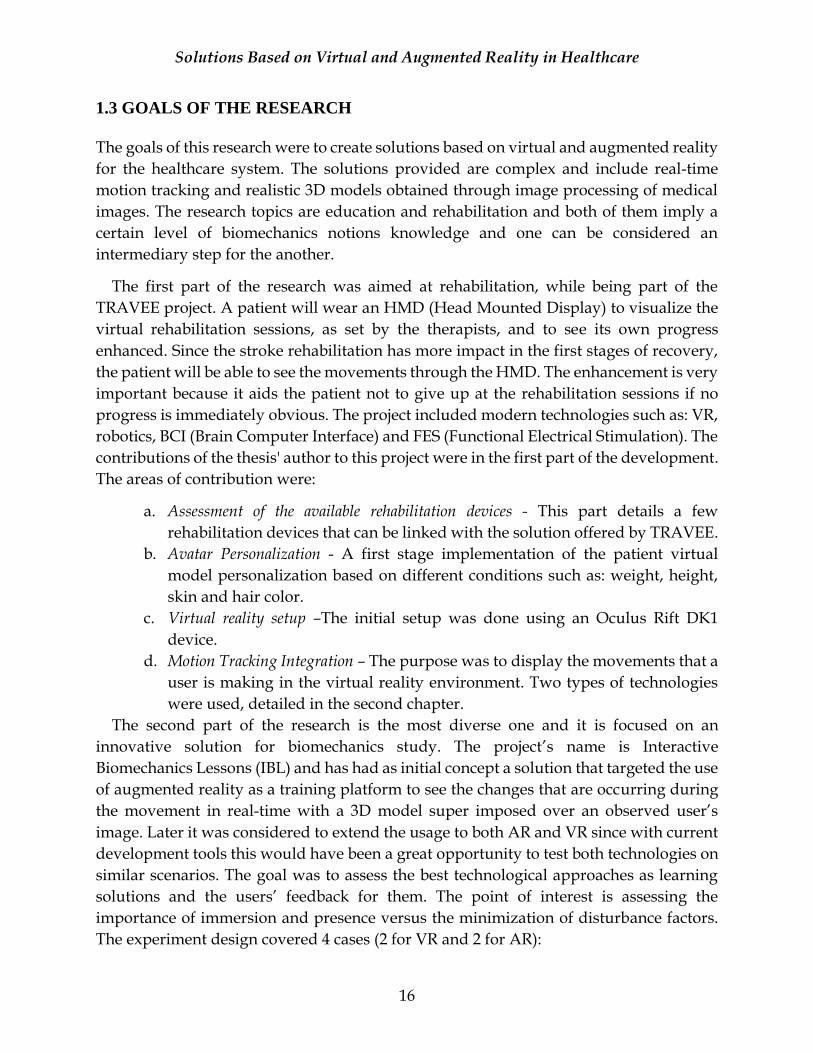
The areas of contribution were:
a. Assessment of the available rehabilitation devices - This part details a few
rehabilitation devices that can be linked with the solution offered by TRAVEE.
b. Avatar Personalization - A first stage implementation of the patient virtual
model personalization based on different conditions such as: weight, height,
skin and hair color.
c. Virtual reality setup –The initial setup was done using an Oculus Rift DK1
device.
d. Motion Tracking Integration – The purpose was to display the movements that a
user is making in the virtual reality environment. Two types of technologies
were used, detailed in the second chapter.
The second part of the research is the most diverse one and it is focused on an
innovative solution for biomechanics study. The project’s name is Interactive
Biomechanics Lessons (IBL) and has had as initial concept a solution that targeted the use
of augmented reality as a training platform to see the changes that are occurring during
the movement in real-time with a 3D model super imposed over an observed user’s
image. Later it was considered to extend the usage to both AR and VR since with current
development tools this would have been a great opportunity to test both technologies on
similar scenarios. The goal was to assess the best technological approaches as learning
solutions and the users’ feedback for them. The point of interest is assessing the
importance of immersion and presence versus the minimization of disturbance factors.
The experiment design covered 4 cases (2 for VR and 2 for AR):
Solutions Based on Virtual and Augmented Reality in Healthcare
17
a. The user wears an HMD and is isolated from external factors. The VR
environment is set into a virtual classroom.
b. The user wears an HMD, is isolated from external factors and the VR
environment is removed leaving an open space. This scenario was added due
to the observed cybersickness.
c. The user uses a marker-based AR application. The virtual lessons have similar
structure with the ones provided in the VR setup.
d. The user uses a markerless AR application. This is more at an experimental
stage where the user can see the bones’ 3D model animated over its image
based on the tracked motion.
The first three scenarios do not include motion tracking, and they are based on mobile
friendly, cost effective learning solutions. These scenarios were part of an in-depth testing
and the results are discussed in the last part of the thesis. This project was developed
from the start till the end. It is detailed in the fourth chapter.
1.4 SCIENTIFIC PUBLICATIONS IN CONNECTION WITH THE THESIS
A significant part of the work involved in this thesis was published in the following
scientific papers (sorted by the publication year):
2018
1. Alexandra Voinea and Florica Moldoveanu, “A Novel Solution Based on Virtual and Augmented Reality for Biomechanics Study” in Scientific Bulletin of UPB, Series C. vol. 80, no.2/2018, ISSN 2286-3540, pp.29-40. WOS:000434342000003.
2017
2. Alexandra Voinea, Florica Moldoveanu and Alin Moldoveanu, “3D Model Generation and Rendering of Human Musculoskeletal System Based on Image Processing” in Proceedings of the 21st International Conference on Control Systems and Computer Science, Bucharest, Romania, pg. 263-270, DOI: 10.1109/CSCS.2017.43, May 2017. (IEEE)
2016
3. Alexandra Voinea, Alin Moldoveanu and Florica Moldoveanu, “Bringing the Augmented Reality Benefits to Biomechanics Study” in Proceedings of the 2016 Workshop
on Multimodal Virtual and Augmented Reality (MVAR 2016), pg. 8757-8764, Tokyo, Japan, DOI: 10.1145/3001959.3001969, ISBN: 978-1-4503-4559-0, November 2016. WOS:000392302900009. 4. Alexandra Voinea, Alin Moldoveanu and Florica Moldoveanu, “Efficient Learning Technique in Medical Education Based on Virtual and Augmented Reality” in Proceedings of
Solutions Based on Virtual and Augmented Reality in Healthcare
18
9th Annual International Conference of Education, Research and Innovation, Seville, Spain, pg. 8757-8764, DOI: 10.21125/iceri.2016.0975, ISBN: 978-84-617-5895-1, November 2016. WOS:000417330208102. 2015
5. Alexandra Voinea, Alin Moldoveanu, Florica Moldoveanu and Oana Ferche, “Motion Detection and Rendering for Upper Limb Post-Stroke Rehabilitation” in Proceedings of the 5th
International Conference on e-Health and Bioengineering – EHB 2015, Iasi, Romania, pg. 811-814, DOI:10.1109/EHB.2015.7391471, ISBN: 978-1-4673-7544-3, November 2015. WOS:000380397900124 6. Alexandra Voinea, Alin Moldoveanu and Florica Moldoveanu “3D Visualization in IT Systems Used for Post Stroke Recovery: Rehabilitation Based on Virtual Reality” in Proceedings of CSCS20: The 20th International Conference on Control Systems and Computer
Science, Bucharest, Romania, pg. 856-862, 10.1109/CSCS.2015.123, ISBN: 978-1-4799-1779-2, May 2015. WOS:000380375200125 7. Alexandra Voinea, Alin Moldoveanu, Florica Moldoveanu and Oana Ferche,” ICT Supported Learning for Neuromotor Rehabilitation - Achievements, Issues and Trends” at The
International Scientific Conference eLearning and Software for Education, Bucharest, Romania, April 2015, Issue 1, pg. 594-601. WOS:000384469000086
8. Oana Maria Ferche, Alin Moldoveanu, Florica Moldoveanu, Alexandra Voinea, Victor Asavei and Ionut Negoi, “Challenges and issues for successfully applying virtual reality in medical rehabilitation” at The International Scientific Conference eLearning and
Software for Education, Bucharest, Romania, April 2015, Issue 1, pg. 494-501. WOS:000384469000073
9. Oana Ferche, Alin Moldoveanu, Delia Cinteza, Corneliu Toader, Florica Moldoveanu, Alexandra Voinea, Cristian Taslitchi, “From Neuromotor Command to Feedback: A survey of techniques for rehabilitation through altered perception” at E-Health and Bioengineering
Conference (EHB), Iasi, Romania, November 2015, pg. 1-4. WOS:00038039790010
1.5 STRUCTURE OF THE THESIS
Chapter 2 contains details regarding the current technologies used in VR and AR
applications. This section contains background information regarding their definitions
and technical details. The chapter continues with a short presentation of a few display
Solutions Based on Virtual and Augmented Reality in Healthcare
19
devices that were of the most interest at the time of the research and continues with a list
of motion tracking devices that were considered to obtain skeletal tracking of an observed
user.
Chapter 3 contains the author’s contributions to the TRAVEE project. The research
focused on solutions used in neuromotor rehabilitations. The literature review of existing
solutions contains two subjects: rehabilitation devices and 3D visualization methods.
Afterwards, implementation details of three areas are provided: avatar personalization,
virtual reality setup and motion tracking.
Chapter 4 contains the details of a novel educational solution named Interactive
Biomechanics Lessons that uses virtual and augmented reality for enhancing the learning
process for biomechanics study of human motion. This section contains the architecture
of the system including devices, sensors and software solutions required for the
implementation. A part of the research was focused on obtaining realistic 3D models of
human bones and muscular systems. Implementations details are provided along with a
few experimental tests on various technologies. Also, this chapter contains the
performance metrics of the developed applications and the results obtained based on
user’s feedback.
Chapter 5 contains the conclusions of the research and summarizes the personal
contributions. A few directions for future research that require additional time, work
capacity or funding are mentioned as well.
Solutions Based on Virtual and Augmented Reality in Healthcare
20
CHAPTER 2
CURRENT TECHNOLOGIES USED IN APPLICATIONS BASED
ON VIRTUAL AND AUGMENTED REALITY
This chapter addresses the basic information about virtual and augmented reality and
continues with their applicability in medical applications. Details about both
technological systems are presented followed by a short revision for some of the most
interesting available devices since the complete list is exhaustive at the moment.
VR and AR are known topics in the scientific community for many years and they had
a significant improvement in the recent years sustained by the media enthusiasm based
on the advancements of both technologies. Nowadays, developing a solution based on
VR and AR is in the best of times as the number of available hardware and software
solutions are growing in a fast pace [AV16b]. The developers, and with them the scientific
community, have many tools that are helping them build better solutions compared with
the ones proposed a few years ago and to overcome a part of the known drawbacks (E.g.
hardware and design changes to minimize the cybersickness). Fortunately, now are
available in high numbers low cost, performant hardware solutions and many system
engines that provide consistent VR and AR support.
Healthcare is one of the beneficiaries of these technological advancements. The new
level of interaction available through these technologies is a good fit for this research’s
topics although there are a few challenges and issues for successfully applying virtual
reality in medical rehabilitation [OMF15]. Two approaches were considered within this
thesis for using VR and AR in healthcare:
a. In the rehabilitation part, where the possibility of executing a large number of
exercises (compared with limited hours of kinesiotherapy) is speeding the
recovering process for the stroke survivors.
b. In the medical education part, the beneficiaries will be especially youth individuals
that are learning biomechanics notions
2.1 BACKGROUND
Virtual and augmented reality are both technological systems created with software and
their goal is to immerse the users into each specific environment. VR displays a fully

Solutions Based on Virtual and Augmented Reality in Healthcare
21
artificial environment where the user should believe that is the real world4. Also, “VR is
a realistic, real-time, 3-dimensional (stereoscopic) computer simulation of physical objects and
space”5. These are the technical definitions of virtual reality and our aim is to use it into
medical applications. According to [JVWR14], along the years there was no clear
consensus about the meaning of VR in medicine. There we find that some6 shared the
same vision of VR in their reviews as they saw the VR as a “collection of technologies that
allow people to interact efficiently with 3D computerized databases in real time using their natural
senses and skills”. Others7, described the VR correlated with the human experience: “a real
or simulated environment in which a perceiver experiences telepresence”. Schultheis (2001)
mentions that VR is “an advanced form of human-computer interface that allows the user to
interact with and become immersed in a computer-generated environment in a naturalistic
fashion”, while Bellani and Fornasari (2011) view VR “as only a simulation of the real world
based on computer graphics”. The definitions mentioned above are underlining two
approaches of VR in healthcare: VR as a simulation tool and VR as an interaction tool
[JVWR14].
On the other side, augmented reality blends real environment and virtual elements
while virtual reality is targeting to display only the virtual environment. AR is adding
virtual elements over the real-world display [IACG15] and according to [RTA97] AR is
based on techniques developed in VR and interacts not only with a virtual world but has
a degree of interdependence with the real world. Reference [OB05] mentions the fact that
AR systems have three major components:
1. Tracking and registration;
2. Display technology;
3. Real-time rendering.
The main motivation of the usage of medical augmented reality lies in the “need of
visualizing medical data and the patient within the same physical space” [MM14].
The advantage of AR is the fact that the users are feeling comfortable because the
presence is highly achieved. This is a consequence of the fact that the users are still
present in the real environment while the virtual elements are superimposed on top of it.
On the other side, in VR the medium (environment) can be fully controlled as opposed
to AR and this factor is important when it comes to minimizing the external disturbing
factors. For example, a user with an HMD and some sound proof headsets can be
4 http://whatis.techtarget.com/definition/virtual-reality
5 http://www.businessdictionary.com/definition/virtual-reality-VR.html 6 Rubino (2002), McCloy and Stone (2001), Szekely and Satava (1999) 7 Riva (2003); Steuer (1992)
Solutions Based on Virtual and Augmented Reality in Healthcare
22
approximately fully isolated by outside factors that can disrupt the attention from the
operations executed in the virtual environment, while this scenario is unachievable in
AR. Both VR and AR are considered reliable methods to simulate realistic experiences
and this makes both technological systems a good fit to reproduce real situations for
training or educational purposes, into a safe environment.
There are two basic AR software implementation types: marker-based and markerless
[MCFM14a]. Results showed that a higher sense of presence was shown for AR invisible
marker system compared with the visible marker one [IACG15]. A marker-based
application solves the problem using visual markers detectable with computer vision
methods (e.g. 2D barcodes) [SS12].
Depth perception issues were noticed with medical AR systems while using
semitransparent structure overlay onto visible surfaces [ZY14]. In AR the virtual elements
are drawn on top of the real environment and besides that, factors like the lighting can
affect the output image and as a solution “seven depth cues are evaluated with rendering using
depth-dependent color and the use of aerial perspective shown to give the best cues.” [ZY14].
Besides VR and AR there is a third notion named Mixed Reality (MR) and often this
one is confused with AR. While AR refers to a system in which an enhanced version of
the real world is available for the user, MR refers to a system that combines real and
virtual objects and information. The enhancement elements are virtual and can include
object and information8. Basically, in Mixed Reality the physical and digital objects co-
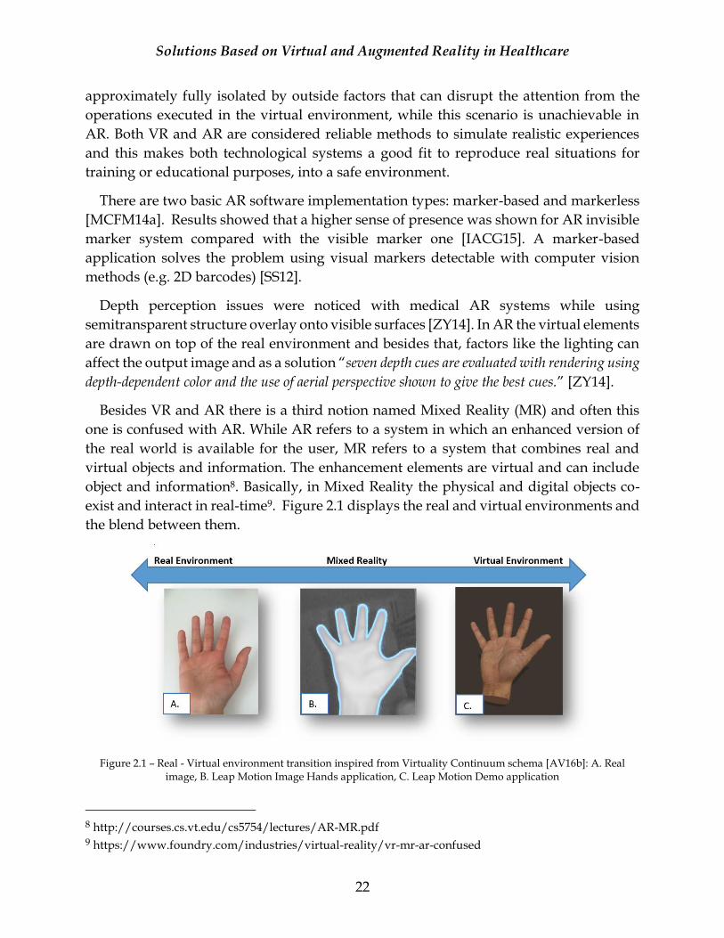
exist and interact in real-time9. Figure 2.1 displays the real and virtual environments and
the blend between them.
Figure 2.1 – Real - Virtual environment transition inspired from Virtuality Continuum schema [AV16b]: A. Real image, B. Leap Motion Image Hands application, C. Leap Motion Demo application
8 http://courses.cs.vt.edu/cs5754/lectures/AR-MR.pdf 9 https://www.foundry.com/industries/virtual-reality/vr-mr-ar-confused
Solutions Based on Virtual and Augmented Reality in Healthcare
23
Another important aspect regarding the AR is the fact that it shouldn’t be limited only
to the graphics while it targets the real environment augmentation. Indeed, the graphical
augmentation is our focus in this thesis thus in a larger scale the augmented reality can
be extended to a multimodal approach [HS16].
After many psychological studies that investigated the adverse consequences of the
incorrectly rendered focus cues in stereoscopic displays it was found that the these might
contribute to the commonly recognized issues, such as: distorted depth perception,
diplopic vision, visual discomfort and fatigue and degradation in oculomotor response
[HH14].
Cybersickness is an important topic while developing for VR and it was noticed that
some design changes can reduce cybersickness as this is not only related with the
hardware specifications. In the following lines a few examples are provided:
1. In virtual environment, the developer should use ramps instead of stairs (based
on a survey from Oculus Rift Tuscany Demo) [JD14].
2. A reduction of cybersickness was noticed if the HUD (Heads-up Display) elements
are blended in the 3D scene, instead of using the classic 2D elements. This was
strongly observed while developing our applications and the positive impact of
this design.
3. FOV and Focus are key elements and they should be properly calibrated. For
example, the human eye is automatically focusing for near or distance. The best
approach will be using an eye tracking device to see where the user is looking into
the scene.
There are two VR software solutions that were considered in the same line with the
topics of this research. First one is the People|Be fearless VR app developed by Samsung
and was available with Samsung Gear VR. It tacked phobias, such as: fear of public
speaking10 and fear of heights11. The users had available a few relevant scenarios to
interact with the fear stimuli. These apps aimed to improve the resistance in real life for
these stimuli to be able to overcome the fear. Another app of interest is Fusion Tech 3D.
This is a project made within the Stanford University that was later acquired by Luminate
Health Systems12 . It is an application available on multiple platforms (mobile, desktop,
VR) that is able to visualize 3D data inside the human body. The devices that supported
this app were iOS, Android, Oculus and zSpace.
10 https://www.oculus.com/experiences/gear-vr/942681562482500/ 11 https://www.oculus.com/experiences/gear-vr/821606624632569/ 12 https://www.luminatehs.com/
Solutions Based on Virtual and Augmented Reality in Healthcare
24
2.2 VIRTUAL AND AUGMENTED REALITY DEVICES
Along the years, the technology had a fast advancement that offered the opportunity to
have a wide range of devices available for development. Depending on their price and
accessibility some of them had a higher success rate compared with others. In the
following are mentioned a few devices that captured our attention and that proved to
have a significant impact for these technologies.
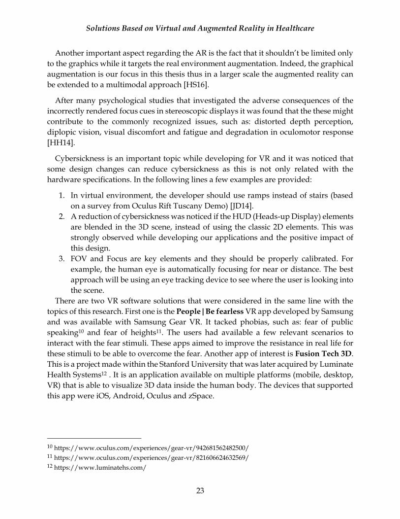
For displaying the VR applications, CAVE (Cave Automatic Virtual Environment) and
HMD devices proved their efficiency in the past years [FT14]. CAVE is a room-sized
virtual reality system and it consists in a series of projectors that are places on the walls
of a room. On the other side, HMD is a device worn on the head where the users cannot
(shouldn’t) see outside the headset while displaying VR. There are a few key differences
between these systems and one of them is related with the fact that with the CAVE system
the user can still see and perceive its body as normal, while using an HMD this is not
available as the user can see most of the time only its hands. In consequence, the
immersion of the user in the virtual environment is total while using CAVE compared
with the usage of HMDs13 thus on other hand the prices and mobility of the HMDs are
better. Figures 2.2 and 2.3. contain images of a well-known HMD – Oculus Rift.
Figure 2.2 – Oculus Rift device 14
Figure 2.3 – Oculus Rift and EEG cap used in
Rehabilitation15
Regarding HMDs, there is a subset of devices named OHMD (Optical Head Mounted Display) that are allowing the users to see through them. We can mention here well-known devices, such as: Google Glass or Microsoft HoloLens. This special category of devices is suited for displaying augmented reality. A consistent number of authors mentioned in their scientific papers the usage of HMD for visualizing AR applications
13 https://www.vrs.org.uk/virtual-reality-environments/cave.html 14 Image source - http://www.cgmagonline.com/2015/05/08/oculus-rift-release-date-announced/ 15 Image source - https://www.theverge.com/2016/8/11/12443026/virtual-reality-exoskeleton-paraplegic-oculus-rift
Solutions Based on Virtual and Augmented Reality in Healthcare
25
[IACG15] [MM14] [CK14] [FC14] [MCFM14a]. However, any device can be utilized for displaying augmented reality if it respects the following rules:
1. The possibility to combine real with virtual; 2. To be interactive in real time; 3. To be registered in 3D space [MM14].
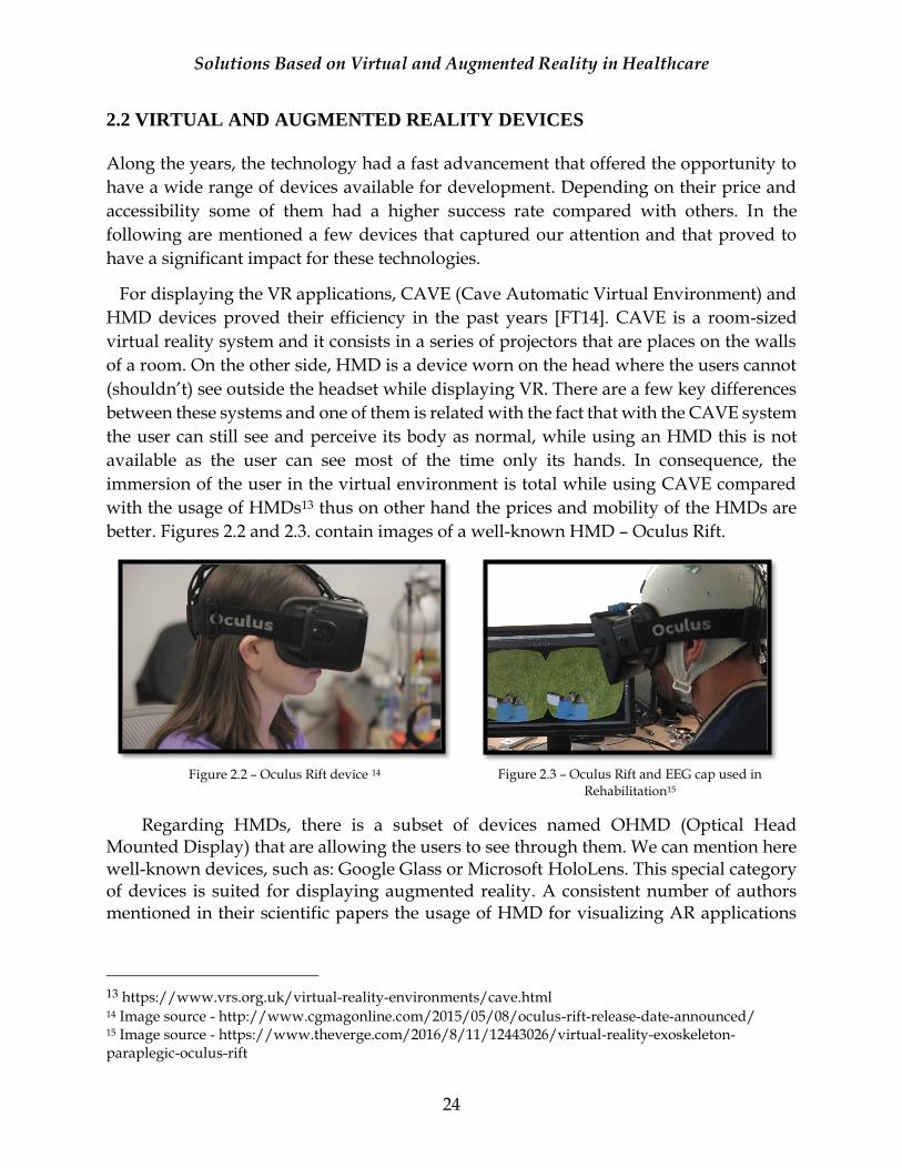
In AR the virtual information is superimposed over the real world and the registration can be interpreted as the accuracy of spatially aligning the virtual elements in the real world. The coordinate system of the real world where the virtual elements are projected should be resolved regardless the environment changes or given time16. A camera is needed to be able to get the real environment information to combine it with the virtual elements. There is a wide list of devices that are a good fit for AR where we can mention: mobile devices (smartphones or tablets), desktop/monitors (used with external web cameras) or HMDs. As stated before, OHMD are a perfect fit for AR systems but their costs are often substantial [AV16b]. Table 2.1 contains a list with devices of interest to display VR or AR along with some basic information about their capabilities and prices [AV16b].
Table 2.1 – Characteristics of top VR and AR devices
Device
Name
Reality
Type
Refresh
Rate [Hz]
FOV
[degrees]
Resolution
[pixels]
Processing
Source
Price
[USD]
Oculus Rift VR 90 110 1080x1200 Computer 599
Samsung
Gear VR
VR
Depends on
the
smartphone
(~60)
96
Depends on the
smartphone
(e.g. Samsung
Galaxy S7 -2560
x1440)
Smartphone
99+17
HTC Vive VR 90 110 1080x1200 Computer 799
Sony VR VR 120 100 960x1080 Game
Console
399
Atheer AiR AR NA18 50 1280x720 Built-in 3950
Microsoft
HoloLens
AR & VR
60
120
1268x720
Built-in
3000
16 http://www.cs.bham.ac.uk/~rjh/courses/ResearchTopicsInHCI/2014-15/Submissions/yan--yan.pdf 17 At the VR headset price is added the smartphone price. This is only an accessory for the compatible smartphones. 18 The refresh rate for this device is not available. However, the available information in this direction is that the device is based on NVIDIA Tegra K1 processor.
Solutions Based on Virtual and Augmented Reality in Healthcare
26
2.3 HUMAN BODY MOTION TRACKING SENSORS
In this subchapter we are discussing about real-time human body motion tracking, more
exactly skeleton tracking, as this feature was present on both projects included in this
research while developing for AR and VR. Following is provided an overview of the
skeletal tracking sensors that were considered during development. The skeleton
movements tracked by the sensors were used to animate a 3D virtual human avatar.
In the next sections are presented the technical details of 3 sensors that can provide
skeletal tracking in VR and AR based applications: Leap Motion Controller, Kinect and
VicoVR. While Leap Motion controller and Kinect were used in our research, VicoVR
sensor is a recently released device that provides performant skeletal tracking suited for
mobile development.
2.3.1 Leap Motion
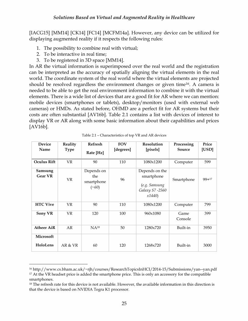
Leap Motion Controller has incorporated two cameras and three infrared lights (Fig.2.5).
The infrared light is outside the visible light spectrum with a wavelength of 850
nanometers. Its viewing range was roughly 60 cm (2 feet) above the device using the
initial version of the software but with their new software (Orion beta) this was extended
to 80 cm (2.6 feet). Fig.2.6 displays the device’s interaction area. The device is connected
to a workstation (PC) via an USB controller (Fig.2.4). After the sensor data is read,
resolution adjustments are performed, if necessary. Data that takes form of a grayscale
stereo image, separated into left and right cameras, is streamed via USB to the tracking
software. As opposed to other solutions, the controller doesn’t generate a depth map but
instead applies advanced algorithms to the raw data provided by the sensor. The
obtained images are analyzed to reconstruct a 3D representation of what is seen and then
the tracking algorithms interpret the 3D data while the position of the occluded objects is
inferred. On top of that, filtering techniques are applied19.
Figure 2.4 – Leap Motion Controller20
Figure 2.5 – Leap Motion
Cameras
Figure 2.6 – Leap Motion Interaction
Area21
19 http://blog.leapmotion.com/hardware-to-software-how-does-the-leap-motion-controller-work 20 Image source : http://www.robotshop.com/blog/en/explore-virtual-reality-with-leap-motion-3d-motion-controller-16806 21 http://blog.leapmotion.com/hardware-to-software-how-does-the-leap-motion-controller-work/
Solutions Based on Virtual and Augmented Reality in Healthcare
27
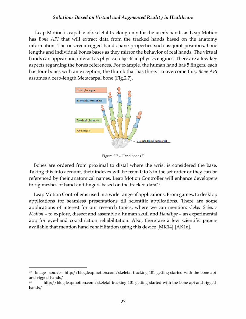
Leap Motion is capable of skeletal tracking only for the user’s hands as Leap Motion
has Bone API that will extract data from the tracked hands based on the anatomy
information. The onscreen rigged hands have properties such as: joint positions, bone
lengths and individual bones bases as they mirror the behavior of real hands. The virtual
hands can appear and interact as physical objects in physics engines. There are a few key
aspects regarding the bones references. For example, the human hand has 5 fingers, each
has four bones with an exception, the thumb that has three. To overcome this, Bone API
assumes a zero-length Metacarpal bone (Fig.2.7).
Figure 2.7 – Hand bones 22
Bones are ordered from proximal to distal where the wrist is considered the base.
Taking this into account, their indexes will be from 0 to 3 in the set order or they can be
referenced by their anatomical names. Leap Motion Controller will enhance developers
to rig meshes of hand and fingers based on the tracked data23.
Leap Motion Controller is used in a wide range of applications. From games, to desktop
applications for seamless presentations till scientific applications. There are some
applications of interest for our research topics, where we can mention: Cyber Science
Motion – to explore, dissect and assemble a human skull and HandEye – an experimental
app for eye-hand coordination rehabilitation. Also, there are a few scientific papers
available that mention hand rehabilitation using this device [MK14] [AK16].
22 Image source: http://blog.leapmotion.com/skeletal-tracking-101-getting-started-with-the-bone-api-and-rigged-hands/ 23 http://blog.leapmotion.com/skeletal-tracking-101-getting-started-with-the-bone-api-and-rigged-hands/
Solutions Based on Virtual and Augmented Reality in Healthcare
28
2.3.2 Kinect
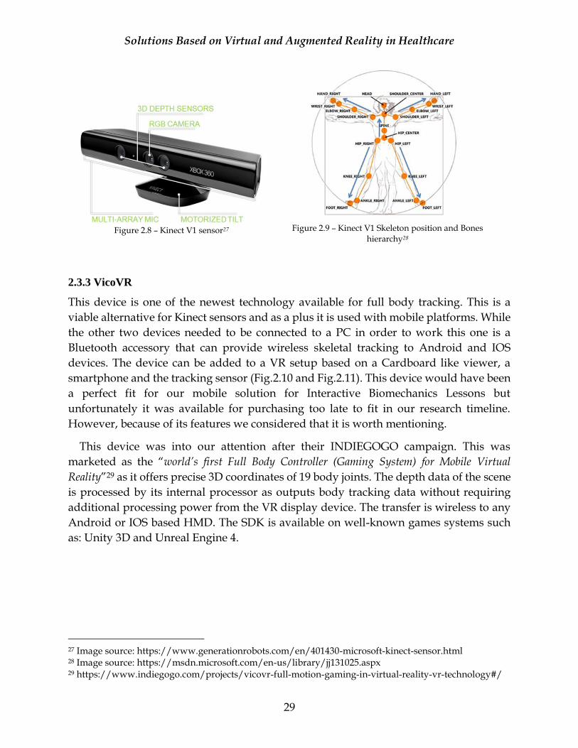
Kinect sensor, developed by Microsoft, is a device with depth sensing technology that
has a built-in color camera, an IR (infrared emitter) and a microphone array (Fig.2.8). This
device can sense the location and movements of people as well as their voices24 and can
be connected to an Xbox console, PC or tablet (Window OS). There were two versions of
this sensor available on the market V1 and V2 at the time of the research. The initial one,
V1, had lower specs regarding the color camera resolution (640x480 vs 1920x1080). Also,
it had a smaller Horizontal and Vertical FOV (57 vs 70 degrees and 43 vs 60 degrees) and
number of skeleton joints defines (20 vs 26 joints)25. While Kinect V1 could have tracked
2 full skeletons, V2 was able to recognize 6 people and track two26.
Kinect sensor can perform whole-body skeletal tracking of an observed user. It can
locate the joints and track their movements in real-time using the IR camera. One of the
advantages is the fact that no specific pose or calibration is necessary for a user to be
tracked. Although Kinect sensor can record the hands movements the quality of the
movements is lower since there is only one bone assigned for the hand (V1). As opposed
with Leap Motion Controller that had a corresponding virtual bone to the anatomical
ones. Indeed, Kinect V2 had better results to track the hands but still didn’t offer the same
quality as the Leap Motion Controller. For the research work of this thesis the most used
sensor was Kinect V1 as it was added in both projects. Fig.2.9 illustrates the tracked joints
while using a Kinect V1 sensor, as well as their hierarchy versus the parent joint (Hip
Center). Until recently, Kinect was the only viable solution available that could perform
skeletal tracking, even with some downsides regarding the tracking errors. A few back-
up solutions were improvised such as: using two Kinect sensors [MCFM15] or using it
with OpenCV [BK15]. This device is by far the most used sensor for skeletal tracking that
was mentioned in the studied scientific papers [DL15] [ZG15] [MCFM14a] [CK14]
[TM14].
24 https://developer.microsoft.com/en-us/windows/kinect/hardware 25 http://zugara.com/how-does-the-kinect-2-compare-to-the-kinect-1 26 https://msdn.microsoft.com/en-us/library/hh973074.aspx
Solutions Based on Virtual and Augmented Reality in Healthcare
29
Figure 2.8 – Kinect V1 sensor27
Figure 2.9 – Kinect V1 Skeleton position and Bones
hierarchy28
2.3.3 VicoVR
This device is one of the newest technology available for full body tracking. This is a
viable alternative for Kinect sensors and as a plus it is used with mobile platforms. While
the other two devices needed to be connected to a PC in order to work this one is a
Bluetooth accessory that can provide wireless skeletal tracking to Android and IOS
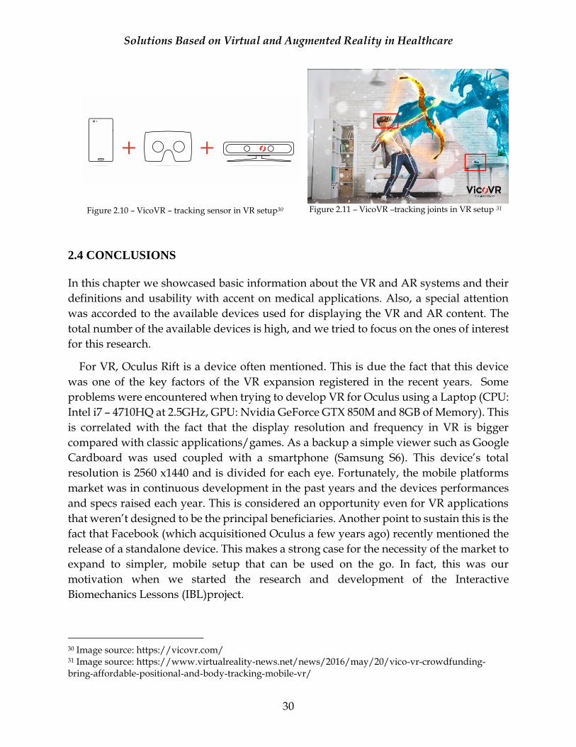
devices. The device can be added to a VR setup based on a Cardboard like viewer, a
smartphone and the tracking sensor (Fig.2.10 and Fig.2.11). This device would have been
a perfect fit for our mobile solution for Interactive Biomechanics Lessons but
unfortunately it was available for purchasing too late to fit in our research timeline.
However, because of its features we considered that it is worth mentioning.
This device was into our attention after their INDIEGOGO campaign. This was
marketed as the “world’s first Full Body Controller (Gaming System) for Mobile Virtual
Reality”29 as it offers precise 3D coordinates of 19 body joints. The depth data of the scene
is processed by its internal processor as outputs body tracking data without requiring
additional processing power from the VR display device. The transfer is wireless to any
Android or IOS based HMD. The SDK is available on well-known games systems such
as: Unity 3D and Unreal Engine 4.
27 Image source: https://www.generationrobots.com/en/401430-microsoft-kinect-sensor.html 28 Image source: https://msdn.microsoft.com/en-us/library/jj131025.aspx 29 https://www.indiegogo.com/projects/vicovr-full-motion-gaming-in-virtual-reality-vr-technology#/
Solutions Based on Virtual and Augmented Reality in Healthcare
30
Figure 2.10 – VicoVR – tracking sensor in VR setup30
Figure 2.11 – VicoVR –tracking joints in VR setup 31
2.4 CONCLUSIONS
In this chapter we showcased basic information about the VR and AR systems and their
definitions and usability with accent on medical applications. Also, a special attention
was accorded to the available devices used for displaying the VR and AR content. The
total number of the available devices is high, and we tried to focus on the ones of interest
for this research.
For VR, Oculus Rift is a device often mentioned. This is due the fact that this device
was one of the key factors of the VR expansion registered in the recent years. Some
problems were encountered when trying to develop VR for Oculus using a Laptop (CPU:
Intel i7 – 4710HQ at 2.5GHz, GPU: Nvidia GeForce GTX 850M and 8GB of Memory). This
is correlated with the fact that the display resolution and frequency in VR is bigger
compared with classic applications/games. As a backup a simple viewer such as Google
Cardboard was used coupled with a smartphone (Samsung S6). This device’s total
resolution is 2560 x1440 and is divided for each eye. Fortunately, the mobile platforms
market was in continuous development in the past years and the devices performances
and specs raised each year. This is considered an opportunity even for VR applications
that weren’t designed to be the principal beneficiaries. Another point to sustain this is the
fact that Facebook (which acquisitioned Oculus a few years ago) recently mentioned the
release of a standalone device. This makes a strong case for the necessity of the market to
expand to simpler, mobile setup that can be used on the go. In fact, this was our
motivation when we started the research and development of the Interactive
Biomechanics Lessons (IBL)project.
30 Image source: https://vicovr.com/ 31 Image source: https://www.virtualreality-news.net/news/2016/may/20/vico-vr-crowdfunding-bring-affordable-positional-and-body-tracking-mobile-vr/
Solutions Based on Virtual and Augmented Reality in Healthcare
31
For AR we initially focused on two types of devices: OHMD (HoloLens) and mobile
devices. The aim was to develop applications to cut edge technology such as Microsoft
HoloLens thus we found some challenges regarding the acquisition of this device and as
a result the focus was shifted on tablets, smartphones and laptops as they met the
requirements to develop AR applications on them. Although HoloLens is unlocking a
new level of realism using holograms the second solution will be available for a much
larger number of users and this will enhance a new level of testing since the targeted pool
of potential users is considerably high. For example, in 2015 there were ~ 1.4 billion
Android users and the market is continuously growing.
The manufacturers improved their devices capabilities to give to their users the best
experiences. The devices have different components and in consequence their prices may
have high variations (as seen in Table 2.1). The scope is to fully immerse the user into the
virtual world, not only with high quality graphics but also without motion sickness. This
is one of the major concerns for VR development nowadays. This can be improved using
a series of special design approaches, but devices capabilities have an important role, too
[ZG15].
This chapter also contains details regarding a few skeletal tracking devices suited for
integration into VR and AR applications. In this research we used two types of sensors:
one for the whole body and another one that tracked only the hand: for the whole body
was used a Kinect sensor and for the hand a Leap Motion controller. Although, the Kinect
sensor can be considered “old” technology it is extremely used and present in many
scientific papers. Recently new devices started to appear on the market with similar
capabilities as some were built especially for VR systems. In this context VicoVR sensor
was briefly presented which is a promising solution compatible with mobile technology,
although it tracks fewer joints compared even with the first version of the Kinect sensor.
The device has its own processor source that does all the computations to avoid the
transfer of the computational burden on the VR display device (a smartphone in this
case).
Solutions Based on Virtual and Augmented Reality in Healthcare
32
CHAPTER 3
ICT SOLUTIONS FOR NEUROMOTOR REHABILITATION
This chapter contains the details of the first part of the research that was focused on
solutions used in neuromotor rehabilitation of stroke survivors. The following contains
the author’s contributions to TRAVEE project and it covers multiple areas such as
assessment of existing rehabilitation devices, patient avatar’s personalization, virtual
reality setup and motion tracking integration.
3.1 RELATED WORK
This section is divided in two parts, where at start a list of rehabilitation devices is
presented and it’s focused more on the hardware part. The mentioned devices are related
with the product TRAVEE. They contain complex technologies such as FES (Functional
Electrical Stimulation) and robotics. The review is continued with an analysis of the
software solutions used in rehabilitation.
3.1.1 Rehabilitation Devices
In rehabilitation, there are a significant number of eLearning solutions that can be used
complementary with the classical therapy, based on kinesiotherapy [AV15c]. The
solutions contain complex technologies such as FES, NMES (Neuromuscular Electrical
Stimulation), EMG (Electromyography), BCI and robotics. Products that target the
patient’s rehabilitation are available on the market and [PM14] has a detailed list with
more than 100 devices used for upper limb rehabilitation. They are classified based on
the following criteria:
a. The joint system they support.
b. The device’s DOF (Degree of Freedom). This is represented by a sum of all
independent movements performed by the joints of the device.
c. The supported movements types, such as: abduction, flexion/extension,
pronation/supination, grip and release, horizontal and vertical displacement,
etc. Also, the movements can be active or passive (with or without external
help to execute a certain movement).
d. The patient’s health condition as neuromotor rehabilitation solutions can be used
for certain conditions such as: stroke, cerebral palsy, essential tremor, multiple
sclerosis, primal cord injuries and traumatic brain injury.
In the following, a few devices of interest, that are linked to TRAVEE project, are shortly
described.
Solutions Based on Virtual and Augmented Reality in Healthcare
33
Music Glove
Music Glove32 is a rehabilitation device that aims to improve the hand function for stroke
survivors and other neuromotor disabilities. The device is connected to a VR module that
displays a music environment similar with the games. In the respective application the
subjects should follow the musical notes that appear on the screen making certain moves.
These types of exercises are helping the subject improve the hand function after a
neuromotor disability. The benefits of using MusicGlove device are supported by a clinical
study [NF14] with patients which suffered a stroke. In that study participated 12 patients
which were diagnosed with mild chronic hemiparesis. They were randomly selected to
use the MusicGlove device in the same time with conventional therapy. Each selected
patient used the device for 6 sessions of one-hour length, three times per week for 2
weeks. At the end of the study was shown that the object grip movement of the selected
subjects improved with a higher rate than traditional therapy.
MIT-Manus and InMotion ARM
InMotion ARM33 device represents a clinical version of the MIT-Manus34 robot. The
clinicians can establish an efficient, personalized therapy for patients with neuromotor
disabilities because the device is based on intelligent, interactive technology which is able
to adapt itself to each patient’s capacity. InMotion covers multiple rehabilitation solutions,
such as:
a. For upper limbs: InMotion ARM Therapy System, InMotion WRIST Interactive
Therapy System and InMotion HAND.
b. For lower limbs: Anklebot InMotion ANKLE Exoskeletal Robot and InMotion
Exoscheletal Arm Robot.
The improvements obtained using the therapy robot were noticed using a controlled,
randomized study [MLA97].
Armeo
This solution uses a VR scenario incorporated with a gravitational compensation system
for the therapy for upper extremities with self-initiated and functional treatment 35. The
included exercises are provided into a game-like setup which helps the patients to
improve their motor abilities and real-time performance through the augmented
performance feedback. In a study [SJH09] that involved stroke patients with mild to
32 https://www.medgadget.com/2014/10/musicglove-hand-rehabilitation-system-now-available-
video.html 33 http://bionikusa.com/healthcarereform/upper-extremity-rehabilitiation/inmotion2-arm/ 34 http://news.mit.edu/2000/manus-0607 35 https://static.hocoma.com/wp-
content/uploads/2016/09/bro_Armeo_160211_en_08_WEB.pdf?x82600
Solutions Based on Virtual and Augmented Reality in Healthcare
34
severe hemiparesis, the patients manifested their preference for this rehabilitation
solution versus the classical therapy. The gravity-supported arm exercises can improve
arm movement ability with a brief 1:1 assistance from a therapist (~ 4 minute per session).
The improvements of rehabilitation based on this solution that contains a 3-dimensional
weight support, instant visual movement feedback and simple VR software, were noticed
even at the 6 months follow up.
Four distinct products are included in the Armeo therapy concept: Armeo Power, Armeo
Spring, Armeo Spring Pediatric and Armeo Boom and each of them are specially conceived
for a certain stage of the recovery process, with one exception the Armeo Spring Pediatric
which is designed to cover children rehabilitation cases.
Bi-Manu-Track
Bi-Manu-Track36 is a robotic device with 2 DOF designed for wrist and forearm region
and works on the principle of bilateral training. According to [ECL11] the device permits
a DOF for the pronation and supination of the forearm. When is utilized in vertical
position the device permits a DOF for dorsiflexion/volar flexion of wrist. The device is
connected to a visual display that shows the number of effectuated cycles and a computer
that collects the data and controls the motors. Bi-Manu-Track can be used in 3 modes:
a. Passive, where the robot assists both upper limbs.
b. Active-Passive, where the movements are effectuated in mirror mode initiated by
the less affected limb.
c. Active-Active, where both upper limbs initiate the movement.
Bi-Manu-Track impact was measured in a study [SH05] where it was found that the
greater number of repetitions and the bilateral approach could have impacted positively
the upper limb motor control and power compared with other techniques based on ES
(Electrical Stimulation).
PowerGrip
The PowerGrip37 device is an EPPO (Electric Powered Prehension Orthosis) that is helpful
for picking up, grasping, holding and manipulating objects. PowerGrip uses switches or
sEMG (surface electromyography) signals to control the input of the device [PM14]. In
the newer versions are used myoelectric sensors that are placed on one or two functional
muscles.
36 http://www.reha-stim.de/cms/index.php?id=60 37 http://www.broadenedhorizons.com/powergrip
Solutions Based on Virtual and Augmented Reality in Healthcare
35
This part of the literature review was in connection with TRAVEE project contribution.
TRAVEE has similar characteristics with the mentioned solutions along with its original
elements, such as:
a. The usage of an interactive virtual environment, as seen in the MusicGlove and Armeo
case. However, in these cases the VR environment is displayed on a computer
monitor instead of an HMD.
b. The existence of personalized treatment related with patient’s health condition for an
appropriate recovery plan.
c. The possibility to effectuate a high number of repetitions for a faster recovery.
3.1.2 3D Visualization Solutions
If in the previous section the focus was on the rehabilitation devices, now our attention will be on software solutions and more exactly the 3D visualization methods. Even tough, some of previously mentioned devices contained a VR module the information presented in this section is more fit to our research goals. The review contains information from two solutions considered representative for neuromotor rehabilitation. They are not functional only based on the rendering part as it is important to review the compatibility of the module, dependent tools and libraries.
Rehabilitation Simulator
A set of rehabilitation applications based on virtual reality and physical-haptic
procedures are proposed by [LDLL14]. The applications have specific tasks to be
performed by patients that suffered a stroke. The applications’ solution contains a haptic
device (Sensable Phantom) that generates force feedback which indicates the interaction
between the patient and a virtual object. It is integrated in the system with Open Haptics
API. The 3D visualization is implemented with OGRE (Object-Oriented Graphics
Rendering Engine) as this software enabled the visualization of virtual elements in real-
time at a high quality [AV15b]. NVIDIA PhysX, a physical engine, was incorporated
complementary with the graphical engine for an accurate physical simulation. 3D
modeling tools such as Blender and 3D Studio were used to create the 3D models used in
this set of applications. They offer two categories of exercises that target the rehabilitation
of upper limbs:
a. Cooking tasks - The displayed scene is a virtual kitchen with various related
elements such as: a pan, potatoes, skaters, a table and a shelf. The scene
complexity is low and based on the rendered models it can be assumed that
there are approximately 10-15 simple 3D models [LDLL14]. The scene has a
first-person perspective and the user can see the virtual model of one arm as
Solutions Based on Virtual and Augmented Reality in Healthcare
36
this model is used to interact with other elements from the virtual kitchen
simulating basic daily living tasks [AV15b].
b. Motor activity of grasping a glass – Another set of tasks that simulate usual daily
activities such as drinking water. This scene is fairly simple containing the
virtual models of a glass, a table, a coaster and the trained arm. Initially the
objective is to grasp the glass, followed by the simulation of the activity of
drinking water and as a final target the user should place the glass on top of a
coaster model.
Personalized Rehabilitation Gaming System
A VR based system named Rehabilitation Gaming System (RGS) is proposed by [MSC10].
The attention is drawn on a certain rehabilitation scenario called Spheroids. Promising
results were reported with a consistent transfer of movements kinematics between the
physical and virtual tasks during trials that involved 21 acute/subacute stroke patients
and 20 controls using a Personalized Training Module (PTM).
The virtual scene is simple and contains a green landscape populated with trees and
with a mountain range background. Along with these elements a virtual model of a
human torso and arms are added in a first-person perspective. A motion tracking system
maps the user’s physical movements into the virtual reality scenarios. The user’s task is
to intercept the spheres that move toward the user and with each successful interception
the user obtains a number of points. The difficulty is set by different parameters such as:
speed, spheres’ appearance interval and the horizontal range of dispersion in the FOV
(Field of View). RGS implements training protocols for neurorehabilitation that allows a
gradual and individualized treatment of deficits of the upper extremities after stroke
[AV15b].
Both solutions have similarities with our contributions and one that is worth
mentioning is the fact that on both projects a rendering engine was used to display the
virtual scenes. Nowadays the well-known rendering engines (e.g. Unity, Unreal) are
integrated into a complete game system and permit to integrate the functionality of
additional components, such as motion tracking. We opted for Unity game system while
the other solution uses OGRE rendering engine. The available rendering solutions were
reviewed multiple times during this research and in each case, it was decided to continue
the usage of Unity. The main advantage was the extended support of various
technologies for motion tracking and display for VR and AR. The other reviewed
solutions were Unreal Engine and CryEngine and we considered that Unity had a wider
support for dependent technologies compared with the other two.
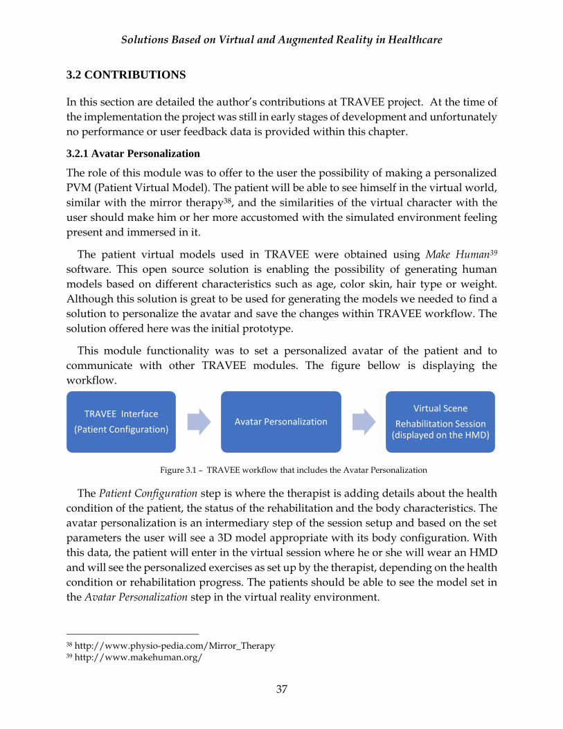
Solutions Based on Virtual and Augmented Reality in Healthcare
37
3.2 CONTRIBUTIONS
In this section are detailed the author’s contributions at TRAVEE project. At the time of
the implementation the project was still in early stages of development and unfortunately
no performance or user feedback data is provided within this chapter.
3.2.1 Avatar Personalization
The role of this module was to offer to the user the possibility of making a personalized
PVM (Patient Virtual Model). The patient will be able to see himself in the virtual world,
similar with the mirror therapy38, and the similarities of the virtual character with the
user should make him or her more accustomed with the simulated environment feeling
present and immersed in it.
The patient virtual models used in TRAVEE were obtained using Make Human39
software. This open source solution is enabling the possibility of generating human
models based on different characteristics such as age, color skin, hair type or weight.
Although this solution is great to be used for generating the models we needed to find a
solution to personalize the avatar and save the changes within TRAVEE workflow. The
solution offered here was the initial prototype.
This module functionality was to set a personalized avatar of the patient and to
communicate with other TRAVEE modules. The figure bellow is displaying the
workflow.
The Patient Configuration step is where the therapist is adding details about the health
condition of the patient, the status of the rehabilitation and the body characteristics. The
avatar personalization is an intermediary step of the session setup and based on the set
parameters the user will see a 3D model appropriate with its body configuration. With
this data, the patient will enter in the virtual session where he or she will wear an HMD
and will see the personalized exercises as set up by the therapist, depending on the health
condition or rehabilitation progress. The patients should be able to see the model set in
the Avatar Personalization step in the virtual reality environment.
38 http://www.physio-pedia.com/Mirror_Therapy 39 http://www.makehuman.org/
TRAVEE Interface
(Patient Configuration)Avatar Personalization
Virtual Scene
Rehabilitation Session (displayed on the HMD)
Figure 3.1 – TRAVEE workflow that includes the Avatar Personalization
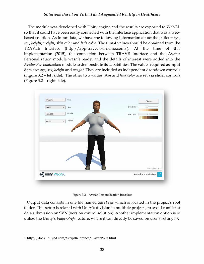
Solutions Based on Virtual and Augmented Reality in Healthcare
38
The module was developed with Unity engine and the results are exported to WebGL
so that it could have been easily connected with the interface application that was a web-
based solution. As input data, we have the following information about the patient: age,
sex, height, weight, skin color and hair color. The first 4 values should be obtained from the
TRAVEE Interface (http://app-travee.osf-demo.com/). At the time of this
implementation (2015), the connection between TRAVE Interface and the Avatar
Personalization module wasn’t ready, and the details of interest were added into the
Avatar Personalization module to demonstrate its capabilities. The values required as input
data are: age, sex, height and weight. They are included as independent dropdown controls
(Figure 3.2 – left side). The other two values: skin and hair color are set via slider controls
(Figure 3.2 – right side).
Figure 3.2 – Avatar Personalization Interface
Output data consists in one file named SavePrefs which is located in the project’s root
folder. This setup is related with Unity’s division in multiple projects, to avoid conflict at
data submission on SVN (version control solution). Another implementation option is to
utilize the Unity’s PlayerPrefs feature, where it can directly be saved on user’s settings40.
40 http://docs.unity3d.com/ScriptReference/PlayerPrefs.html
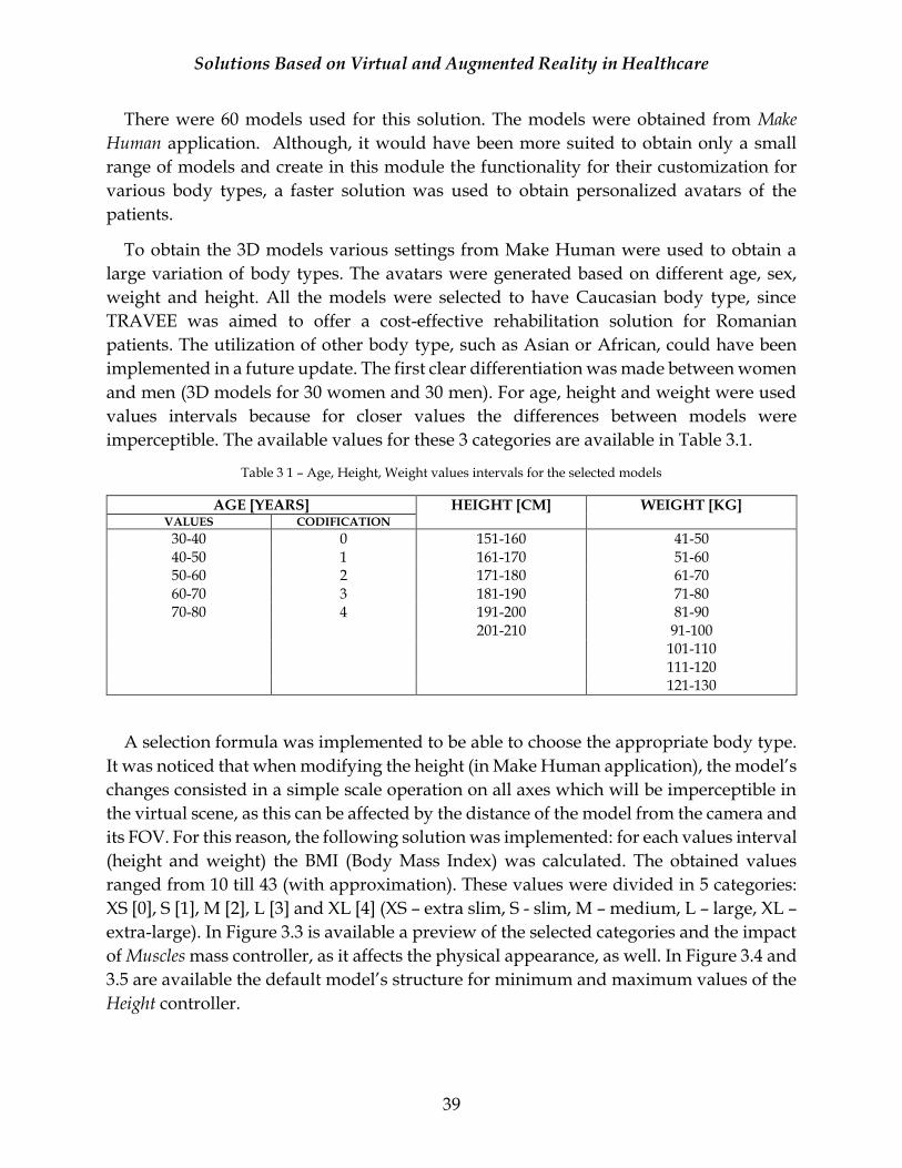
Solutions Based on Virtual and Augmented Reality in Healthcare
39
There were 60 models used for this solution. The models were obtained from Make
Human application. Although, it would have been more suited to obtain only a small
range of models and create in this module the functionality for their customization for
various body types, a faster solution was used to obtain personalized avatars of the
patients.
To obtain the 3D models various settings from Make Human were used to obtain a
large variation of body types. The avatars were generated based on different age, sex,
weight and height. All the models were selected to have Caucasian body type, since
TRAVEE was aimed to offer a cost-effective rehabilitation solution for Romanian
patients. The utilization of other body type, such as Asian or African, could have been
implemented in a future update. The first clear differentiation was made between women
and men (3D models for 30 women and 30 men). For age, height and weight were used
values intervals because for closer values the differences between models were
imperceptible. The available values for these 3 categories are available in Table 3.1.
Table 3 1 – Age, Height, Weight values intervals for the selected models
AGE [YEARS] HEIGHT [CM] WEIGHT [KG] VALUES CODIFICATION
30-40 0 151-160 41-50 40-50 1 161-170 51-60 50-60 2 171-180 61-70 60-70 3 181-190 71-80 70-80 4 191-200 81-90
201-210 91-100 101-110 111-120 121-130
A selection formula was implemented to be able to choose the appropriate body type.
It was noticed that when modifying the height (in Make Human application), the model’s
changes consisted in a simple scale operation on all axes which will be imperceptible in
the virtual scene, as this can be affected by the distance of the model from the camera and
its FOV. For this reason, the following solution was implemented: for each values interval
(height and weight) the BMI (Body Mass Index) was calculated. The obtained values
ranged from 10 till 43 (with approximation). These values were divided in 5 categories:
XS [0], S [1], M [2], L [3] and XL [4] (XS – extra slim, S - slim, M – medium, L – large, XL –
extra-large). In Figure 3.3 is available a preview of the selected categories and the impact
of Muscles mass controller, as it affects the physical appearance, as well. In Figure 3.4 and
3.5 are available the default model’s structure for minimum and maximum values of the
Height controller.
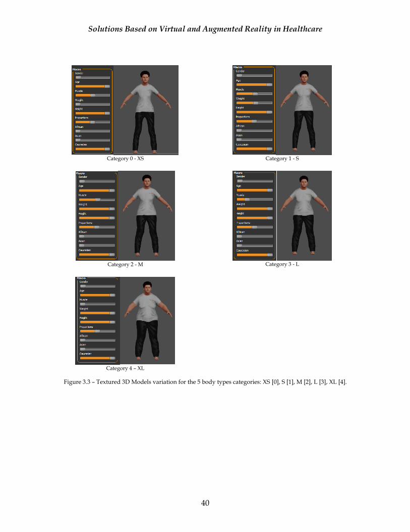
Solutions Based on Virtual and Augmented Reality in Healthcare
40
Category 0 - XS
Category 1 - S
Category 2 - M
Category 3 - L
Category 4 – XL
Figure 3.3 – Textured 3D Models variation for the 5 body types categories: XS [0], S [1], M [2], L [3], XL [4].
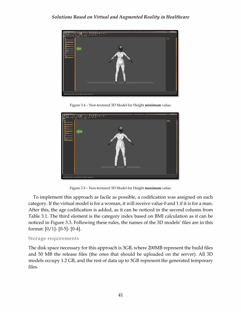
Solutions Based on Virtual and Augmented Reality in Healthcare
41
Figure 3.4 – Non-textured 3D Model for Height minimum value.
Figure 3.5 – Non-textured 3D Model for Height maximum value.
To implement this approach as facile as possible, a codification was assigned on each
category. If the virtual model is for a woman, it will receive value 0 and 1 if it is for a man.
After this, the age codification is added, as it can be noticed in the second column from
Table 3.1. The third element is the category index based on BMI calculation as it can be
noticed in Figure 3.3. Following these rules, the names of the 3D models’ files are in this
format: [0/1]- [0-5]- [0-4].
Storage requirements
The disk space necessary for this approach is 3GB, where 200MB represent the build files
and 50 MB the release files (the ones that should be uploaded on the server). All 3D
models occupy 1.2 GB, and the rest of data up to 3GB represent the generated temporary
files.
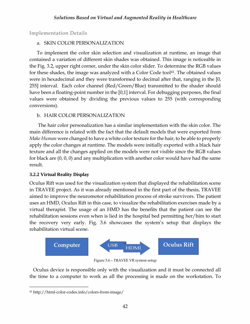
Solutions Based on Virtual and Augmented Reality in Healthcare
42
Implementation Details
a. SKIN COLOR PERSONALIZATION
To implement the color skin selection and visualization at runtime, an image that
contained a variation of different skin shades was obtained. This image is noticeable in
the Fig. 3.2, upper right corner, under the skin color slider. To determine the RGB values
for these shades, the image was analyzed with a Color Code tool41. The obtained values
were in hexadecimal and they were transformed to decimal after that, ranging in the [0,
255] interval. Each color channel (Red/Green/Blue) transmitted to the shader should
have been a floating-point number in the [0,1] interval. For debugging purposes, the final
values were obtained by dividing the previous values to 255 (with corresponding
conversions).
b. HAIR COLOR PERSONALIZATION
The hair color personalization has a similar implementation with the skin color. The
main difference is related with the fact that the default models that were exported from
Make Human were changed to have a white color texture for the hair, to be able to properly
apply the color changes at runtime. The models were initially exported with a black hair
texture and all the changes applied on the models were not visible since the RGB values
for black are (0, 0, 0) and any multiplication with another color would have had the same
result.
3.2.2 Virtual Reality Display
Oculus Rift was used for the visualization system that displayed the rehabilitation scene
in TRAVEE project. As it was already mentioned in the first part of the thesis, TRAVEE
aimed to improve the neuromotor rehabilitation process of stroke survivors. The patient
uses an HMD, Oculus Rift in this case, to visualize the rehabilitation exercises made by a
virtual therapist. The usage of an HMD has the benefits that the patient can see the
rehabilitation sessions even when is lied in the hospital bed permitting her/him to start
the recovery very early. Fig. 3.6 showcases the system’s setup that displays the
rehabilitation virtual scene.
Figure 3.6 – TRAVEE VR system setup
Oculus device is responsible only with the visualization and it must be connected all
the time to a computer to work as all the processing is made on the workstation. To
41 http://html-color-codes.info/colors-from-image/
Computer Oculus Rift
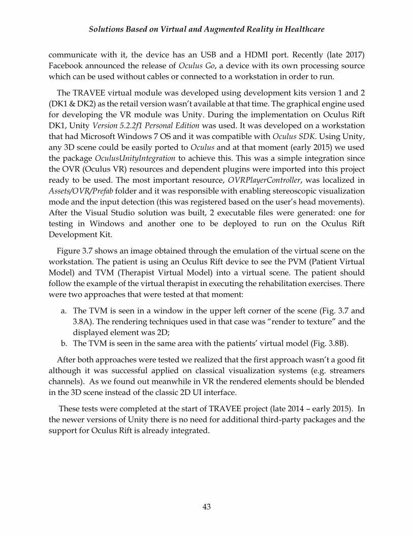
Solutions Based on Virtual and Augmented Reality in Healthcare
43
communicate with it, the device has an USB and a HDMI port. Recently (late 2017)
Facebook announced the release of Oculus Go, a device with its own processing source
which can be used without cables or connected to a workstation in order to run.
The TRAVEE virtual module was developed using development kits version 1 and 2
(DK1 & DK2) as the retail version wasn’t available at that time. The graphical engine used
for developing the VR module was Unity. During the implementation on Oculus Rift
DK1, Unity Version 5.2.2f1 Personal Edition was used. It was developed on a workstation
that had Microsoft Windows 7 OS and it was compatible with Oculus SDK. Using Unity,
any 3D scene could be easily ported to Oculus and at that moment (early 2015) we used
the package OculusUnityIntegration to achieve this. This was a simple integration since
the OVR (Oculus VR) resources and dependent plugins were imported into this project
ready to be used. The most important resource, OVRPlayerController, was localized in
Assets/OVR/Prefab folder and it was responsible with enabling stereoscopic visualization
mode and the input detection (this was registered based on the user’s head movements).
After the Visual Studio solution was built, 2 executable files were generated: one for
testing in Windows and another one to be deployed to run on the Oculus Rift
Development Kit.
Figure 3.7 shows an image obtained through the emulation of the virtual scene on the
workstation. The patient is using an Oculus Rift device to see the PVM (Patient Virtual
Model) and TVM (Therapist Virtual Model) into a virtual scene. The patient should
follow the example of the virtual therapist in executing the rehabilitation exercises. There
were two approaches that were tested at that moment:
a. The TVM is seen in a window in the upper left corner of the scene (Fig. 3.7 and
3.8A). The rendering techniques used in that case was “render to texture” and the
displayed element was 2D;
b. The TVM is seen in the same area with the patients’ virtual model (Fig. 3.8B).
After both approaches were tested we realized that the first approach wasn’t a good fit
although it was successful applied on classical visualization systems (e.g. streamers
channels). As we found out meanwhile in VR the rendered elements should be blended
in the 3D scene instead of the classic 2D UI interface.
These tests were completed at the start of TRAVEE project (late 2014 – early 2015). In
the newer versions of Unity there is no need for additional third-party packages and the
support for Oculus Rift is already integrated.
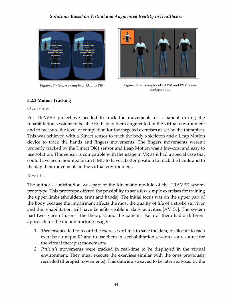
Solutions Based on Virtual and Augmented Reality in Healthcare
44
Figure 3.7 – Scene example on Oculus Rift
Figure 3.8 – Examples of a TVM and PVM scene configuration.
3.2.3 Motion Tracking
Overview
For TRAVEE project we needed to track the movements of a patient during the
rehabilitation sessions to be able to display them augmented in the virtual environment
and to measure the level of completion for the targeted exercises as set by the therapists.
This was achieved with a Kinect sensor to track the body’s skeleton and a Leap Motion
device to track the hands and fingers movements. The fingers movements weren’t
properly tracked by the Kinect DK1 sensor and Leap Motion was a low-cost and easy to
use solution. This sensor is compatible with the usage in VR as it had a special case that
could have been mounted on an HMD to have a better position to track the hands and to
display their movements in the virtual environment.
Results
The author’s contribution was part of the kinematic module of the TRAVEE system
prototype. This prototype offered the possibility to set a few simple exercises for training
the upper limbs (shoulders, arms and hands). The initial focus was on the upper part of
the body because the impairment affects the most the quality of life of a stroke survivor
and the rehabilitation will have benefits visible in daily activities [AV15c]. The system
had two types of users: the therapist and the patient. Each of them had a different
approach for the motion tracking usage:
1. Therapist needed to record the exercises offline, to save the data, to allocate to each
exercise a unique ID and to use them in a rehabilitation session as a resource for
the virtual therapist movements.
2. Patient’s movements were tracked in real-time to be displayed in the virtual
environment. They must execute the exercises similar with the ones previously
recorded (therapist movements). This data is also saved to be later analyzed by the
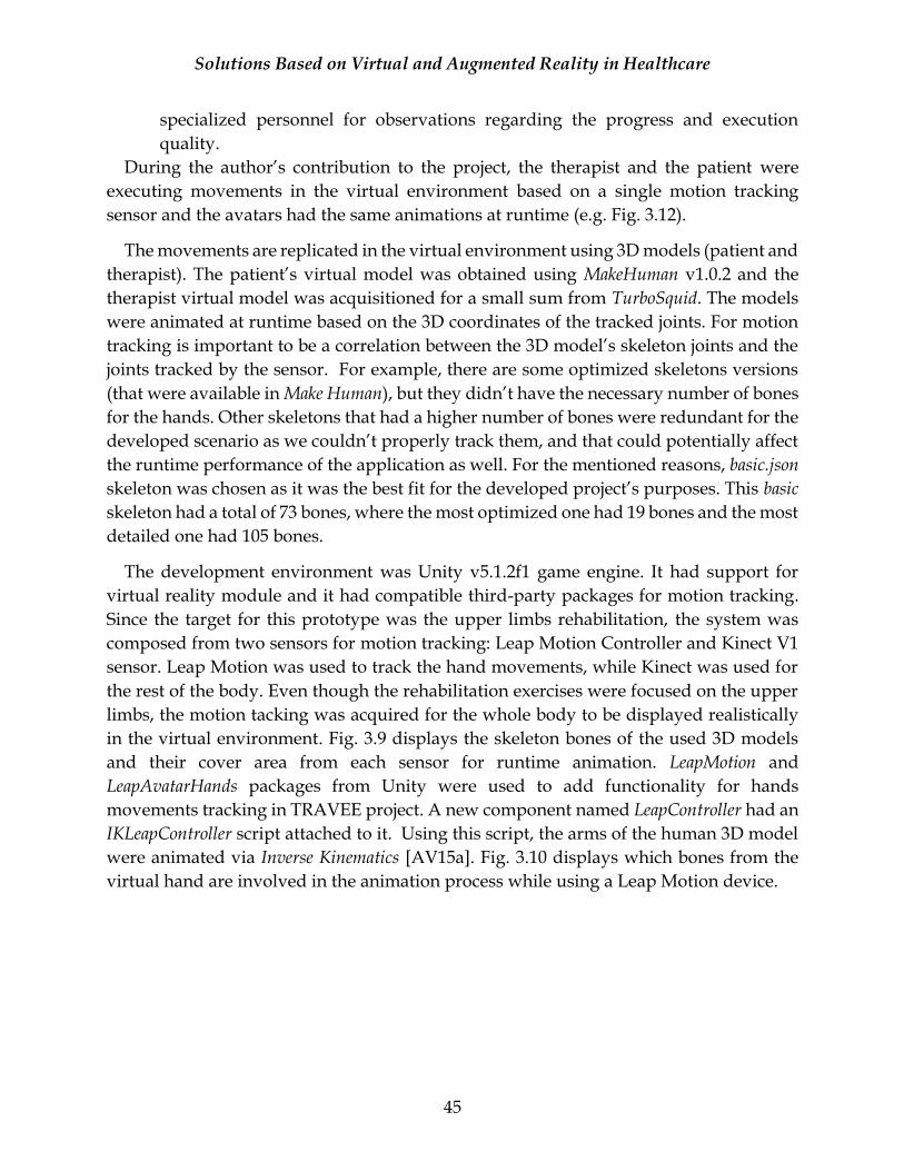
Solutions Based on Virtual and Augmented Reality in Healthcare
45
specialized personnel for observations regarding the progress and execution
quality.
During the author’s contribution to the project, the therapist and the patient were
executing movements in the virtual environment based on a single motion tracking
sensor and the avatars had the same animations at runtime (e.g. Fig. 3.12).
The movements are replicated in the virtual environment using 3D models (patient and
therapist). The patient’s virtual model was obtained using MakeHuman v1.0.2 and the
therapist virtual model was acquisitioned for a small sum from TurboSquid. The models
were animated at runtime based on the 3D coordinates of the tracked joints. For motion
tracking is important to be a correlation between the 3D model’s skeleton joints and the
joints tracked by the sensor. For example, there are some optimized skeletons versions
(that were available in Make Human), but they didn’t have the necessary number of bones
for the hands. Other skeletons that had a higher number of bones were redundant for the
developed scenario as we couldn’t properly track them, and that could potentially affect
the runtime performance of the application as well. For the mentioned reasons, basic.json
skeleton was chosen as it was the best fit for the developed project’s purposes. This basic
skeleton had a total of 73 bones, where the most optimized one had 19 bones and the most
detailed one had 105 bones.
The development environment was Unity v5.1.2f1 game engine. It had support for
virtual reality module and it had compatible third-party packages for motion tracking.
Since the target for this prototype was the upper limbs rehabilitation, the system was
composed from two sensors for motion tracking: Leap Motion Controller and Kinect V1
sensor. Leap Motion was used to track the hand movements, while Kinect was used for
the rest of the body. Even though the rehabilitation exercises were focused on the upper
limbs, the motion tacking was acquired for the whole body to be displayed realistically
in the virtual environment. Fig. 3.9 displays the skeleton bones of the used 3D models
and their cover area from each sensor for runtime animation. LeapMotion and
LeapAvatarHands packages from Unity were used to add functionality for hands
movements tracking in TRAVEE project. A new component named LeapController had an
IKLeapController script attached to it. Using this script, the arms of the human 3D model
were animated via Inverse Kinematics [AV15a]. Fig. 3.10 displays which bones from the
virtual hand are involved in the animation process while using a Leap Motion device.
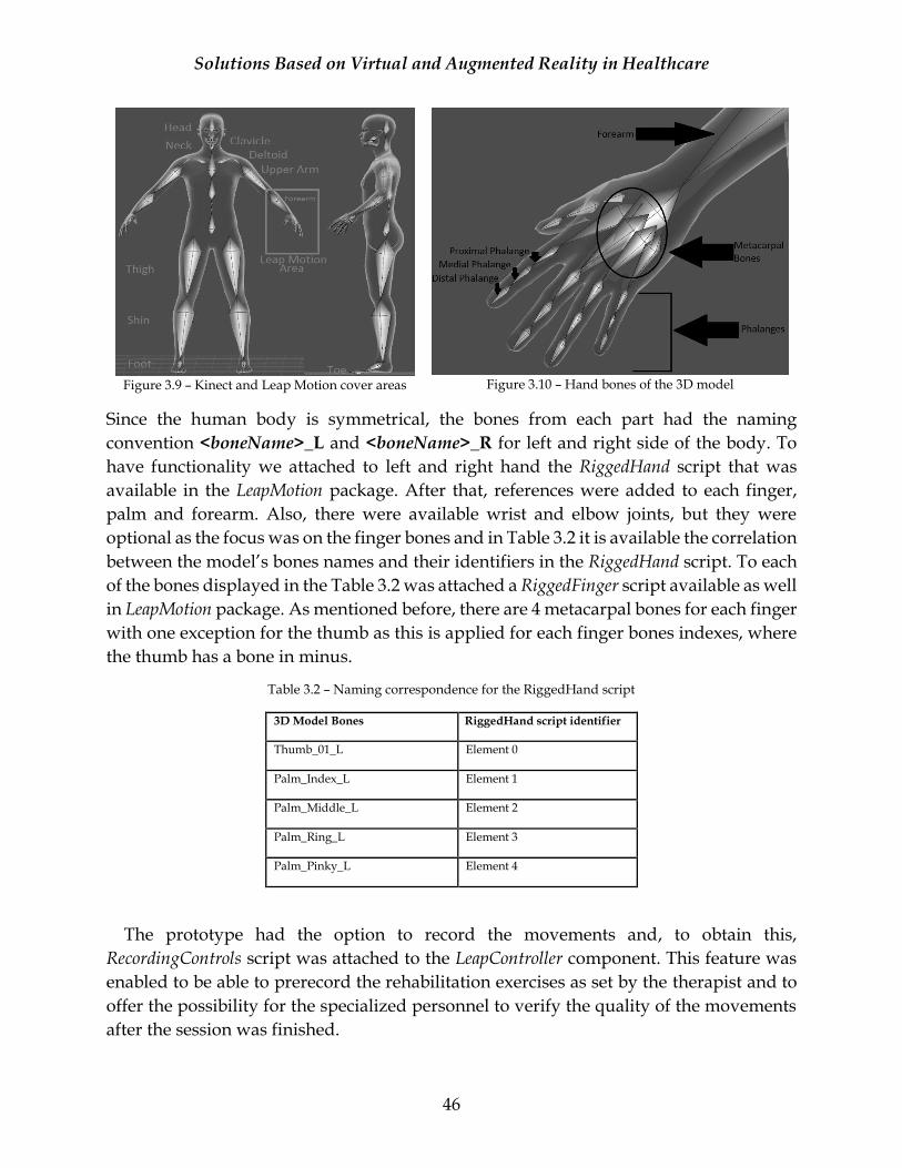
Solutions Based on Virtual and Augmented Reality in Healthcare
46
Figure 3.9 – Kinect and Leap Motion cover areas
Figure 3.10 – Hand bones of the 3D model
Since the human body is symmetrical, the bones from each part had the naming
convention <boneName>_L and <boneName>_R for left and right side of the body. To
have functionality we attached to left and right hand the RiggedHand script that was
available in the LeapMotion package. After that, references were added to each finger,
palm and forearm. Also, there were available wrist and elbow joints, but they were
optional as the focus was on the finger bones and in Table 3.2 it is available the correlation
between the model’s bones names and their identifiers in the RiggedHand script. To each
of the bones displayed in the Table 3.2 was attached a RiggedFinger script available as well
in LeapMotion package. As mentioned before, there are 4 metacarpal bones for each finger
with one exception for the thumb as this is applied for each finger bones indexes, where
the thumb has a bone in minus.
Table 3.2 – Naming correspondence for the RiggedHand script
3D Model Bones RiggedHand script identifier
Thumb_01_L Element 0
Palm_Index_L Element 1
Palm_Middle_L Element 2
Palm_Ring_L Element 3
Palm_Pinky_L Element 4
The prototype had the option to record the movements and, to obtain this,
RecordingControls script was attached to the LeapController component. This feature was
enabled to be able to prerecord the rehabilitation exercises as set by the therapist and to
offer the possibility for the specialized personnel to verify the quality of the movements
after the session was finished.
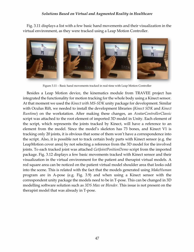
Solutions Based on Virtual and Augmented Reality in Healthcare
47
Fig. 3.11 displays a list with a few basic hand movements and their visualization in the
virtual environment, as they were tracked using a Leap Motion Controller.
Figure 3.11 – Basic hand movements tracked in real-time with Leap Motion Controller
Besides a Leap Motion device, the kinematics module from TRAVEE project has
integrated the functionality for motion tracking for the whole body using a Kinect sensor.
At that moment we used the Kinect with MS-SDK unity package for development. Similar
with Oculus Rift, we needed to install the development libraries (Kinect SDK and Kinect
Runtime) on the workstation. After making these changes, an AvatarControllerClassic
script was attached to the root element of imported 3D model in Unity. Each element of
the script, which represents the joints tracked by Kinect, will have a reference to an
element from the model. Since the model’s skeleton has 73 bones, and Kinect V1 is
tracking only 20 joints, it is obvious that some of them won’t have a correspondence into
the script. Also, it is possible not to track certain body parts with Kinect sensor (e.g. the
LeapMotion cover area) by not selecting a reference from the 3D model for the involved
joints. To each tracked joint was attached GetJointPositionDemo script from the imported
package. Fig. 3.12 displays a few basic movements tracked with Kinect sensor and their
visualization in the virtual environment for the patient and therapist virtual models. A
red square area can be noticed on the patient virtual model shoulder area that looks odd
into the scene. This is related with the fact that the models generated using MakeHuman
program are in A-pose (e.g. Fig. 3.9) and when using a Kinect sensor with the
correspondent unity package the models need to be in T-pose. This can be changed in 3D
modelling software solution such as 3DS Max or Blender. This issue is not present on the
therapist model that was already in T-pose.
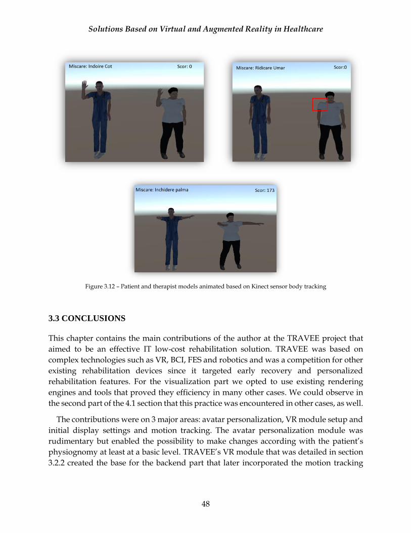
Solutions Based on Virtual and Augmented Reality in Healthcare
48
Figure 3.12 – Patient and therapist models animated based on Kinect sensor body tracking
3.3 CONCLUSIONS
This chapter contains the main contributions of the author at the TRAVEE project that
aimed to be an effective IT low-cost rehabilitation solution. TRAVEE was based on
complex technologies such as VR, BCI, FES and robotics and was a competition for other
existing rehabilitation devices since it targeted early recovery and personalized
rehabilitation features. For the visualization part we opted to use existing rendering
engines and tools that proved they efficiency in many other cases. We could observe in
the second part of the 4.1 section that this practice was encountered in other cases, as well.
The contributions were on 3 major areas: avatar personalization, VR module setup and
initial display settings and motion tracking. The avatar personalization module was
rudimentary but enabled the possibility to make changes according with the patient’s
physiognomy at least at a basic level. TRAVEE’s VR module that was detailed in section
3.2.2 created the base for the backend part that later incorporated the motion tracking

Solutions Based on Virtual and Augmented Reality in Healthcare
49
features. The third-party packages that were used in Unity had some tracking errors, but
they were a real help into developing faster these features.
These contributions can be considered an intermediary step for the research as the next
project (Interactive Biomechanics Lessons) used common elements such as VR and
motion tracking to provide a novel IT educational solution for biomechanics study.
Solutions Based on Virtual and Augmented Reality in Healthcare
50
CHAPTER 4
AUGMENTED AND VIRTUAL REALITY IN MEDICAL
EDUCATION This chapter contains the second part of the research and the most extensive one. Our aim
was to continue the research of VR and AR usability in Healthcare. We considered an
opportunity to develop applications that contain a few interactive biomechanics lessons.
The related work section contains a brief literature review for existing AR and VR
solutions used in Healthcare. It was considered an opportunity to focus on a novel
approach that aimed the usage of interactive technologies in medical education.
4.1 RELATED WORK
This subchapter contains an overview of the areas of interest regarding the usage of
virtual and augmented reality in healthcare. The research’s goal was to comprehend the
state of the art and was completed in early 2016. The target was to focus especially on
applications and scientific papers published after 01.01.2014 to have a clear picture of the
newest technologies and approaches. We are aware that a few additional solutions and
research topics that could be of interest might have been published meanwhile. In any
case, the ideas of our own approach were tested through audience feedback during
presentations at various conferences. The Interactive Biomechanics Lessons project
relates to the data presented in this section and its idea was built based on the information
extracted from the literature review and the available solutions.
The initial batch of reviewed scientific papers contained an extensive period to make
sure we weren’t missing interesting topics that might be implemented differently or
better with the current technology as opposed with what was available a few years ago,
although the aim was to look mainly at newer research topics. We gathered a total of 77
scientific papers, where 47 are published starting with 2014 and the other 30 were
published in the between 1999-2013. More details are available in Figure 4.1 regarding
the topics related with this literature review.
From the categories and subcategories mentioned in the Figure 4.1, we considered to
detail the ones that contained the most important information for our topic. Also, a part
of the data extracted from the General/Overview category and Cybersickness is mentioned
in the second chapter as they include some general insights regarding VR and AR. The
ones excluded were considered unfit for our research topic. However, from all the papers
reviewed there was overlapping information between them. Note that the rehabilitation
related solutions were excluded from the search.
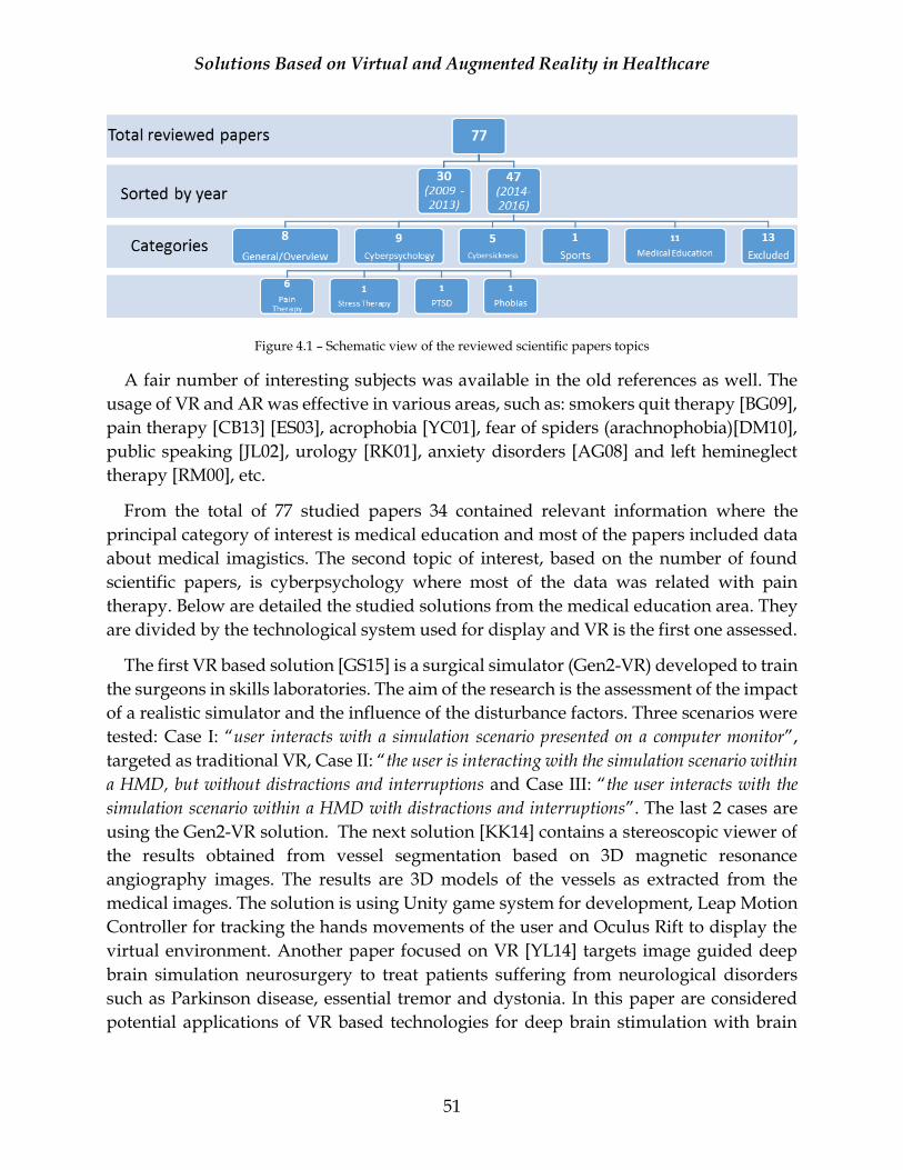
Solutions Based on Virtual and Augmented Reality in Healthcare
51
Figure 4.1 – Schematic view of the reviewed scientific papers topics
A fair number of interesting subjects was available in the old references as well. The
usage of VR and AR was effective in various areas, such as: smokers quit therapy [BG09],
pain therapy [CB13] [ES03], acrophobia [YC01], fear of spiders (arachnophobia)[DM10],
public speaking [JL02], urology [RK01], anxiety disorders [AG08] and left hemineglect
therapy [RM00], etc.
From the total of 77 studied papers 34 contained relevant information where the
principal category of interest is medical education and most of the papers included data
about medical imagistics. The second topic of interest, based on the number of found
scientific papers, is cyberpsychology where most of the data was related with pain
therapy. Below are detailed the studied solutions from the medical education area. They
are divided by the technological system used for display and VR is the first one assessed.
The first VR based solution [GS15] is a surgical simulator (Gen2-VR) developed to train
the surgeons in skills laboratories. The aim of the research is the assessment of the impact
of a realistic simulator and the influence of the disturbance factors. Three scenarios were
tested: Case I: “user interacts with a simulation scenario presented on a computer monitor”,
targeted as traditional VR, Case II: “the user is interacting with the simulation scenario within
a HMD, but without distractions and interruptions and Case III: “the user interacts with the
simulation scenario within a HMD with distractions and interruptions”. The last 2 cases are
using the Gen2-VR solution. The next solution [KK14] contains a stereoscopic viewer of
the results obtained from vessel segmentation based on 3D magnetic resonance
angiography images. The results are 3D models of the vessels as extracted from the
medical images. The solution is using Unity game system for development, Leap Motion
Controller for tracking the hands movements of the user and Oculus Rift to display the
virtual environment. Another paper focused on VR [YL14] targets image guided deep
brain simulation neurosurgery to treat patients suffering from neurological disorders
such as Parkinson disease, essential tremor and dystonia. In this paper are considered
potential applications of VR based technologies for deep brain stimulation with brain
Solutions Based on Virtual and Augmented Reality in Healthcare
52
magnetic resonance imagining data. The last paper [SL15] mentions the usage of 3D
technologies and stereoscopic visualization in medical endoscopic teleoperation.
The AR related papers are in majority related with medical imagistics. Reference
[CK14] contains three examples of AR solutions used in medical training:
a. Visualizing human anatomical structure. The mentioned solution augments CT
data onto the body of a user. The project is named magic mirror “Miracle” and
displays the training sessions on a TV while the user movements are tracked
using a Kinect device.
b. Visualizing human 3D lung dynamics. The solution is based on a system that
allows real-time visualization of the lung dynamics superimposed on a patient
in the OR (Operating Room).
c. Laparoscopy skills training. The laparoscopy environments based on augmented
reality offer realistic haptic feedback crucial for the development of the
necessary skills.
A video see-through solution [FC14] uses the AR HMD in the medical procedures, such
as: maxillofacial surgical clinical study, orthopedic surgical clinical study and AR
magnetic guidance of endovascular device. Another system that contains the usage of a
HMD [SY14] uses a vison finger tracking technique applied to medical education.
Reference [ZY15] contains a review of AR in OR applications, more precisely the
augmented visualization in surgical navigation. Based on the medical imagistics
modality, 4 situations of augmented guidance were approached:
a. Augmented X-ray guidance.
b. Augmented ultrasound guidance.
c. Augmented video and SPECT (Single-photon emission computer
tomography) guidance.
d. Augmented endoscopic video guidance.
The last 3 scientific papers [MCFM14a] [MCFM14b] [MCFM15] refer to different areas
of development for an AR based solution for in-situ visualization of the craniofacial
region. The augmentation is done using medical images such as MRI (Magnetic
Resonance Imagistics) or CT (Computed Tomography). Regarding the augmentation
tracking, initially a semi-automatic markerless augmented reality approach was
considered [MCFM14a] and later a markerless AR environment was implemented
[MCFM15]. The markerless live tracking was completed based on the registration
between a 3D reference model and the 3D model captured with the tracking sensor.
Besides the initial assessment, during our research we discovered another application
that served as example. Reference [BP14] contains details regarding a solution dedicated
for biomechanics study by visualizing the lower limbs muscles activity in real time. The
Solutions Based on Virtual and Augmented Reality in Healthcare
53
presented material targets a small set of lower limb movements such as knee flexion and
extension. It showcases in real-time the muscles activations on a 3D avatar based on the
movements of an observed user. The muscles activation data was acquired separately
(preliminary) using a Biopac MP150 device with non-invasive EMG and was added into
a database while assigning the tracked movements type. This operation was effectuated
separately to improve the runtime performance by minimizing the required
computations. The movement of the observed user was registered using a Kinect sensor,
NiTE and OpenNi SDK42. Fifteen joints are tracked by this system while the 3D models of
the lower limbs are imported from MyCorporisFabrica43. C++ was used for framework
development and the main application runs on a Laptop PC while the feedback is
provided on the computer’s screen. All the tests were completed in the same room to
preserve similar lighting conditions. As mentioned by the authors, the AR visual
feedback needed improvements as it shown separately the animated muscles models.
If we look at the resources available for learning, we can observe that they are diverse.
The basic selection criteria, when it comes to educational applications, is based on the
quality of the information, the setup difficulty and the cost. Based on the scientific papers
studied we considered an opportunity to develop a solution that targets biomechanics
study of the human movements. It has similarities with the previous topic (TRAVEE
project) hence the thesis could maintain the same research note. The project is complex,
so practices found in the related literature were applied during its development. For
example, a part of the 3D models used in the applications are obtained from medical
images. Two approaches for augmented reality display were considered and 4 different
scenarios were developed for both AR and VR [AV18].
4.2 CONTRIBUTIONS
In this subchapter we present the results of our research. After making an in-depth
analysis of the existing solutions based on virtual and augmented reality in healthcare
we observed the opportunity of developing a solution that targets medical education and
more exactly to improve the learning process of biomechanics study. We want to
demonstrate that the usage of VR and AR in medical education can be a plus. We consider
that today’s technology can aid the educational process as we have the opportunity to
unlock a new visualization method that can add details on the fly to the observed
environment.
42 NiTE and Open NI online page: http://openni.ru/files/nite/ 43 MyCorporisFabrica: http://www.mycorporisfabrica.org/.
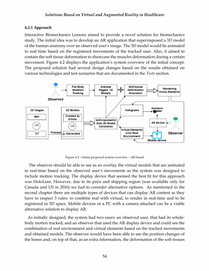
Solutions Based on Virtual and Augmented Reality in Healthcare
54
4.2.1 Approach
Interactive Biomechanics Lessons aimed to provide a novel solution for biomechanics
study. The initial idea was to develop an AR application that superimposed a 3D model
of the human anatomy over an observed user’s image. The 3D model would be animated
in real time based on the registered movements of the tracked user. Also, it aimed to
contain the soft tissue deformation to showcase the muscles deformation during a certain
movement. Figure 4.2 displays the application’s system overview of the initial concept.
The proposed solution had several design changes based on the results obtained on
various technologies and test scenarios that are documented in the Tests section.
Figure 4.2 – Initial proposed system overview – AR based
The observer should be able to see as an overlay the virtual models that are animated
in real-time based on the observed user’s movements as the system was designed to
include motion tracking. The display device that seemed the best fit for this approach
was HoloLens. However, due to its price and shipping region (was available only for
Canada and US in 2016) we had to consider alternative options. As mentioned in the
second chapter there are multiple types of devices that can display AR content as they
have to respect 3 rules: to combine real with virtual, to render in real-time and to be
registered in 3D space. Mobile devices or a PC with a camera attached can be a viable
alternative solution to display AR.
As initially designed, the system had two users: an observed user, that had its whole-
body motion tracked, and an observer that used the AR display device and could see the
combination of real environment and virtual elements based on the tracked movements
and obtained models. The observer would have been able to see the position changes of
the bones and, on top of that, as an extra information, the deformation of the soft tissues
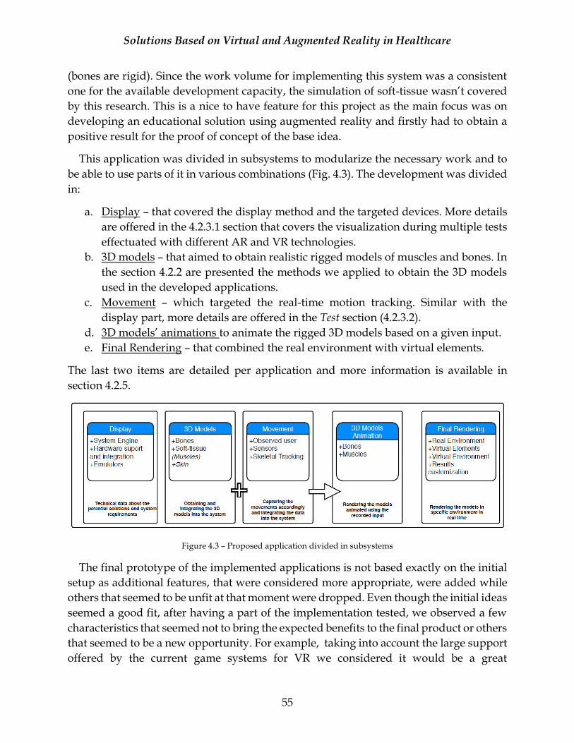
Solutions Based on Virtual and Augmented Reality in Healthcare
55
(bones are rigid). Since the work volume for implementing this system was a consistent
one for the available development capacity, the simulation of soft-tissue wasn’t covered
by this research. This is a nice to have feature for this project as the main focus was on
developing an educational solution using augmented reality and firstly had to obtain a
positive result for the proof of concept of the base idea.
This application was divided in subsystems to modularize the necessary work and to
be able to use parts of it in various combinations (Fig. 4.3). The development was divided
in:
a. Display – that covered the display method and the targeted devices. More details
are offered in the 4.2.3.1 section that covers the visualization during multiple tests
effectuated with different AR and VR technologies.
b. 3D models – that aimed to obtain realistic rigged models of muscles and bones. In
the section 4.2.2 are presented the methods we applied to obtain the 3D models
used in the developed applications.
c. Movement – which targeted the real-time motion tracking. Similar with the
display part, more details are offered in the Test section (4.2.3.2).
d. 3D models’ animations to animate the rigged 3D models based on a given input.
e. Final Rendering – that combined the real environment with virtual elements.
The last two items are detailed per application and more information is available in
section 4.2.5.
Figure 4.3 – Proposed application divided in subsystems
The final prototype of the implemented applications is not based exactly on the initial
setup as additional features, that were considered more appropriate, were added while
others that seemed to be unfit at that moment were dropped. Even though the initial ideas
seemed a good fit, after having a part of the implementation tested, we observed a few
characteristics that seemed not to bring the expected benefits to the final product or others
that seemed to be a new opportunity. For example, taking into account the large support
offered by the current game systems for VR we considered it would be a great
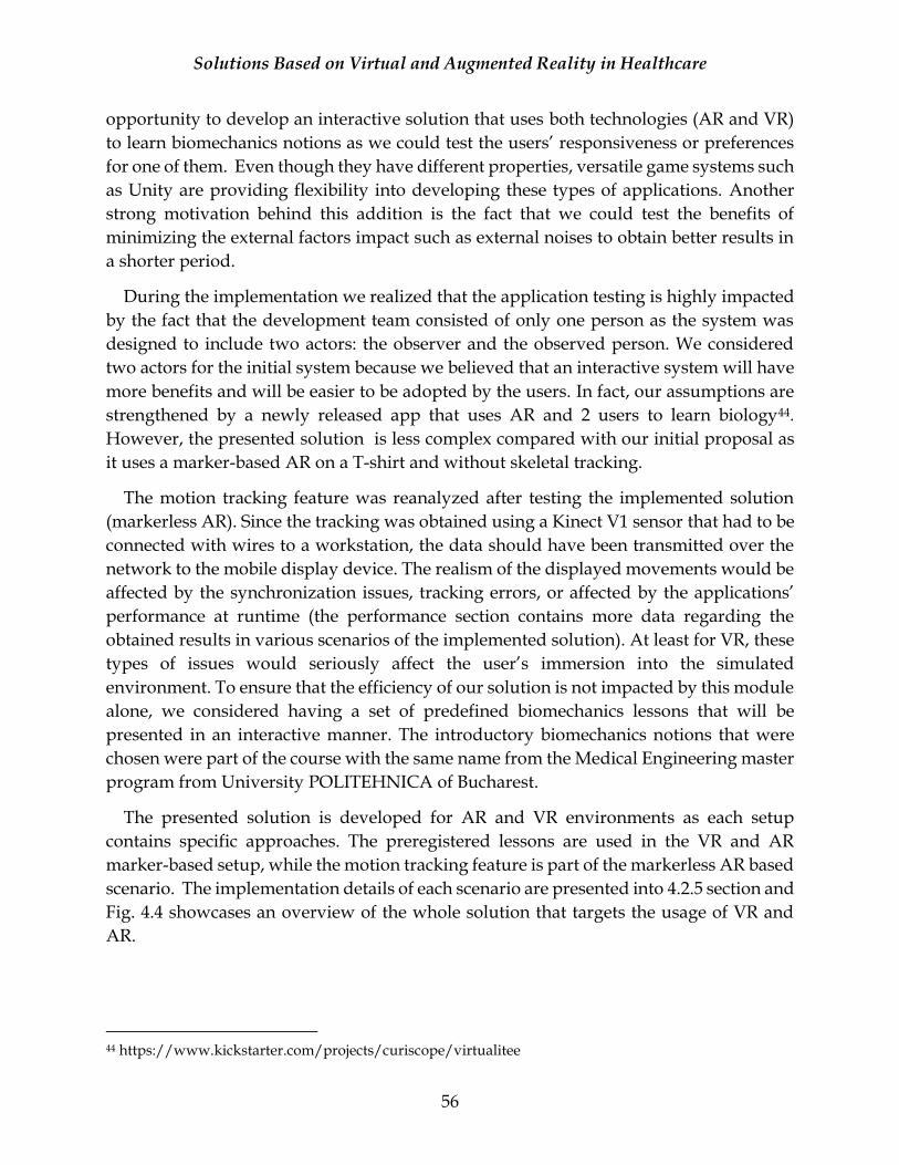
Solutions Based on Virtual and Augmented Reality in Healthcare
56
opportunity to develop an interactive solution that uses both technologies (AR and VR)
to learn biomechanics notions as we could test the users’ responsiveness or preferences
for one of them. Even though they have different properties, versatile game systems such
as Unity are providing flexibility into developing these types of applications. Another
strong motivation behind this addition is the fact that we could test the benefits of
minimizing the external factors impact such as external noises to obtain better results in
a shorter period.
During the implementation we realized that the application testing is highly impacted
by the fact that the development team consisted of only one person as the system was
designed to include two actors: the observer and the observed person. We considered
two actors for the initial system because we believed that an interactive system will have
more benefits and will be easier to be adopted by the users. In fact, our assumptions are
strengthened by a newly released app that uses AR and 2 users to learn biology44.
However, the presented solution is less complex compared with our initial proposal as
it uses a marker-based AR on a T-shirt and without skeletal tracking.
The motion tracking feature was reanalyzed after testing the implemented solution
(markerless AR). Since the tracking was obtained using a Kinect V1 sensor that had to be
connected with wires to a workstation, the data should have been transmitted over the
network to the mobile display device. The realism of the displayed movements would be
affected by the synchronization issues, tracking errors, or affected by the applications’
performance at runtime (the performance section contains more data regarding the
obtained results in various scenarios of the implemented solution). At least for VR, these
types of issues would seriously affect the user’s immersion into the simulated
environment. To ensure that the efficiency of our solution is not impacted by this module
alone, we considered having a set of predefined biomechanics lessons that will be
presented in an interactive manner. The introductory biomechanics notions that were
chosen were part of the course with the same name from the Medical Engineering master
program from University POLITEHNICA of Bucharest.
The presented solution is developed for AR and VR environments as each setup
contains specific approaches. The preregistered lessons are used in the VR and AR
marker-based setup, while the motion tracking feature is part of the markerless AR based
scenario. The implementation details of each scenario are presented into 4.2.5 section and
Fig. 4.4 showcases an overview of the whole solution that targets the usage of VR and
AR.
44 https://www.kickstarter.com/projects/curiscope/virtualitee
Solutions Based on Virtual and Augmented Reality in Healthcare
57
Figure 4.4 – System overview to support VR and AR
4.2.2 3D Models
A part of the contributions is represented by the methods we used to obtain realistic 3D
models that were integrated into the applications. These models serve as avatars for the
designed applications and they are characters in the VR and AR scenes. A cost-effective
solution should be implemented since the level of realism needs to be at an acceptable
standard and since perfect faithfulness is impossible [JVWR14].
This subchapter showcases how we obtained the 3D models of the human
musculoskeletal system and skin. Two options were considered to obtain these models:
a. To purchase them from an online 3D models store.
b. To generate them, since the models that have a high degree of realism and details
usually have a high cost.
The cost of these types of models varied from 500-600$ for a rigged model of bones and
muscles and went up to 2000$ for the high-quality ones. However, these models weren’t
perfect as there were reviews that mentioned the clear differences between them and the
biological data or the fact that the abdominal muscles meshes were a simple texture
[AV16a]. Some solutions that targeted medical education used raw 3D images that were

Solutions Based on Virtual and Augmented Reality in Healthcare
58
rendered as virtual elements in an AR environment [MCFM15] [TB12] [CK14]. Others,
such as the one from [KK14] used 3D models of blood vessels generated from 3D
magnetic resonance angiography images. However, the level of complexity of those
models is much smaller compared with the ones presented in this section.
The first set of models were obtained from medical images for two reasons: realism
and cost efficiency. For specialists that are not graduated in art and 3D modelling,
creating a human musculoskeletal system from nothing is a very challenging task. The
methods we applied for obtaining 3D models from medical images are detailed in the
next section. Due to some implementation limitations we continued searching for
relevant 3D models suited for our research topic. We managed to find alternative options
that became available during the implementation period.
4.2.2.1 3D Models Obtained from Medical Images
MRI (Magnetic Resonance Imaging) or CT (Computed Tomography) datasets can be used
to generate the 3D models. To obtain the needed data a professional software such as
SimpleWare Scan IP45 or 3D Doctor46 can be used. Both solutions aren’t free, but we
managed to obtain trial access to SimpleWare Scan IP software for a limited period in
which we could proceed fast to obtain our data. This means that additional issues that
were discovered after the access was removed couldn’t have been modified but the
quality of this solution was far superior to others. These models will be animated at
runtime and they need to have a bone hierarchy attached and to be skinned. The initial
results are static meshes that cannot be animated at runtime without additional changes
regarding rigging and skinning.
Although the focus is on the human musculoskeletal system, we generated the skin
layer as well to be able to test an alternative scenario regarding the immersion of the users
as this will make the users see initially a realistic human body at the first interaction with
the application.
As previously mentioned, the quality of the models and their fidelity with the
biological data is important for the developed application. However, these types of
models tend to be very complex, with a large number of vertices that can overload the
application and can impact the users’ experience. To be able to improve the quality of the
resulted models a significant additional time was necessary, hence a balance between the
realism, cost effectiveness and development time had to be chosen.
45 https://www.simpleware.com/software/scanip/ 46 http://www.ablesw.com/3d-doctor/
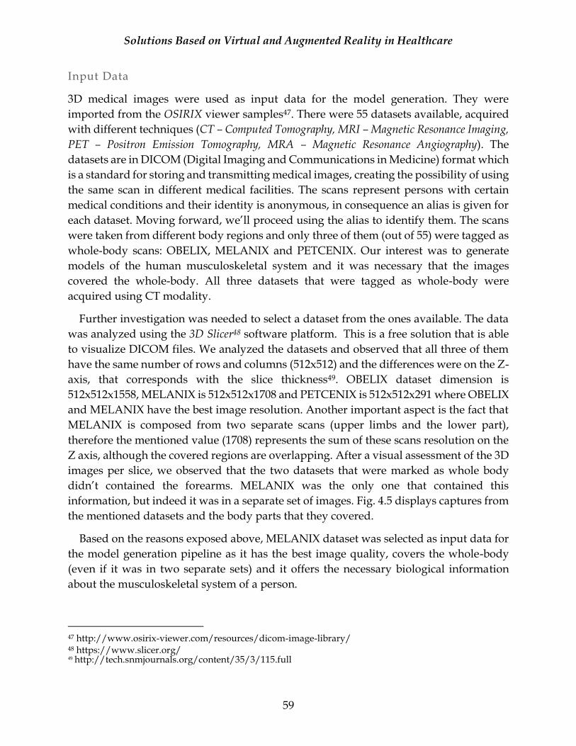
Solutions Based on Virtual and Augmented Reality in Healthcare
59
Input Data
3D medical images were used as input data for the model generation. They were
imported from the OSIRIX viewer samples47. There were 55 datasets available, acquired
with different techniques (CT – Computed Tomography, MRI – Magnetic Resonance Imaging,
PET – Positron Emission Tomography, MRA – Magnetic Resonance Angiography). The
datasets are in DICOM (Digital Imaging and Communications in Medicine) format which
is a standard for storing and transmitting medical images, creating the possibility of using
the same scan in different medical facilities. The scans represent persons with certain
medical conditions and their identity is anonymous, in consequence an alias is given for
each dataset. Moving forward, we’ll proceed using the alias to identify them. The scans
were taken from different body regions and only three of them (out of 55) were tagged as
whole-body scans: OBELIX, MELANIX and PETCENIX. Our interest was to generate
models of the human musculoskeletal system and it was necessary that the images
covered the whole-body. All three datasets that were tagged as whole-body were
acquired using CT modality.
Further investigation was needed to select a dataset from the ones available. The data
was analyzed using the 3D Slicer48 software platform. This is a free solution that is able
to visualize DICOM files. We analyzed the datasets and observed that all three of them
have the same number of rows and columns (512x512) and the differences were on the Z-
axis, that corresponds with the slice thickness49. OBELIX dataset dimension is
512x512x1558, MELANIX is 512x512x1708 and PETCENIX is 512x512x291 where OBELIX
and MELANIX have the best image resolution. Another important aspect is the fact that
MELANIX is composed from two separate scans (upper limbs and the lower part),
therefore the mentioned value (1708) represents the sum of these scans resolution on the
Z axis, although the covered regions are overlapping. After a visual assessment of the 3D
images per slice, we observed that the two datasets that were marked as whole body
didn’t contained the forearms. MELANIX was the only one that contained this
information, but indeed it was in a separate set of images. Fig. 4.5 displays captures from
the mentioned datasets and the body parts that they covered.
Based on the reasons exposed above, MELANIX dataset was selected as input data for
the model generation pipeline as it has the best image quality, covers the whole-body
(even if it was in two separate sets) and it offers the necessary biological information
about the musculoskeletal system of a person.
47 http://www.osirix-viewer.com/resources/dicom-image-library/ 48 https://www.slicer.org/ 49 http://tech.snmjournals.org/content/35/3/115.full
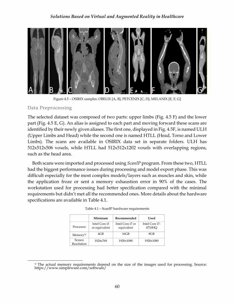
Solutions Based on Virtual and Augmented Reality in Healthcare
60
Figure 4.5 – OSIRIX samples: OBELIX [A, B], PETCENIX [C, D], MELANIX [E, F, G]
Data Preprocessing
The selected dataset was composed of two parts: upper limbs (Fig. 4.5 F) and the lower
part (Fig. 4.5 E, G). An alias is assigned to each part and moving forward these scans are
identified by their newly given aliases. The first one, displayed in Fig. 4.5F, is named ULH
(Upper Limbs and Head) while the second one is named HTLL (Head, Torso and Lower
Limbs). The scans are available in OSIRIX data set in separate folders. ULH has
512x512x506 voxels, while HTLL had 512x512x1202 voxels with overlapping regions,
such as the head area.
Both scans were imported and processed using ScanIP program. From these two, HTLL
had the biggest performance issues during processing and model export phase. This was
difficult especially for the most complex models/layers such as muscles and skin, while
the application froze or sent a memory exhaustion error in 90% of the cases. The
workstation used for processing had better specification compared with the minimal
requirements but didn’t met all the recommended ones. More details about the hardware
specifications are available in Table 4.1.
Table 4.1 – ScanIP hardware requirements
Minimum Recommended Used
Processor Intel Core i3 or equivalent
Intel Core i7 or equivalent
Intel Core i7-4710HQ
Memory50 4GB 16GB 8GB
Screen Resolution
1024x768 1920x1080 1920x1080
50 The actual memory requirements depend on the size of the images used for processing. Source: https://www.simpleware.com/software/
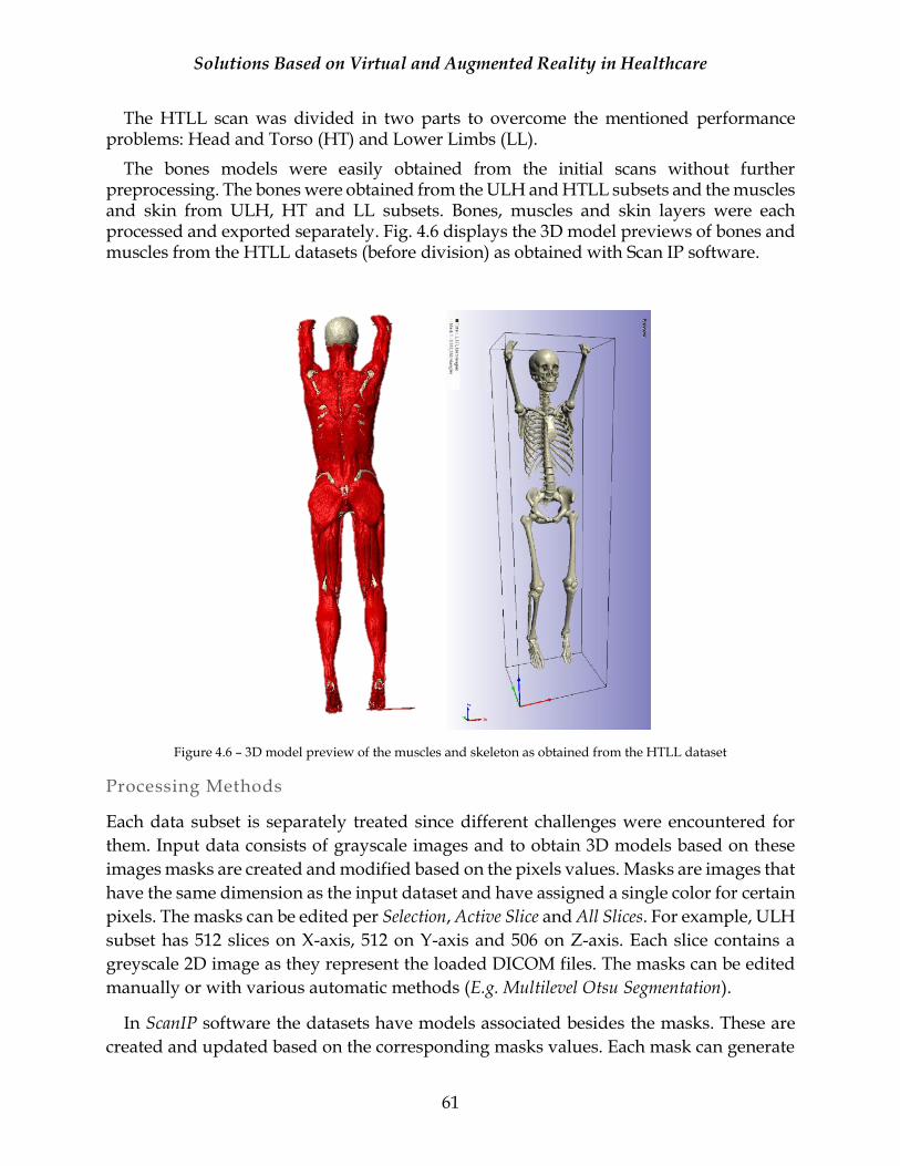
Solutions Based on Virtual and Augmented Reality in Healthcare
61
The HTLL scan was divided in two parts to overcome the mentioned performance problems: Head and Torso (HT) and Lower Limbs (LL).
The bones models were easily obtained from the initial scans without further preprocessing. The bones were obtained from the ULH and HTLL subsets and the muscles and skin from ULH, HT and LL subsets. Bones, muscles and skin layers were each processed and exported separately. Fig. 4.6 displays the 3D model previews of bones and muscles from the HTLL datasets (before division) as obtained with Scan IP software.
Figure 4.6 – 3D model preview of the muscles and skeleton as obtained from the HTLL dataset
Processing Methods
Each data subset is separately treated since different challenges were encountered for
them. Input data consists of grayscale images and to obtain 3D models based on these
images masks are created and modified based on the pixels values. Masks are images that
have the same dimension as the input dataset and have assigned a single color for certain
pixels. The masks can be edited per Selection, Active Slice and All Slices. For example, ULH
subset has 512 slices on X-axis, 512 on Y-axis and 506 on Z-axis. Each slice contains a
greyscale 2D image as they represent the loaded DICOM files. The masks can be edited
manually or with various automatic methods (E.g. Multilevel Otsu Segmentation).
In ScanIP software the datasets have models associated besides the masks. These are
created and updated based on the corresponding masks values. Each mask can generate
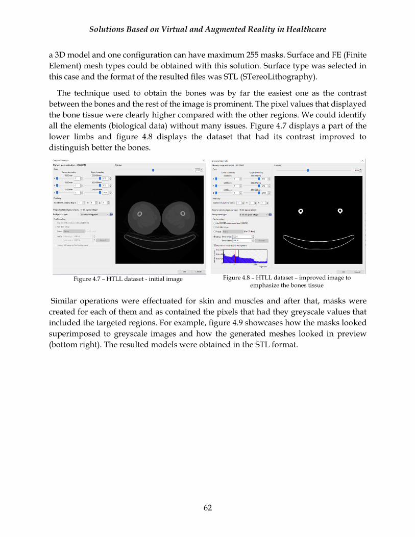
Solutions Based on Virtual and Augmented Reality in Healthcare
62
a 3D model and one configuration can have maximum 255 masks. Surface and FE (Finite
Element) mesh types could be obtained with this solution. Surface type was selected in
this case and the format of the resulted files was STL (STereoLithography).
The technique used to obtain the bones was by far the easiest one as the contrast
between the bones and the rest of the image is prominent. The pixel values that displayed
the bone tissue were clearly higher compared with the other regions. We could identify
all the elements (biological data) without many issues. Figure 4.7 displays a part of the
lower limbs and figure 4.8 displays the dataset that had its contrast improved to
distinguish better the bones.
Figure 4.7 – HTLL dataset - initial image
Figure 4.8 – HTLL dataset – improved image to
emphasize the bones tissue
Similar operations were effectuated for skin and muscles and after that, masks were
created for each of them and as contained the pixels that had they greyscale values that
included the targeted regions. For example, figure 4.9 showcases how the masks looked
superimposed to greyscale images and how the generated meshes looked in preview
(bottom right). The resulted models were obtained in the STL format.
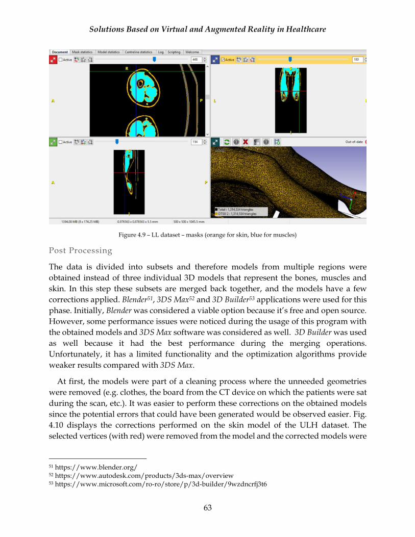
Solutions Based on Virtual and Augmented Reality in Healthcare
63
Figure 4.9 – LL dataset – masks (orange for skin, blue for muscles)
Post Processing
The data is divided into subsets and therefore models from multiple regions were
obtained instead of three individual 3D models that represent the bones, muscles and
skin. In this step these subsets are merged back together, and the models have a few
corrections applied. Blender51, 3DS Max52 and 3D Builder53 applications were used for this
phase. Initially, Blender was considered a viable option because it’s free and open source.
However, some performance issues were noticed during the usage of this program with
the obtained models and 3DS Max software was considered as well. 3D Builder was used
as well because it had the best performance during the merging operations.
Unfortunately, it has a limited functionality and the optimization algorithms provide
weaker results compared with 3DS Max.
At first, the models were part of a cleaning process where the unneeded geometries
were removed (e.g. clothes, the board from the CT device on which the patients were sat
during the scan, etc.). It was easier to perform these corrections on the obtained models
since the potential errors that could have been generated would be observed easier. Fig.
4.10 displays the corrections performed on the skin model of the ULH dataset. The
selected vertices (with red) were removed from the model and the corrected models were
51 https://www.blender.org/ 52 https://www.autodesk.com/products/3ds-max/overview 53 https://www.microsoft.com/ro-ro/store/p/3d-builder/9wzdncrfj3t6
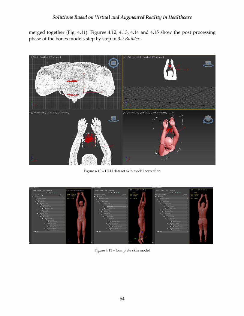
Solutions Based on Virtual and Augmented Reality in Healthcare
64
merged together (Fig. 4.11). Figures 4.12, 4.13, 4.14 and 4.15 show the post processing
phase of the bones models step by step in 3D Builder.
Figure 4.10 – ULH dataset skin model correction
Figure 4.11 – Complete skin model
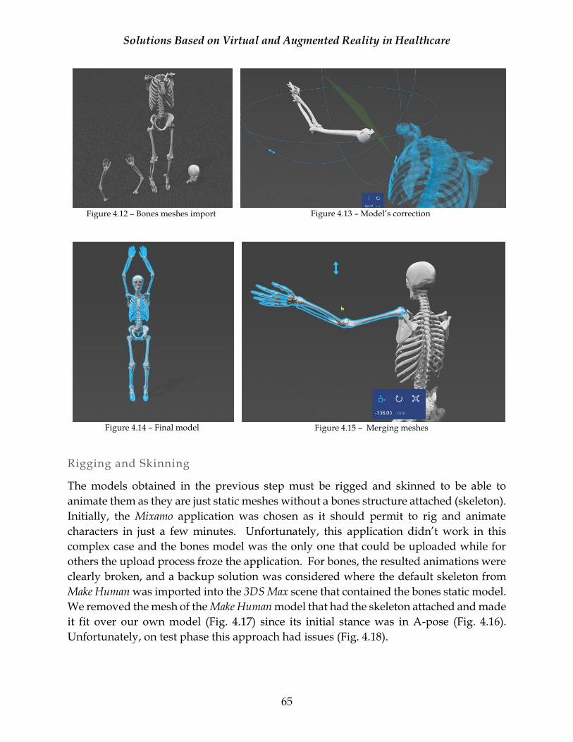
Solutions Based on Virtual and Augmented Reality in Healthcare
65
Figure 4.12 – Bones meshes import
Figure 4.13 – Model’s correction
Figure 4.14 – Final model
Figure 4.15 – Merging meshes
Rigging and Skinning
The models obtained in the previous step must be rigged and skinned to be able to
animate them as they are just static meshes without a bones structure attached (skeleton).
Initially, the Mixamo application was chosen as it should permit to rig and animate
characters in just a few minutes. Unfortunately, this application didn’t work in this
complex case and the bones model was the only one that could be uploaded while for
others the upload process froze the application. For bones, the resulted animations were
clearly broken, and a backup solution was considered where the default skeleton from
Make Human was imported into the 3DS Max scene that contained the bones static model.
We removed the mesh of the Make Human model that had the skeleton attached and made
it fit over our own model (Fig. 4.17) since its initial stance was in A-pose (Fig. 4.16).
Unfortunately, on test phase this approach had issues (Fig. 4.18).
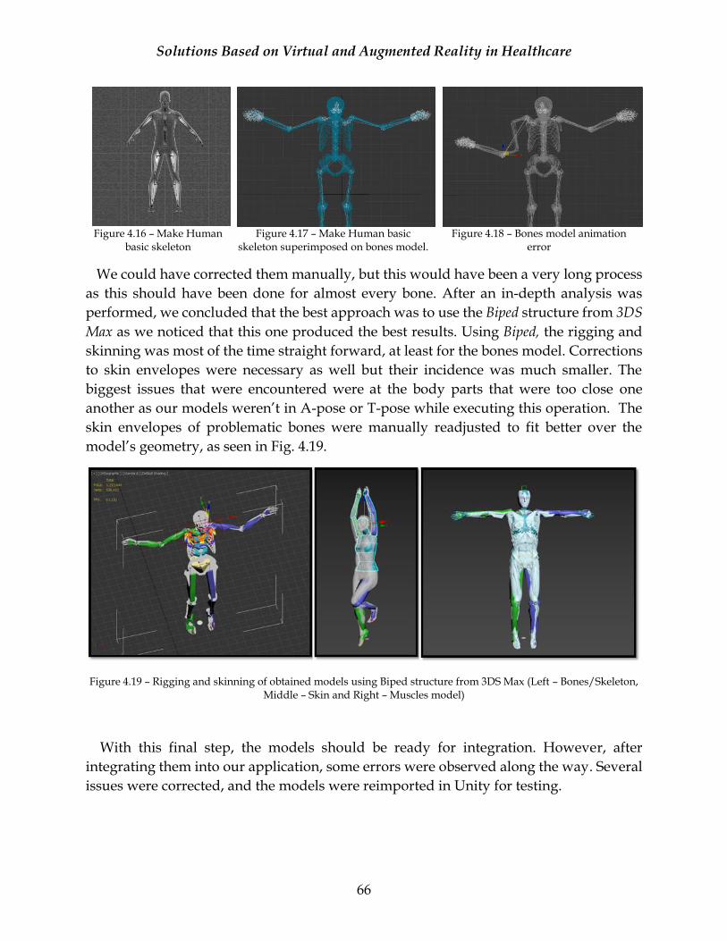
Solutions Based on Virtual and Augmented Reality in Healthcare
66
Figure 4.16 – Make Human
basic skeleton
Figure 4.17 – Make Human basic
skeleton superimposed on bones model.
Figure 4.18 – Bones model animation
error
We could have corrected them manually, but this would have been a very long process
as this should have been done for almost every bone. After an in-depth analysis was
performed, we concluded that the best approach was to use the Biped structure from 3DS
Max as we noticed that this one produced the best results. Using Biped, the rigging and
skinning was most of the time straight forward, at least for the bones model. Corrections
to skin envelopes were necessary as well but their incidence was much smaller. The
biggest issues that were encountered were at the body parts that were too close one
another as our models weren’t in A-pose or T-pose while executing this operation. The
skin envelopes of problematic bones were manually readjusted to fit better over the
model’s geometry, as seen in Fig. 4.19.
Figure 4.19 – Rigging and skinning of obtained models using Biped structure from 3DS Max (Left – Bones/Skeleton, Middle – Skin and Right – Muscles model)
With this final step, the models should be ready for integration. However, after
integrating them into our application, some errors were observed along the way. Several
issues were corrected, and the models were reimported in Unity for testing.
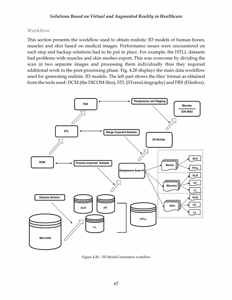
Solutions Based on Virtual and Augmented Reality in Healthcare
67
Workflow
This section presents the workflow used to obtain realistic 3D models of human bones,
muscles and skin based on medical images. Performance issues were encountered on
each step and backup solutions had to be put in place. For example, the HTLL datasets
had problems with muscles and skin meshes export. This was overcome by dividing the
scan in two separate images and processing them individually thus they required
additional work in the post-processing phase. Fig. 4.20 displays the main data workflow
used for generating realistic 3D models. The left part shows the files’ format as obtained
from the tools used: DCM (the DICOM files), STL (STereoLitography) and FBX (Filmbox).
Figure 4.20 – 3D Model Generation workflow
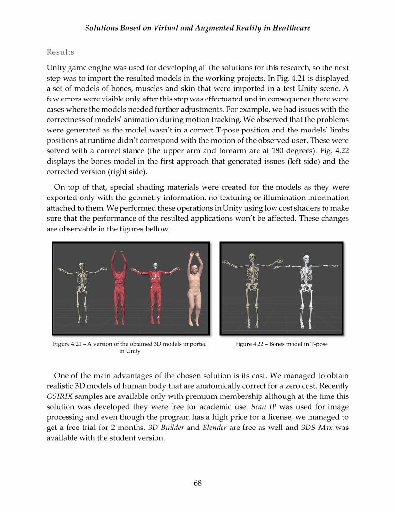
Solutions Based on Virtual and Augmented Reality in Healthcare
68
Results
Unity game engine was used for developing all the solutions for this research, so the next
step was to import the resulted models in the working projects. In Fig. 4.21 is displayed
a set of models of bones, muscles and skin that were imported in a test Unity scene. A
few errors were visible only after this step was effectuated and in consequence there were
cases where the models needed further adjustments. For example, we had issues with the
correctness of models’ animation during motion tracking. We observed that the problems
were generated as the model wasn’t in a correct T-pose position and the models’ limbs
positions at runtime didn’t correspond with the motion of the observed user. These were
solved with a correct stance (the upper arm and forearm are at 180 degrees). Fig. 4.22
displays the bones model in the first approach that generated issues (left side) and the
corrected version (right side).
On top of that, special shading materials were created for the models as they were
exported only with the geometry information, no texturing or illumination information
attached to them. We performed these operations in Unity using low cost shaders to make
sure that the performance of the resulted applications won’t be affected. These changes
are observable in the figures bellow.
Figure 4.21 – A version of the obtained 3D models imported
in Unity
Figure 4.22 – Bones model in T-pose
One of the main advantages of the chosen solution is its cost. We managed to obtain
realistic 3D models of human body that are anatomically correct for a zero cost. Recently
OSIRIX samples are available only with premium membership although at the time this
solution was developed they were free for academic use. Scan IP was used for image
processing and even though the program has a high price for a license, we managed to
get a free trial for 2 months. 3D Builder and Blender are free as well and 3DS Max was
available with the student version.
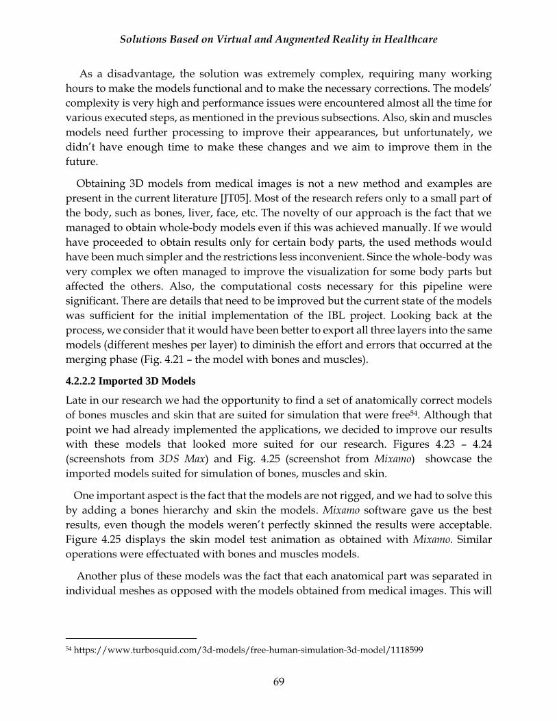
Solutions Based on Virtual and Augmented Reality in Healthcare
69
As a disadvantage, the solution was extremely complex, requiring many working
hours to make the models functional and to make the necessary corrections. The models’
complexity is very high and performance issues were encountered almost all the time for
various executed steps, as mentioned in the previous subsections. Also, skin and muscles
models need further processing to improve their appearances, but unfortunately, we
didn’t have enough time to make these changes and we aim to improve them in the
future.
Obtaining 3D models from medical images is not a new method and examples are
present in the current literature [JT05]. Most of the research refers only to a small part of
the body, such as bones, liver, face, etc. The novelty of our approach is the fact that we
managed to obtain whole-body models even if this was achieved manually. If we would
have proceeded to obtain results only for certain body parts, the used methods would
have been much simpler and the restrictions less inconvenient. Since the whole-body was
very complex we often managed to improve the visualization for some body parts but
affected the others. Also, the computational costs necessary for this pipeline were
significant. There are details that need to be improved but the current state of the models
was sufficient for the initial implementation of the IBL project. Looking back at the
process, we consider that it would have been better to export all three layers into the same
models (different meshes per layer) to diminish the effort and errors that occurred at the
merging phase (Fig. 4.21 – the model with bones and muscles).
4.2.2.2 Imported 3D Models
Late in our research we had the opportunity to find a set of anatomically correct models
of bones muscles and skin that are suited for simulation that were free54. Although that
point we had already implemented the applications, we decided to improve our results
with these models that looked more suited for our research. Figures 4.23 – 4.24
(screenshots from 3DS Max) and Fig. 4.25 (screenshot from Mixamo) showcase the
imported models suited for simulation of bones, muscles and skin.
One important aspect is the fact that the models are not rigged, and we had to solve this
by adding a bones hierarchy and skin the models. Mixamo software gave us the best
results, even though the models weren’t perfectly skinned the results were acceptable.
Figure 4.25 displays the skin model test animation as obtained with Mixamo. Similar
operations were effectuated with bones and muscles models.
Another plus of these models was the fact that each anatomical part was separated in
individual meshes as opposed with the models obtained from medical images. This will
54 https://www.turbosquid.com/3d-models/free-human-simulation-3d-model/1118599
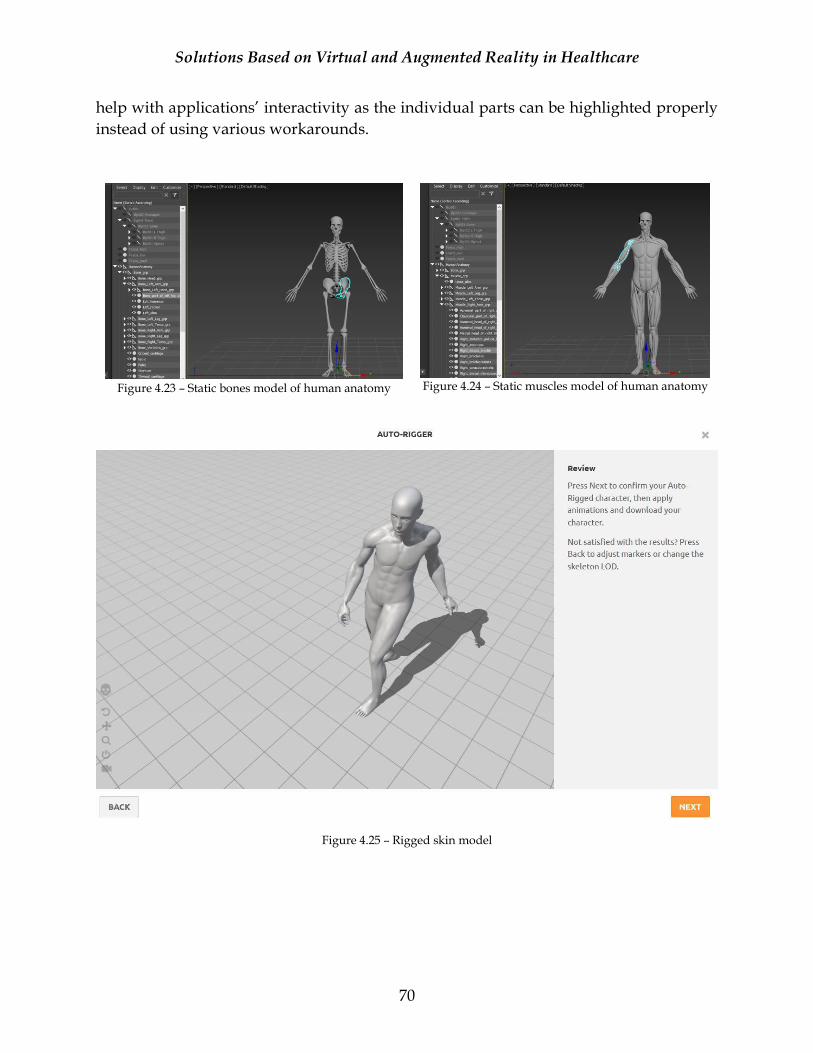
Solutions Based on Virtual and Augmented Reality in Healthcare
70
help with applications’ interactivity as the individual parts can be highlighted properly
instead of using various workarounds.
Figure 4.23 – Static bones model of human anatomy
Figure 4.24 – Static muscles model of human anatomy
Figure 4.25 – Rigged skin model
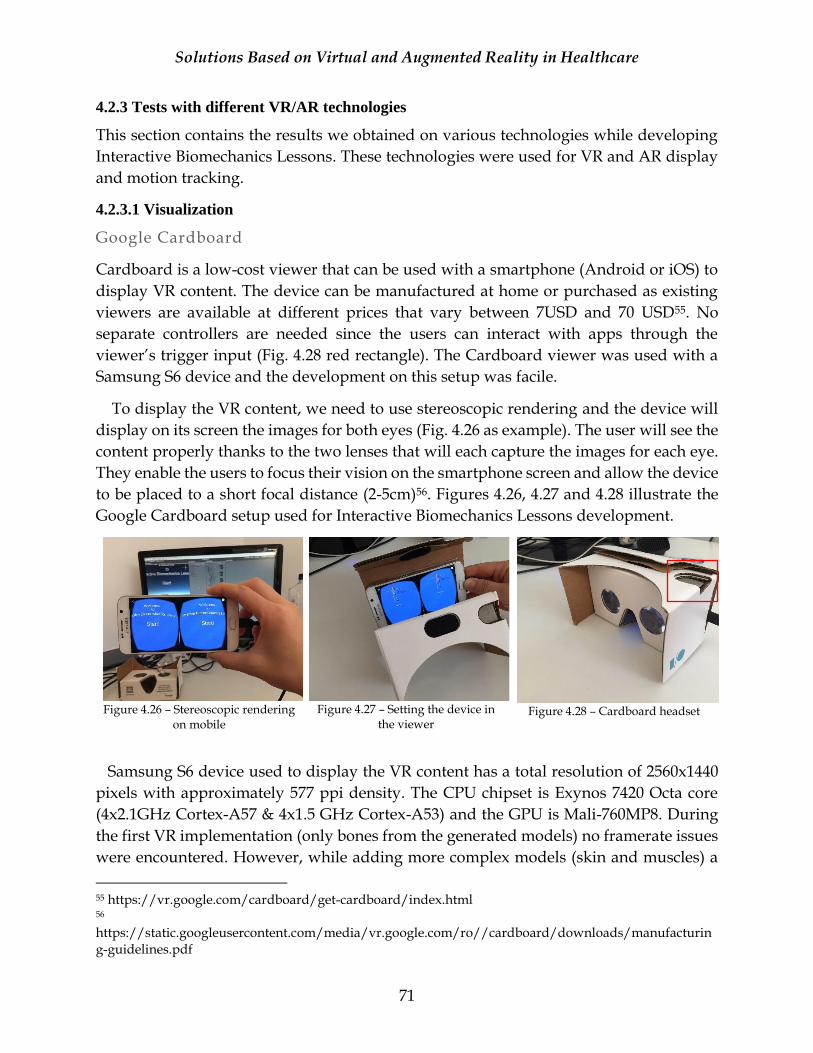
Solutions Based on Virtual and Augmented Reality in Healthcare
71
4.2.3 Tests with different VR/AR technologies
This section contains the results we obtained on various technologies while developing
Interactive Biomechanics Lessons. These technologies were used for VR and AR display
and motion tracking.
4.2.3.1 Visualization
Google Cardboard
Cardboard is a low-cost viewer that can be used with a smartphone (Android or iOS) to
display VR content. The device can be manufactured at home or purchased as existing
viewers are available at different prices that vary between 7USD and 70 USD55. No
separate controllers are needed since the users can interact with apps through the
viewer’s trigger input (Fig. 4.28 red rectangle). The Cardboard viewer was used with a
Samsung S6 device and the development on this setup was facile.
To display the VR content, we need to use stereoscopic rendering and the device will
display on its screen the images for both eyes (Fig. 4.26 as example). The user will see the
content properly thanks to the two lenses that will each capture the images for each eye.
They enable the users to focus their vision on the smartphone screen and allow the device
to be placed to a short focal distance (2-5cm)56. Figures 4.26, 4.27 and 4.28 illustrate the
Google Cardboard setup used for Interactive Biomechanics Lessons development.
Figure 4.26 – Stereoscopic rendering
on mobile
Figure 4.27 – Setting the device in
the viewer
Figure 4.28 – Cardboard headset
Samsung S6 device used to display the VR content has a total resolution of 2560x1440
pixels with approximately 577 ppi density. The CPU chipset is Exynos 7420 Octa core
(4x2.1GHz Cortex-A57 & 4x1.5 GHz Cortex-A53) and the GPU is Mali-760MP8. During
the first VR implementation (only bones from the generated models) no framerate issues
were encountered. However, while adding more complex models (skin and muscles) a
55 https://vr.google.com/cardboard/get-cardboard/index.html 56 https://static.googleusercontent.com/media/vr.google.com/ro//cardboard/downloads/manufacturing-guidelines.pdf
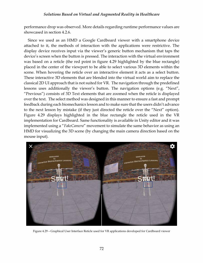
Solutions Based on Virtual and Augmented Reality in Healthcare
72
performance drop was observed. More details regarding runtime performance values are
showcased in section 4.2.6.
Since we used as an HMD a Google Cardboard viewer with a smartphone device
attached to it, the methods of interaction with the applications were restrictive. The
display device receives input via the viewer’s generic button mechanism that taps the
device’s screen when the button is pressed. The interaction with the virtual environment
was based on a reticle (the red point in figure 4.29 highlighted by the blue rectangle)
placed in the center of the viewport to be able to select various 3D elements within the
scene. When hovering the reticle over an interactive element it acts as a select button.
These interactive 3D elements that are blended into the virtual world aim to replace the
classical 2D UI approach that is not suited for VR. The navigation through the predefined
lessons uses additionally the viewer’s button. The navigation options (e.g. “Next”,
“Previous”) consists of 3D Text elements that are zoomed when the reticle is displayed
over the text. The select method was designed in this manner to ensure a fast and prompt
feedback during each biomechanics lesson and to make sure that the users didn’t advance
to the next lesson by mistake (if they just directed the reticle over the “Next” option).
Figure 4.29 displays highlighted in the blue rectangle the reticle used in the VR
implementation for Cardboard. Same functionality is available in Unity editor and it was
implemented using a “FakeCamera” movement to simulate the same behavior as using an
HMD for visualizing the 3D scene (by changing the main camera direction based on the
mouse input).
Figure 4.29 – Graphical User Interface Reticle used for VR applications developed for Cardboard viewer
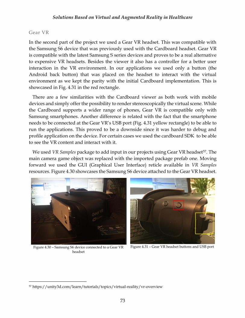
Solutions Based on Virtual and Augmented Reality in Healthcare
73
Gear VR
In the second part of the project we used a Gear VR headset. This was compatible with
the Samsung S6 device that was previously used with the Cardboard headset. Gear VR
is compatible with the latest Samsung S series devices and proves to be a real alternative
to expensive VR headsets. Besides the viewer it also has a controller for a better user
interaction in the VR environment. In our applications we used only a button (the
Android back button) that was placed on the headset to interact with the virtual
environment as we kept the parity with the initial Cardboard implementation. This is
showcased in Fig. 4.31 in the red rectangle.
There are a few similarities with the Cardboard viewer as both work with mobile
devices and simply offer the possibility to render stereoscopically the virtual scene. While
the Cardboard supports a wider range of phones, Gear VR is compatible only with
Samsung smartphones. Another difference is related with the fact that the smartphone
needs to be connected at the Gear VR’s USB port (Fig. 4.31 yellow rectangle) to be able to
run the applications. This proved to be a downside since it was harder to debug and
profile application on the device. For certain cases we used the cardboard SDK to be able
to see the VR content and interact with it.
We used VR Samples package to add input in our projects using Gear VR headset57. The
main camera game object was replaced with the imported package prefab one. Moving
forward we used the GUI (Graphical User Interface) reticle available in VR Samples
resources. Figure 4.30 showcases the Samsung S6 device attached to the Gear VR headset.
Figure 4.30 – Samsung S6 device connected to a Gear VR
headset
Figure 4.31 – Gear VR headset buttons and USB port
57 https://unity3d.com/learn/tutorials/topics/virtual-reality/vr-overview
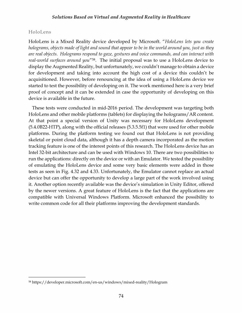
Solutions Based on Virtual and Augmented Reality in Healthcare
74
HoloLens
HoloLens is a Mixed Reality device developed by Microsoft. “HoloLens lets you create
holograms, objects made of light and sound that appear to be in the world around you, just as they
are real objects. Holograms respond to gaze, gestures and voice commands, and can interact with
real-world surfaces around you”58. The initial proposal was to use a HoloLens device to
display the Augmented Reality, but unfortunately, we couldn’t manage to obtain a device
for development and taking into account the high cost of a device this couldn’t be
acquisitioned. However, before renouncing at the idea of using a HoloLens device we
started to test the possibility of developing on it. The work mentioned here is a very brief
proof of concept and it can be extended in case the opportunity of developing on this
device is available in the future.
These tests were conducted in mid-2016 period. The development was targeting both
HoloLens and other mobile platforms (tablets) for displaying the holograms/AR content.
At that point a special version of Unity was necessary for HoloLens development
(5.4.0B22-HTP), along with the official releases (5.3.5.5f1) that were used for other mobile
platforms. During the platform testing we found out that HoloLens is not providing
skeletal or point cloud data, although it has a depth camera incorporated as the motion
tracking feature is one of the interest points of this research. The HoloLens device has an
Intel 32-bit architecture and can be used with Windows 10. There are two possibilities to
run the applications: directly on the device or with an Emulator. We tested the possibility
of emulating the HoloLens device and some very basic elements were added in those
tests as seen in Fig. 4.32 and 4.33. Unfortunately, the Emulator cannot replace an actual
device but can offer the opportunity to develop a large part of the work involved using
it. Another option recently available was the device’s simulation in Unity Editor, offered
by the newer versions. A great feature of HoloLens is the fact that the applications are
compatible with Universal Windows Platform. Microsoft enhanced the possibility to
write common code for all their platforms improving the development standards.
58 https://developer.microsoft.com/en-us/windows/mixed-reality/Hologram
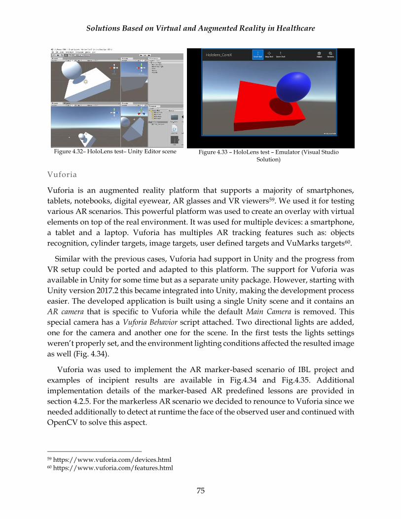
Solutions Based on Virtual and Augmented Reality in Healthcare
75
Figure 4.32– HoloLens test– Unity Editor scene
Figure 4.33 – HoloLens test – Emulator (Visual Studio
Solution)
Vuforia
Vuforia is an augmented reality platform that supports a majority of smartphones,
tablets, notebooks, digital eyewear, AR glasses and VR viewers59. We used it for testing
various AR scenarios. This powerful platform was used to create an overlay with virtual
elements on top of the real environment. It was used for multiple devices: a smartphone,
a tablet and a laptop. Vuforia has multiples AR tracking features such as: objects
recognition, cylinder targets, image targets, user defined targets and VuMarks targets60.
Similar with the previous cases, Vuforia had support in Unity and the progress from
VR setup could be ported and adapted to this platform. The support for Vuforia was
available in Unity for some time but as a separate unity package. However, starting with
Unity version 2017.2 this became integrated into Unity, making the development process
easier. The developed application is built using a single Unity scene and it contains an
AR camera that is specific to Vuforia while the default Main Camera is removed. This
special camera has a Vuforia Behavior script attached. Two directional lights are added,
one for the camera and another one for the scene. In the first tests the lights settings
weren’t properly set, and the environment lighting conditions affected the resulted image
as well (Fig. 4.34).
Vuforia was used to implement the AR marker-based scenario of IBL project and
examples of incipient results are available in Fig.4.34 and Fig.4.35. Additional
implementation details of the marker-based AR predefined lessons are provided in
section 4.2.5. For the markerless AR scenario we decided to renounce to Vuforia since we
needed additionally to detect at runtime the face of the observed user and continued with
OpenCV to solve this aspect.
59 https://www.vuforia.com/devices.html 60 https://www.vuforia.com/features.html
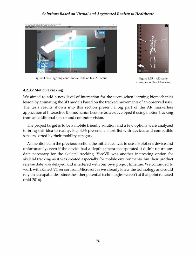
Solutions Based on Virtual and Augmented Reality in Healthcare
76
Figure 4.34 – Lighting conditions effects on test AR scene
Figure 4.35 – AR scene example - without tracking
4.2.3.2 Motion Tracking
We aimed to add a new level of interaction for the users when learning biomechanics
lesson by animating the 3D models based on the tracked movements of an observed user.
The tests results shown into this section present a big part of the AR markerless
application of Interactive Biomechanics Lessons as we developed it using motion tracking
from an additional sensor and computer vision.
The project target is to be a mobile friendly solution and a few options were analyzed
to bring this idea to reality. Fig. 4.36 presents a short list with devices and compatible
sensors sorted by their mobility category.
As mentioned in the previous section, the initial idea was to use a HoloLens device and
unfortunately, even if the device had a depth camera incorporated it didn’t return any
data necessary for the skeletal tracking. VicoVR was another interesting option for
skeletal tracking as it was created especially for mobile environments, but their product
release date was delayed and interfered with our own project timeline. We continued to
work with Kinect V1 sensor from Microsoft as we already knew the technology and could
rely on its capabilities, since the other potential technologies weren’t at that point released
(mid 2016).
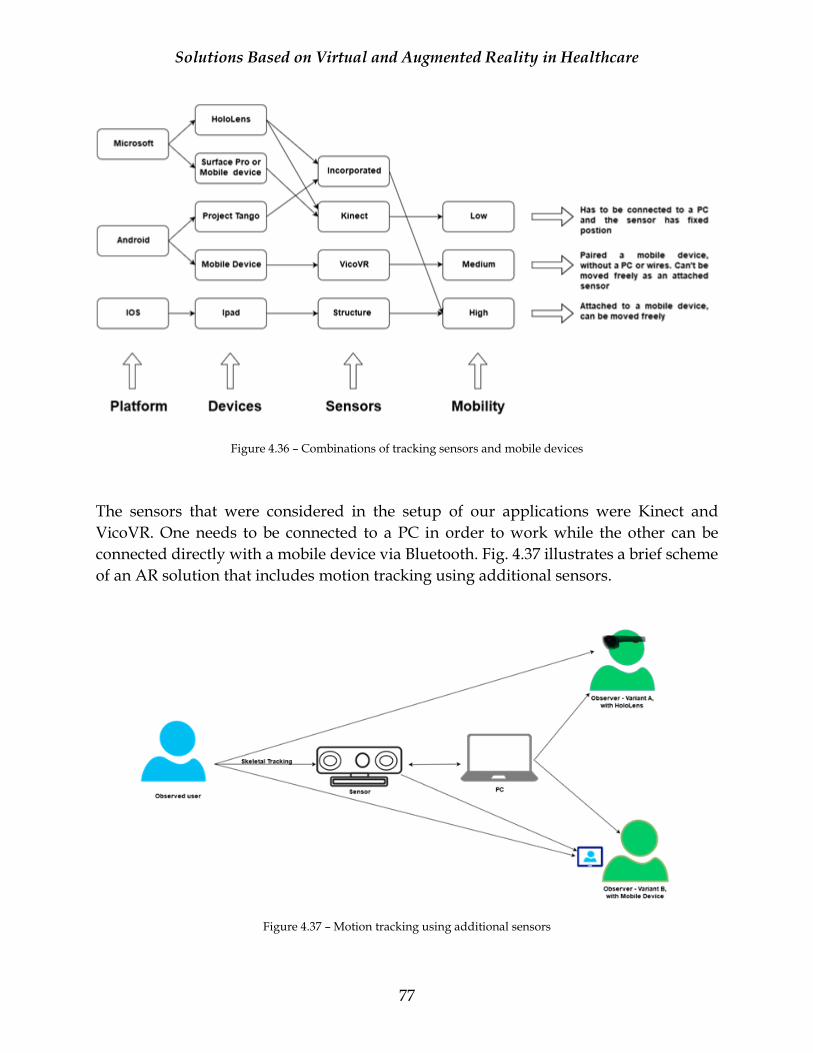
Solutions Based on Virtual and Augmented Reality in Healthcare
77
Figure 4.36 – Combinations of tracking sensors and mobile devices
The sensors that were considered in the setup of our applications were Kinect and
VicoVR. One needs to be connected to a PC in order to work while the other can be
connected directly with a mobile device via Bluetooth. Fig. 4.37 illustrates a brief scheme
of an AR solution that includes motion tracking using additional sensors.
Figure 4.37 – Motion tracking using additional sensors
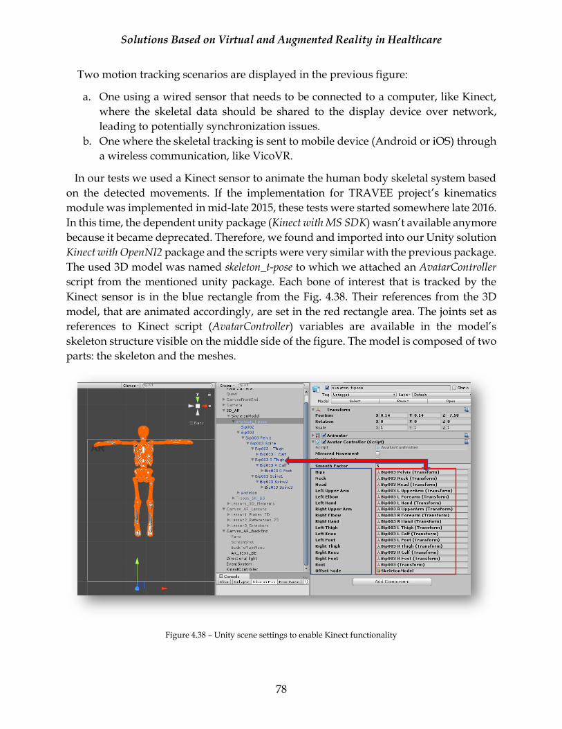
Solutions Based on Virtual and Augmented Reality in Healthcare
78
Two motion tracking scenarios are displayed in the previous figure:
a. One using a wired sensor that needs to be connected to a computer, like Kinect,
where the skeletal data should be shared to the display device over network,
leading to potentially synchronization issues.
b. One where the skeletal tracking is sent to mobile device (Android or iOS) through
a wireless communication, like VicoVR.
In our tests we used a Kinect sensor to animate the human body skeletal system based
on the detected movements. If the implementation for TRAVEE project’s kinematics
module was implemented in mid-late 2015, these tests were started somewhere late 2016.
In this time, the dependent unity package (Kinect with MS SDK) wasn’t available anymore
because it became deprecated. Therefore, we found and imported into our Unity solution
Kinect with OpenNI2 package and the scripts were very similar with the previous package.
The used 3D model was named skeleton_t-pose to which we attached an AvatarController
script from the mentioned unity package. Each bone of interest that is tracked by the
Kinect sensor is in the blue rectangle from the Fig. 4.38. Their references from the 3D
model, that are animated accordingly, are set in the red rectangle area. The joints set as
references to Kinect script (AvatarController) variables are available in the model’s
skeleton structure visible on the middle side of the figure. The model is composed of two
parts: the skeleton and the meshes.
Figure 4.38 – Unity scene settings to enable Kinect functionality
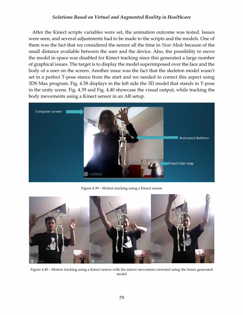
Solutions Based on Virtual and Augmented Reality in Healthcare
79
After the Kinect scripts variables were set, the animation outcome was tested. Issues
were seen, and several adjustments had to be made to the scripts and the models. One of
them was the fact that we considered the sensor all the time in Near Mode because of the
small distance available between the user and the device. Also, the possibility to move
the model in space was disabled for Kinect tracking since this generated a large number
of graphical issues. The target is to display the model superimposed over the face and the
body of a user on the screen. Another issue was the fact that the skeleton model wasn’t
set in a perfect T-pose stance from the start and we needed to correct this aspect using
3DS Max program. Fig. 4.38 displays in the left side the 3D model that stands in T-pose
in the unity scene. Fig. 4.39 and Fig. 4.40 showcase the visual output, while tracking the
body movements using a Kinect sensor in an AR setup.
Figure 4.39 – Motion tracking using a Kinect sensor
Figure 4.40 – Motion tracking using a Kinect sensor with the mirror movement corrected using the bones generated model
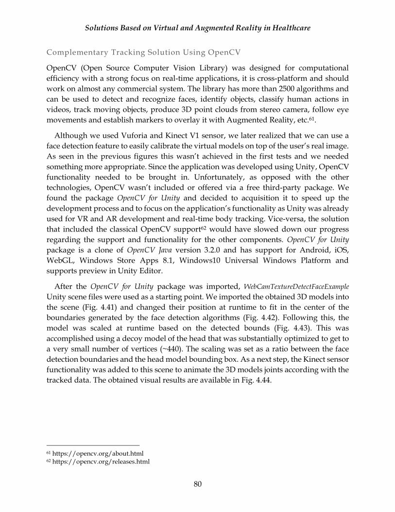
Solutions Based on Virtual and Augmented Reality in Healthcare
80
Complementary Tracking Solution Using OpenCV
OpenCV (Open Source Computer Vision Library) was designed for computational
efficiency with a strong focus on real-time applications, it is cross-platform and should
work on almost any commercial system. The library has more than 2500 algorithms and
can be used to detect and recognize faces, identify objects, classify human actions in
videos, track moving objects, produce 3D point clouds from stereo camera, follow eye
movements and establish markers to overlay it with Augmented Reality, etc.61.
Although we used Vuforia and Kinect V1 sensor, we later realized that we can use a
face detection feature to easily calibrate the virtual models on top of the user’s real image.
As seen in the previous figures this wasn’t achieved in the first tests and we needed
something more appropriate. Since the application was developed using Unity, OpenCV
functionality needed to be brought in. Unfortunately, as opposed with the other
technologies, OpenCV wasn’t included or offered via a free third-party package. We
found the package OpenCV for Unity and decided to acquisition it to speed up the
development process and to focus on the application’s functionality as Unity was already
used for VR and AR development and real-time body tracking. Vice-versa, the solution
that included the classical OpenCV support62 would have slowed down our progress
regarding the support and functionality for the other components. OpenCV for Unity
package is a clone of OpenCV Java version 3.2.0 and has support for Android, iOS,
WebGL, Windows Store Apps 8.1, Windows10 Universal Windows Platform and
supports preview in Unity Editor.
After the OpenCV for Unity package was imported, WebCamTextureDetectFaceExample
Unity scene files were used as a starting point. We imported the obtained 3D models into
the scene (Fig. 4.41) and changed their position at runtime to fit in the center of the
boundaries generated by the face detection algorithms (Fig. 4.42). Following this, the
model was scaled at runtime based on the detected bounds (Fig. 4.43). This was
accomplished using a decoy model of the head that was substantially optimized to get to
a very small number of vertices (~440). The scaling was set as a ratio between the face
detection boundaries and the head model bounding box. As a next step, the Kinect sensor
functionality was added to this scene to animate the 3D models joints according with the
tracked data. The obtained visual results are available in Fig. 4.44.
61 https://opencv.org/about.html 62 https://opencv.org/releases.html
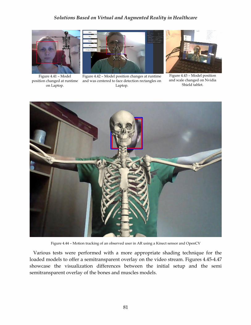
Solutions Based on Virtual and Augmented Reality in Healthcare
81
Figure 4.41 – Model
position changed at runtime on Laptop.
Figure 4.42 – Model position changes at runtime and was centered to face detection rectangles on
Laptop.
Figure 4.43 – Model position and scale changed on Nvidia
Shield tablet.
Figure 4.44 – Motion tracking of an observed user in AR using a Kinect sensor and OpenCV
Various tests were performed with a more appropriate shading technique for the
loaded models to offer a semitransparent overlay on the video stream. Figures 4.45-4.47
showcase the visualization differences between the initial setup and the semi
semitransparent overlay of the bones and muscles models.
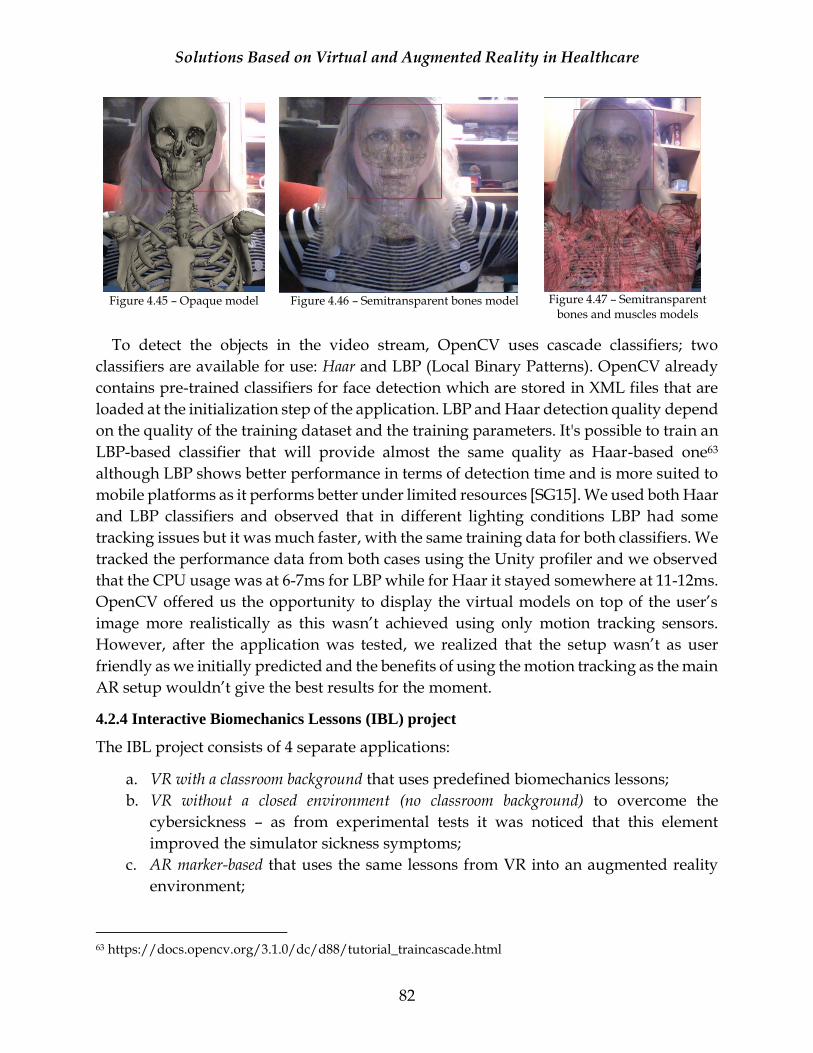
Solutions Based on Virtual and Augmented Reality in Healthcare
82
Figure 4.45 – Opaque model
Figure 4.46 – Semitransparent bones model
Figure 4.47 – Semitransparent
bones and muscles models
To detect the objects in the video stream, OpenCV uses cascade classifiers; two
classifiers are available for use: Haar and LBP (Local Binary Patterns). OpenCV already
contains pre-trained classifiers for face detection which are stored in XML files that are
loaded at the initialization step of the application. LBP and Haar detection quality depend
on the quality of the training dataset and the training parameters. It's possible to train an
LBP-based classifier that will provide almost the same quality as Haar-based one63
although LBP shows better performance in terms of detection time and is more suited to
mobile platforms as it performs better under limited resources [SG15]. We used both Haar
and LBP classifiers and observed that in different lighting conditions LBP had some
tracking issues but it was much faster, with the same training data for both classifiers. We
tracked the performance data from both cases using the Unity profiler and we observed
that the CPU usage was at 6-7ms for LBP while for Haar it stayed somewhere at 11-12ms.
OpenCV offered us the opportunity to display the virtual models on top of the user’s
image more realistically as this wasn’t achieved using only motion tracking sensors.
However, after the application was tested, we realized that the setup wasn’t as user
friendly as we initially predicted and the benefits of using the motion tracking as the main
AR setup wouldn’t give the best results for the moment.
4.2.4 Interactive Biomechanics Lessons (IBL) project
The IBL project consists of 4 separate applications:
a. VR with a classroom background that uses predefined biomechanics lessons;
b. VR without a closed environment (no classroom background) to overcome the
cybersickness – as from experimental tests it was noticed that this element
improved the simulator sickness symptoms;
c. AR marker-based that uses the same lessons from VR into an augmented reality
environment;
63 https://docs.opencv.org/3.1.0/dc/d88/tutorial_traincascade.html
Solutions Based on Virtual and Augmented Reality in Healthcare
83
d. AR markerless that uses motion tracking sensors to animate in real time the virtual
3D models superimposed over the user’ image.
The VR applications were displayed on a low-cost HMD composed of a Samsung S6
device, model SM-G920F, with a Google Cardboard viewer, initially, and later with a
Gear VR viewer. The built scenes were developed using Unity version 5.6.0b.10. The
cardboard viewer functionality was included in that specific Unity version and no
additional packages were required to add the functionality for VR display with it. We
later added VR Samples package while making the applications compatible with Gear VR.
The AR applications had separate setups, as the markerless one included additional
tracking sensors while the marker-based one had a simpler structure, similar with the VR
ones. Vuforia was used for the AR marker-based specific features and, in order to include
its functionality without additional support packages, we used Unity version 2017.2. The
AR markerless application was built using Unity version 5.6.0b.10. The setup contains a
Kinect V1 sensor for motion tracking that is connected to a Laptop with this
configuration: CPU: Intel Core i7 4710HQ @ 2.50 GHz, GPU: Nvidia GeForce 850M,
Memory: 8GB with Windows 10 Pro OS. The tracking is complemented with OpenCV
face detection functionality to improve the superimposing of the 3D models over the
user’s image. The functionality is brought into this project using OpenCV with Unity
package while the Kinect functionality is imported from Kinect with OpenNi2 unity
package. The final prototype of this scenario was functional on the mentioned laptop
while several tests were done with OpenCV on mobile devices such as a smartphone and
a tablet. The smartphone is the one used in the VR setup while the tablet is NVIDIA Shield
tablet with a screen resolution of 1200x1920 pixels, 2.2GHz quad-core and Nvidia Tegra
K1 graphics card running with Android 6.0.1 OS. These setup details are correlated with
the performance data presented in section 4.2.6.
4.2.5 Implementation Details
This section presents the implementation details of the developed applications. As we
already mentioned, the AR marker-based one and the VR ones have a similar setup and
are based on predefined biomechanics lessons while the AR markerless was kept more at
a prototype status due to the intermediary results obtained. Since the AR markerless
setup was consistently described in a previous section (4.2.3.2) in the following lines we’ll
focus on the implementation details of the other three scenarios which have a common
background.
The pre-defined lessons can be considered a proof of concept for the targeted subject and further additions can be applied. The main applications contain 4 simple biomechanics lessons that cover different areas:
• Lesson I contains information about the human anatomy.
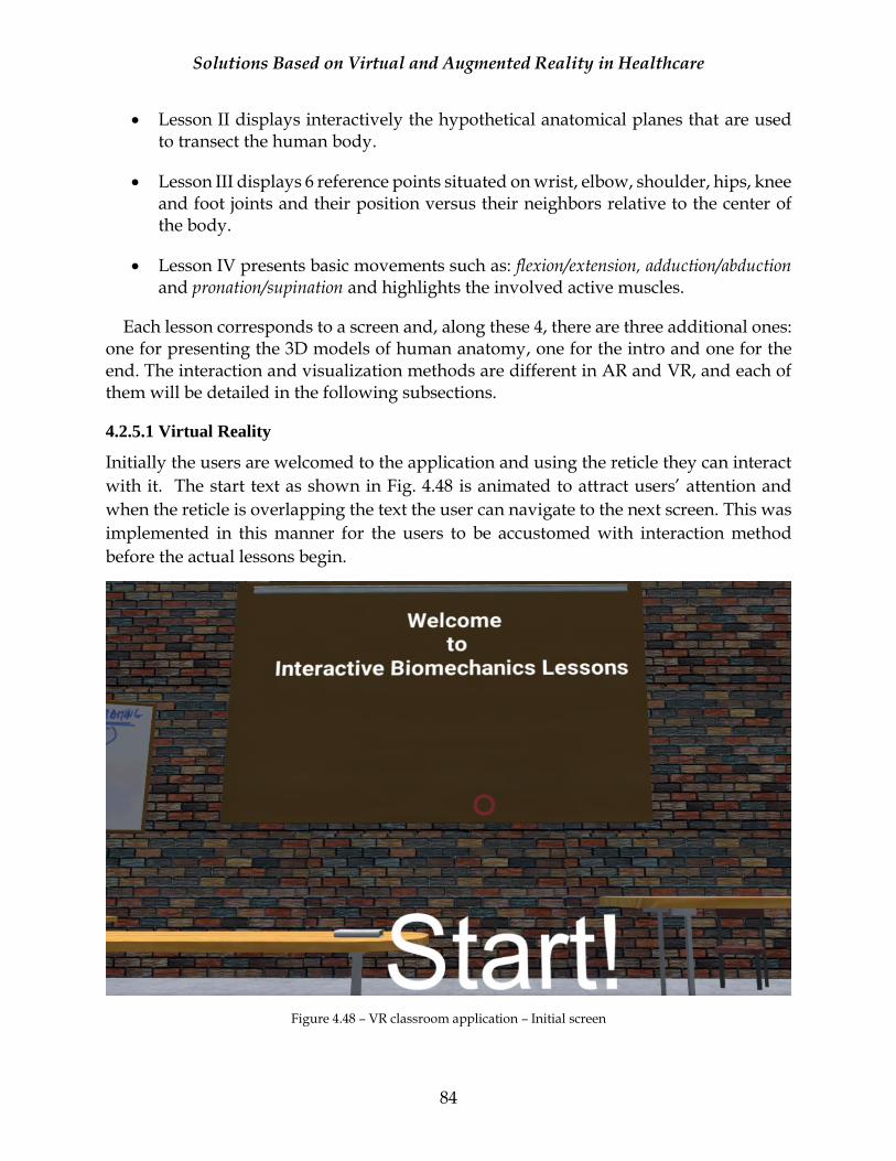
Solutions Based on Virtual and Augmented Reality in Healthcare
84
• Lesson II displays interactively the hypothetical anatomical planes that are used to transect the human body.
• Lesson III displays 6 reference points situated on wrist, elbow, shoulder, hips, knee and foot joints and their position versus their neighbors relative to the center of the body.
• Lesson IV presents basic movements such as: flexion/extension, adduction/abduction and pronation/supination and highlights the involved active muscles.
Each lesson corresponds to a screen and, along these 4, there are three additional ones: one for presenting the 3D models of human anatomy, one for the intro and one for the end. The interaction and visualization methods are different in AR and VR, and each of them will be detailed in the following subsections.
4.2.5.1 Virtual Reality
Initially the users are welcomed to the application and using the reticle they can interact
with it. The start text as shown in Fig. 4.48 is animated to attract users’ attention and
when the reticle is overlapping the text the user can navigate to the next screen. This was
implemented in this manner for the users to be accustomed with interaction method
before the actual lessons begin.
Figure 4.48 – VR classroom application – Initial screen
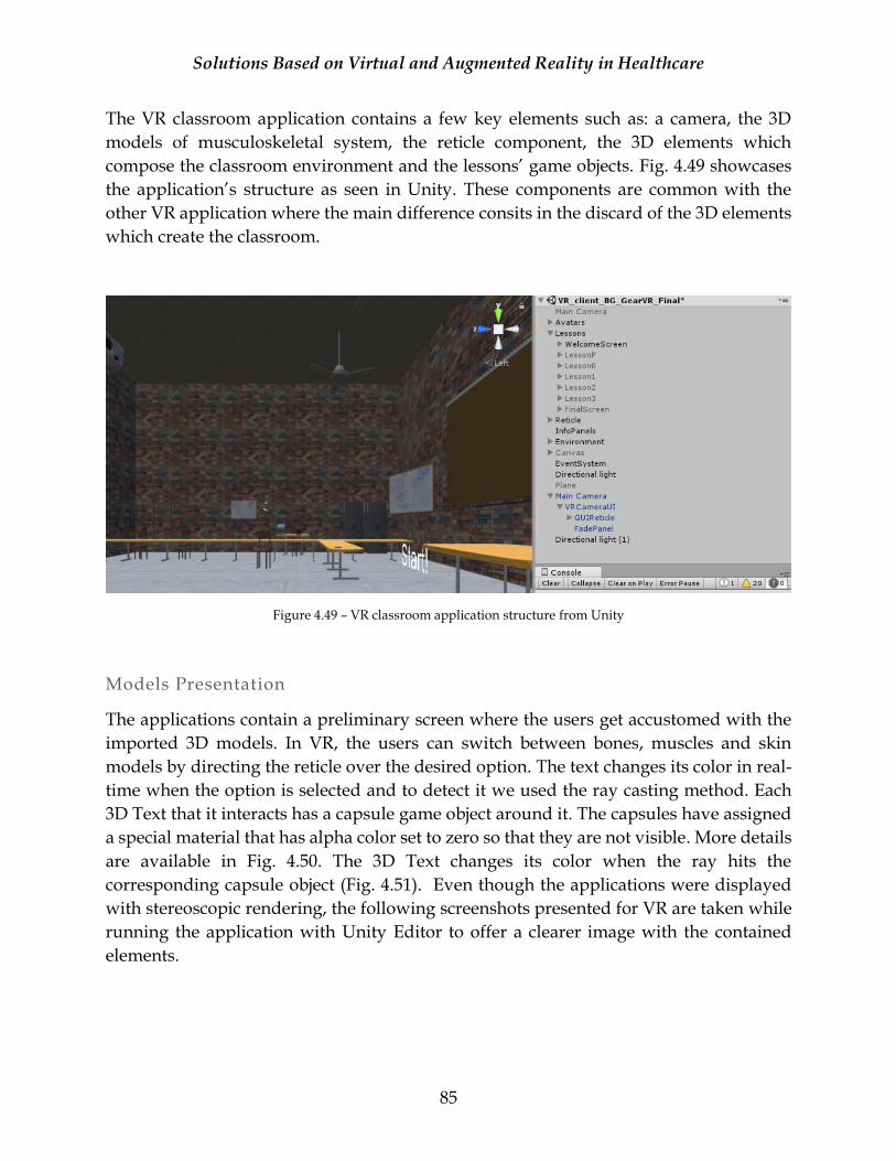
Solutions Based on Virtual and Augmented Reality in Healthcare
85
The VR classroom application contains a few key elements such as: a camera, the 3D
models of musculoskeletal system, the reticle component, the 3D elements which
compose the classroom environment and the lessons’ game objects. Fig. 4.49 showcases
the application’s structure as seen in Unity. These components are common with the
other VR application where the main difference consits in the discard of the 3D elements
which create the classroom.
Figure 4.49 – VR classroom application structure from Unity
Models Presentation
The applications contain a preliminary screen where the users get accustomed with the
imported 3D models. In VR, the users can switch between bones, muscles and skin
models by directing the reticle over the desired option. The text changes its color in real-
time when the option is selected and to detect it we used the ray casting method. Each
3D Text that it interacts has a capsule game object around it. The capsules have assigned
a special material that has alpha color set to zero so that they are not visible. More details
are available in Fig. 4.50. The 3D Text changes its color when the ray hits the
corresponding capsule object (Fig. 4.51). Even though the applications were displayed
with stereoscopic rendering, the following screenshots presented for VR are taken while
running the application with Unity Editor to offer a clearer image with the contained
elements.
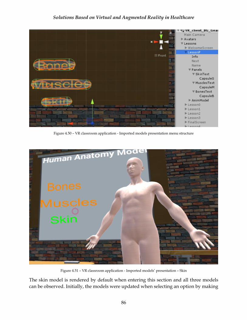
Solutions Based on Virtual and Augmented Reality in Healthcare
86
Figure 4.50 – VR classroom application - Imported models presentation menu structure
Figure 4.51 – VR classroom application - Imported models’ presentation – Skin
The skin model is rendered by default when entering this section and all three models
can be observed. Initially, the models were updated when selecting an option by making
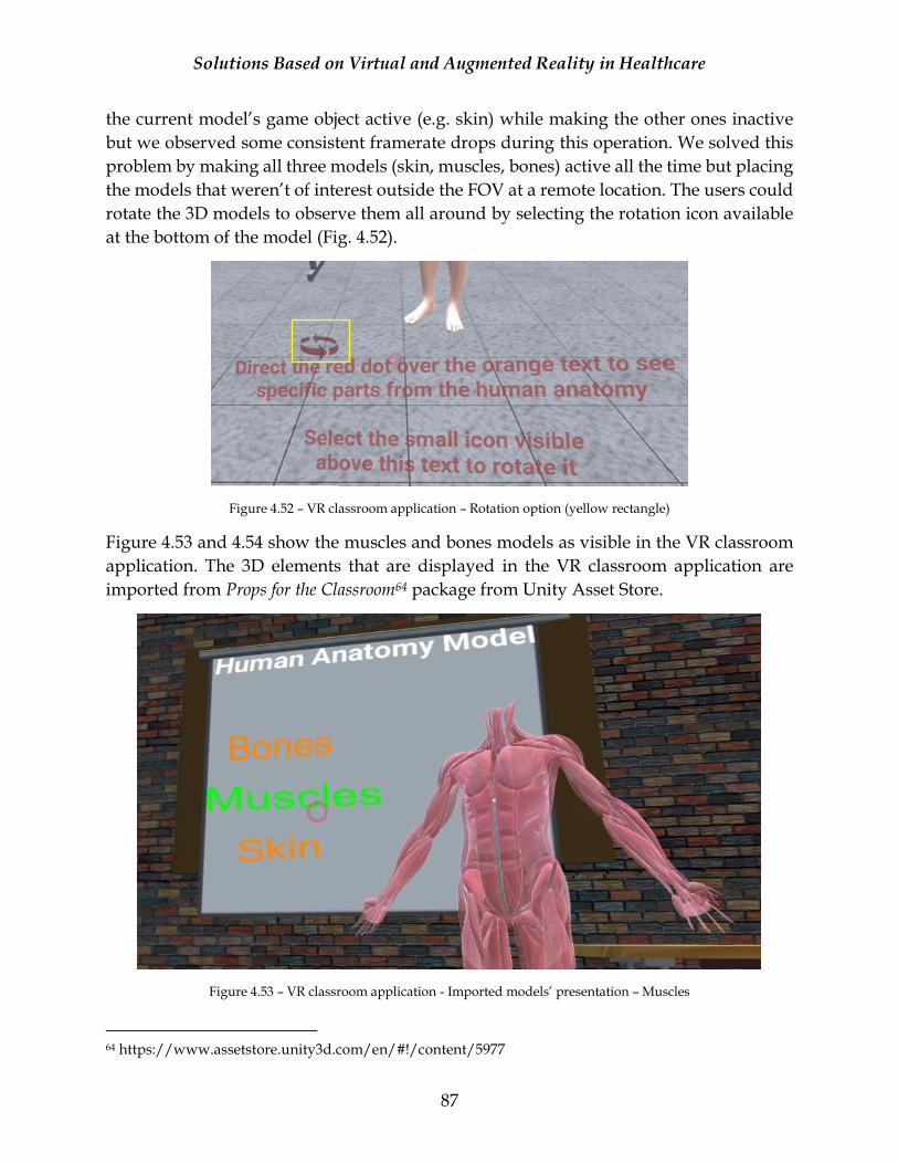
Solutions Based on Virtual and Augmented Reality in Healthcare
87
the current model’s game object active (e.g. skin) while making the other ones inactive
but we observed some consistent framerate drops during this operation. We solved this
problem by making all three models (skin, muscles, bones) active all the time but placing
the models that weren’t of interest outside the FOV at a remote location. The users could
rotate the 3D models to observe them all around by selecting the rotation icon available
at the bottom of the model (Fig. 4.52).
Figure 4.52 – VR classroom application – Rotation option (yellow rectangle)
Figure 4.53 and 4.54 show the muscles and bones models as visible in the VR classroom
application. The 3D elements that are displayed in the VR classroom application are
imported from Props for the Classroom64 package from Unity Asset Store.
Figure 4.53 – VR classroom application - Imported models’ presentation – Muscles
64 https://www.assetstore.unity3d.com/en/#!/content/5977
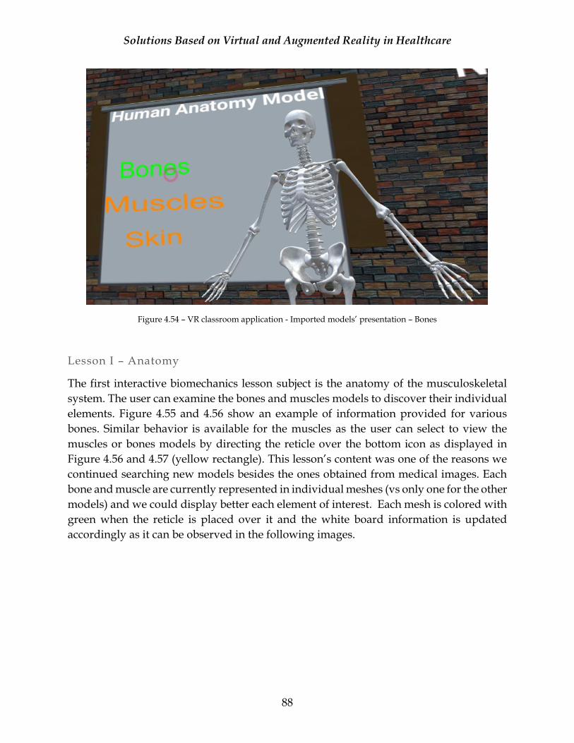
Solutions Based on Virtual and Augmented Reality in Healthcare
88
Figure 4.54 – VR classroom application - Imported models’ presentation – Bones
Lesson I – Anatomy
The first interactive biomechanics lesson subject is the anatomy of the musculoskeletal
system. The user can examine the bones and muscles models to discover their individual
elements. Figure 4.55 and 4.56 show an example of information provided for various
bones. Similar behavior is available for the muscles as the user can select to view the
muscles or bones models by directing the reticle over the bottom icon as displayed in
Figure 4.56 and 4.57 (yellow rectangle). This lesson’s content was one of the reasons we
continued searching new models besides the ones obtained from medical images. Each
bone and muscle are currently represented in individual meshes (vs only one for the other
models) and we could display better each element of interest. Each mesh is colored with
green when the reticle is placed over it and the white board information is updated
accordingly as it can be observed in the following images.
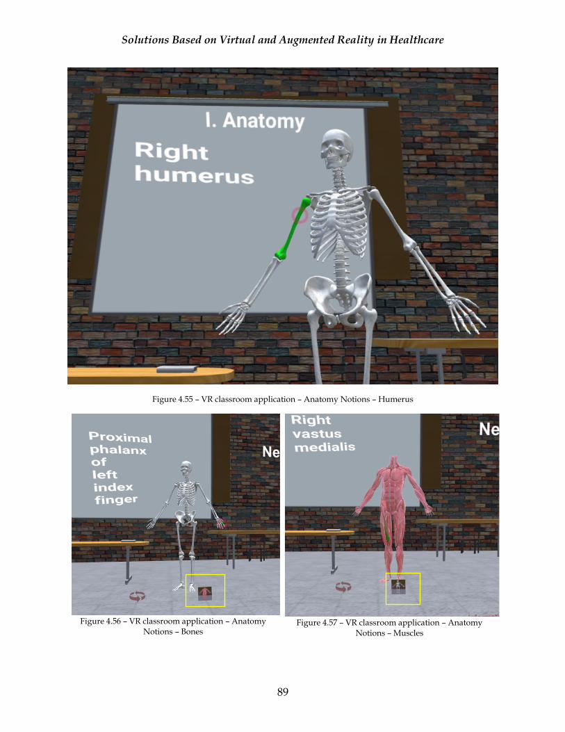
Solutions Based on Virtual and Augmented Reality in Healthcare
89
Figure 4.55 – VR classroom application – Anatomy Notions – Humerus
Figure 4.56 – VR classroom application – Anatomy
Notions – Bones
Figure 4.57 – VR classroom application – Anatomy
Notions – Muscles
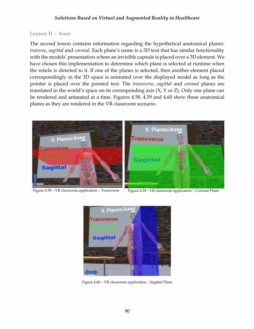
Solutions Based on Virtual and Augmented Reality in Healthcare
90
Lesson II – Axes
The second lesson contains information regarding the hypothetical anatomical planes:
traverse, sagittal and coronal. Each plane’s name is a 3D text that has similar functionality
with the models’ presentation where an invisible capsule is placed over a 3D element. We
have chosen this implementation to determine which plane is selected at runtime when
the reticle is directed to it. If one of the planes is selected, then another element placed
correspondingly in the 3D space is animated over the displayed model as long as the
pointer is placed over the pointed text. The transverse, sagittal and coronal planes are
translated in the world’s space on its corresponding axis (X, Y or Z). Only one plane can
be rendered and animated at a time. Figures 4.58, 4.59 and 4.60 show these anatomical
planes as they are rendered in the VR classroom scenario.
Figure 4.58 – VR classroom application – Transverse
Figure 4.59 - VR classroom application – Coronal Plane
Figure 4.60 – VR classroom application – Sagittal Plane
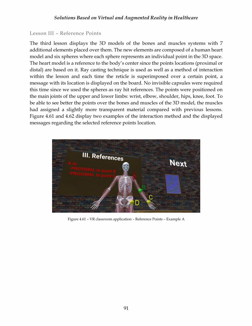
Solutions Based on Virtual and Augmented Reality in Healthcare
91
Lesson III – Reference Points
The third lesson displays the 3D models of the bones and muscles systems with 7
additional elements placed over them. The new elements are composed of a human heart
model and six spheres where each sphere represents an individual point in the 3D space.
The heart model is a reference to the body’s center since the points locations (proximal or
distal) are based on it. Ray casting technique is used as well as a method of interaction
within the lesson and each time the reticle is superimposed over a certain point, a
message with its location is displayed on the board. No invisible capsules were required
this time since we used the spheres as ray hit references. The points were positioned on
the main joints of the upper and lower limbs: wrist, elbow, shoulder, hips, knee, foot. To
be able to see better the points over the bones and muscles of the 3D model, the muscles
had assigned a slightly more transparent material compared with previous lessons.
Figure 4.61 and 4.62 display two examples of the interaction method and the displayed
messages regarding the selected reference points location.
Figure 4.61 – VR classroom application – Reference Points – Example A
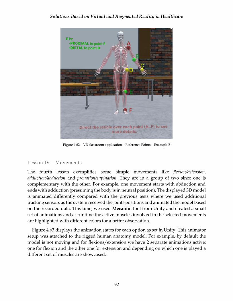
Solutions Based on Virtual and Augmented Reality in Healthcare
92
Figure 4.62 – VR classroom application – Reference Points – Example B
Lesson IV – Movements
The fourth lesson exemplifies some simple movements like flexion/extension,
adduction/abduction and pronation/supination. They are in a group of two since one is
complementary with the other. For example, one movement starts with abduction and
ends with adduction (presuming the body is in neutral position). The displayed 3D model
is animated differently compared with the previous tests where we used additional
tracking sensors as the system received the joints positions and animated the model based
on the recorded data. This time, we used Mecanim tool from Unity and created a small
set of animations and at runtime the active muscles involved in the selected movements
are highlighted with different colors for a better observation.
Figure 4.63 displays the animation states for each option as set in Unity. This animator
setup was attached to the rigged human anatomy model. For example, by default the
model is not moving and for flexions/extension we have 2 separate animations active:
one for flexion and the other one for extension and depending on which one is played a
different set of muscles are showcased.
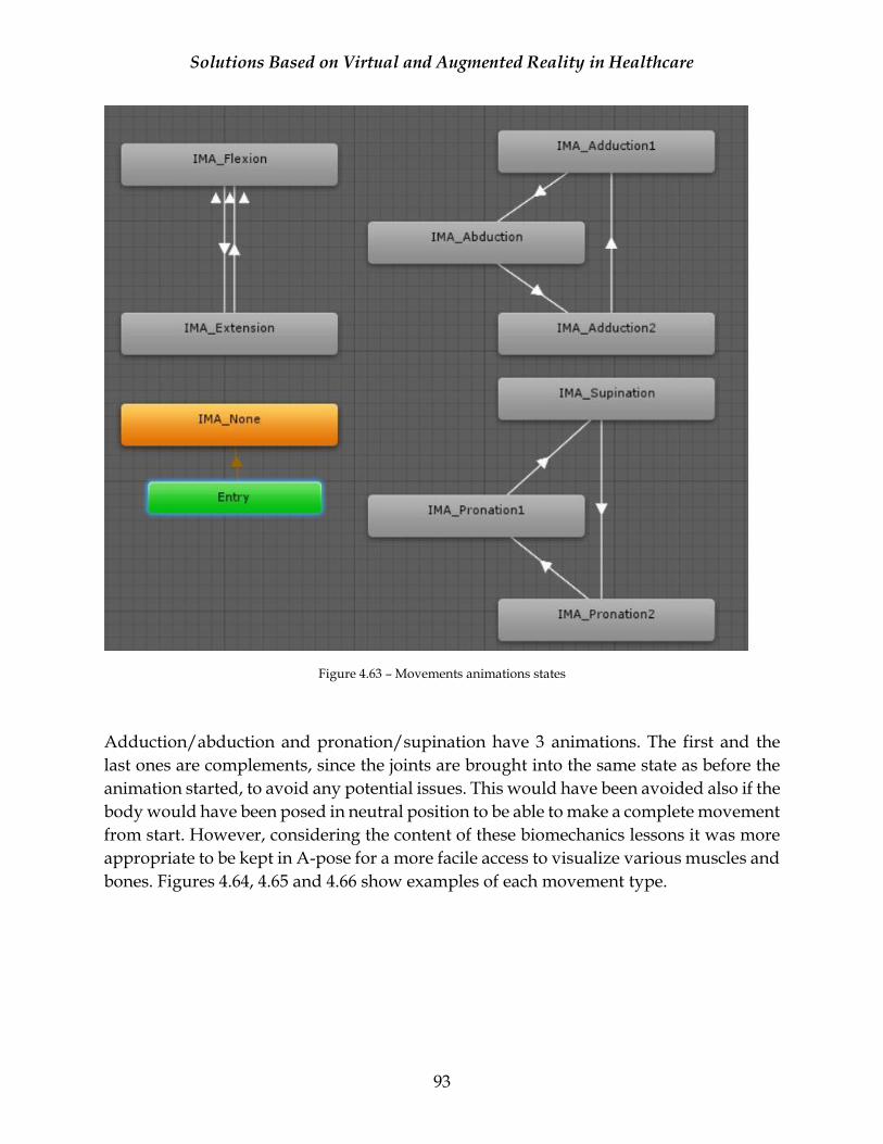
Solutions Based on Virtual and Augmented Reality in Healthcare
93
Figure 4.63 – Movements animations states
Adduction/abduction and pronation/supination have 3 animations. The first and the
last ones are complements, since the joints are brought into the same state as before the
animation started, to avoid any potential issues. This would have been avoided also if the
body would have been posed in neutral position to be able to make a complete movement
from start. However, considering the content of these biomechanics lessons it was more
appropriate to be kept in A-pose for a more facile access to visualize various muscles and
bones. Figures 4.64, 4.65 and 4.66 show examples of each movement type.
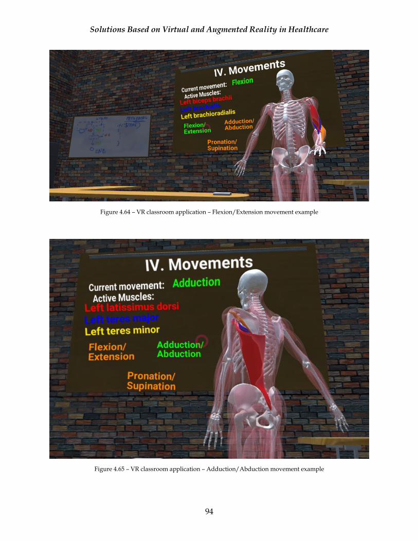
Solutions Based on Virtual and Augmented Reality in Healthcare
94
Figure 4.64 – VR classroom application – Flexion/Extension movement example
Figure 4.65 – VR classroom application – Adduction/Abduction movement example
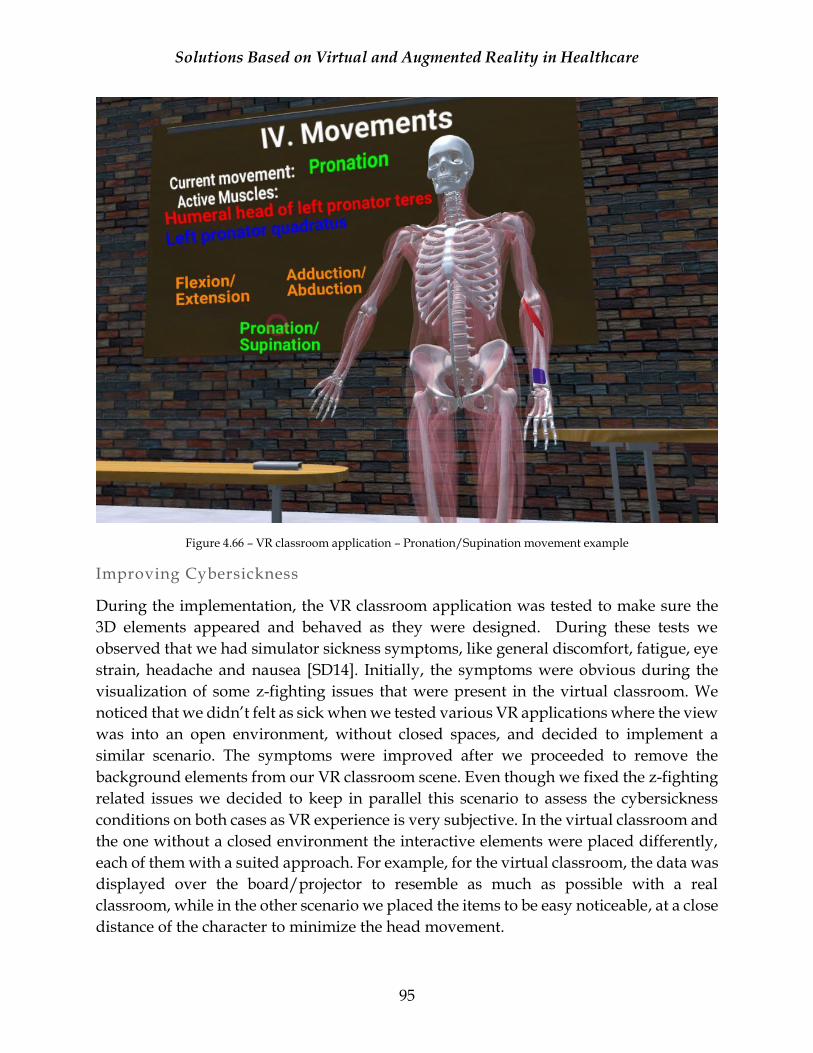
Solutions Based on Virtual and Augmented Reality in Healthcare
95
Figure 4.66 – VR classroom application – Pronation/Supination movement example
Improving Cybersickness
During the implementation, the VR classroom application was tested to make sure the
3D elements appeared and behaved as they were designed. During these tests we
observed that we had simulator sickness symptoms, like general discomfort, fatigue, eye
strain, headache and nausea [SD14]. Initially, the symptoms were obvious during the
visualization of some z-fighting issues that were present in the virtual classroom. We
noticed that we didn’t felt as sick when we tested various VR applications where the view
was into an open environment, without closed spaces, and decided to implement a
similar scenario. The symptoms were improved after we proceeded to remove the
background elements from our VR classroom scene. Even though we fixed the z-fighting
related issues we decided to keep in parallel this scenario to assess the cybersickness
conditions on both cases as VR experience is very subjective. In the virtual classroom and
the one without a closed environment the interactive elements were placed differently,
each of them with a suited approach. For example, for the virtual classroom, the data was
displayed over the board/projector to resemble as much as possible with a real
classroom, while in the other scenario we placed the items to be easy noticeable, at a close
distance of the character to minimize the head movement.
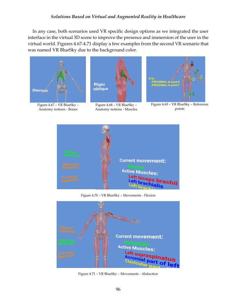
Solutions Based on Virtual and Augmented Reality in Healthcare
96
In any case, both scenarios used VR specific design options as we integrated the user
interface in the virtual 3D scene to improve the presence and immersion of the user in the
virtual world. Figures 4.67-4.71 display a few examples from the second VR scenario that
was named VR BlueSky due to the background color.
Figure 4.67 – VR BlueSky – Anatomy notions - Bones
Figure 4.68 – VR BlueSky – Anatomy notions - Muscles
Figure 4.69 – VR BlueSky – Reference
points
Figure 4.70 – VR BlueSky – Movements - Flexion
Figure 4.71 – VR BlueSky – Movements - Abduction
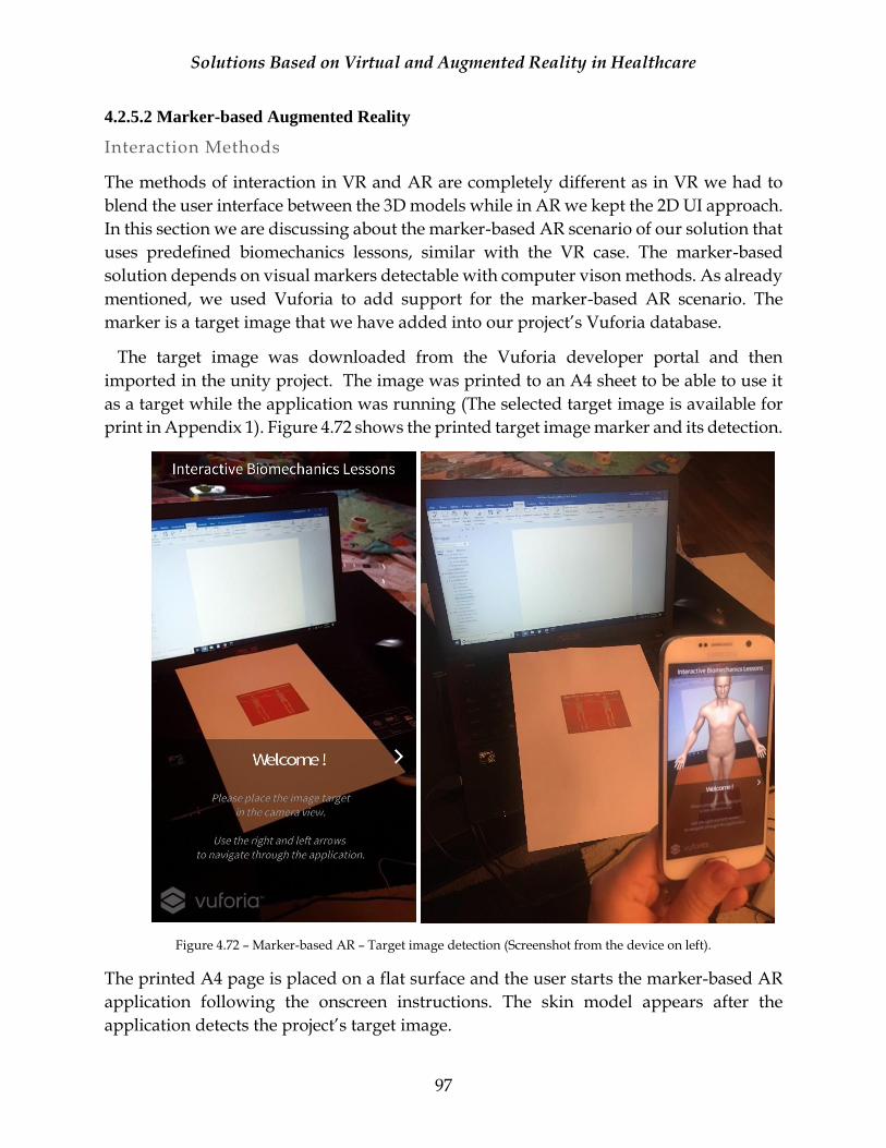
Solutions Based on Virtual and Augmented Reality in Healthcare
97
4.2.5.2 Marker-based Augmented Reality
Interaction Methods
The methods of interaction in VR and AR are completely different as in VR we had to
blend the user interface between the 3D models while in AR we kept the 2D UI approach.
In this section we are discussing about the marker-based AR scenario of our solution that
uses predefined biomechanics lessons, similar with the VR case. The marker-based
solution depends on visual markers detectable with computer vison methods. As already
mentioned, we used Vuforia to add support for the marker-based AR scenario. The
marker is a target image that we have added into our project’s Vuforia database.
The target image was downloaded from the Vuforia developer portal and then
imported in the unity project. The image was printed to an A4 sheet to be able to use it
as a target while the application was running (The selected target image is available for
print in Appendix 1). Figure 4.72 shows the printed target image marker and its detection.
Figure 4.72 – Marker-based AR – Target image detection (Screenshot from the device on left).
The printed A4 page is placed on a flat surface and the user starts the marker-based AR
application following the onscreen instructions. The skin model appears after the
application detects the project’s target image.
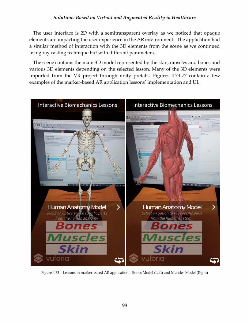
Solutions Based on Virtual and Augmented Reality in Healthcare
98
The user interface is 2D with a semitransparent overlay as we noticed that opaque
elements are impacting the user experience in the AR environment. The application had
a similar method of interaction with the 3D elements from the scene as we continued
using ray casting technique but with different parameters.
The scene contains the main 3D model represented by the skin, muscles and bones and
various 3D elements depending on the selected lesson. Many of the 3D elements were
imported from the VR project through unity prefabs. Figures 4.73-77 contain a few
examples of the marker-based AR application lessons’ implementation and UI.
Figure 4.73 – Lessons in marker-based AR application – Bones Model (Left) and Muscles Model (Right)
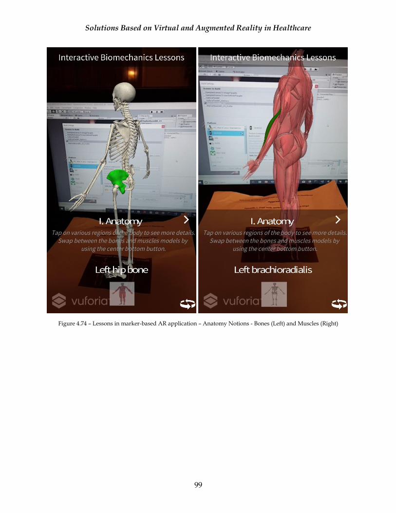
Solutions Based on Virtual and Augmented Reality in Healthcare
99
Figure 4.74 – Lessons in marker-based AR application – Anatomy Notions - Bones (Left) and Muscles (Right)
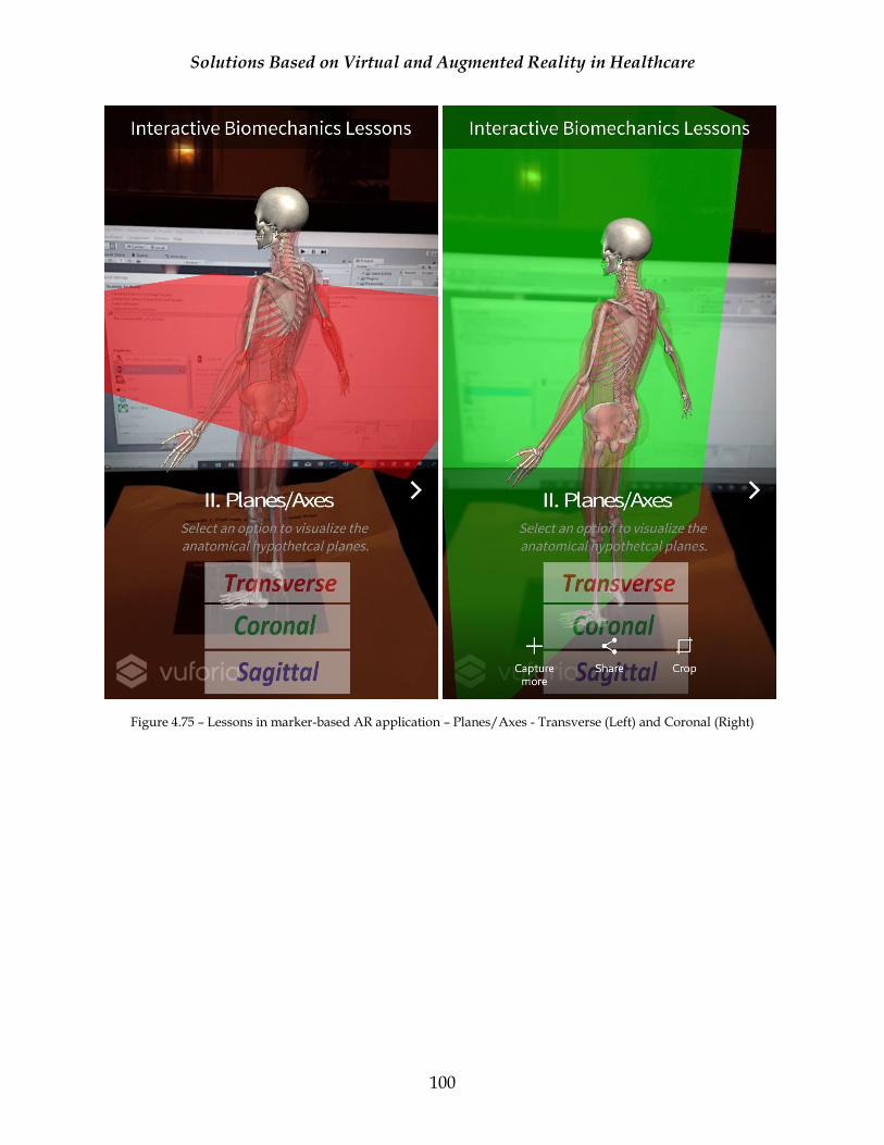
Solutions Based on Virtual and Augmented Reality in Healthcare
100
Figure 4.75 – Lessons in marker-based AR application – Planes/Axes - Transverse (Left) and Coronal (Right)
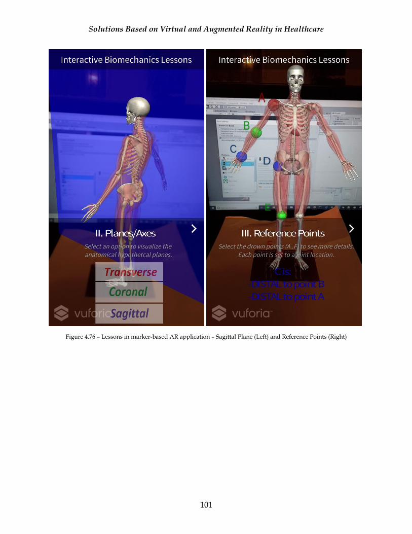
Solutions Based on Virtual and Augmented Reality in Healthcare
101
Figure 4.76 – Lessons in marker-based AR application – Sagittal Plane (Left) and Reference Points (Right)
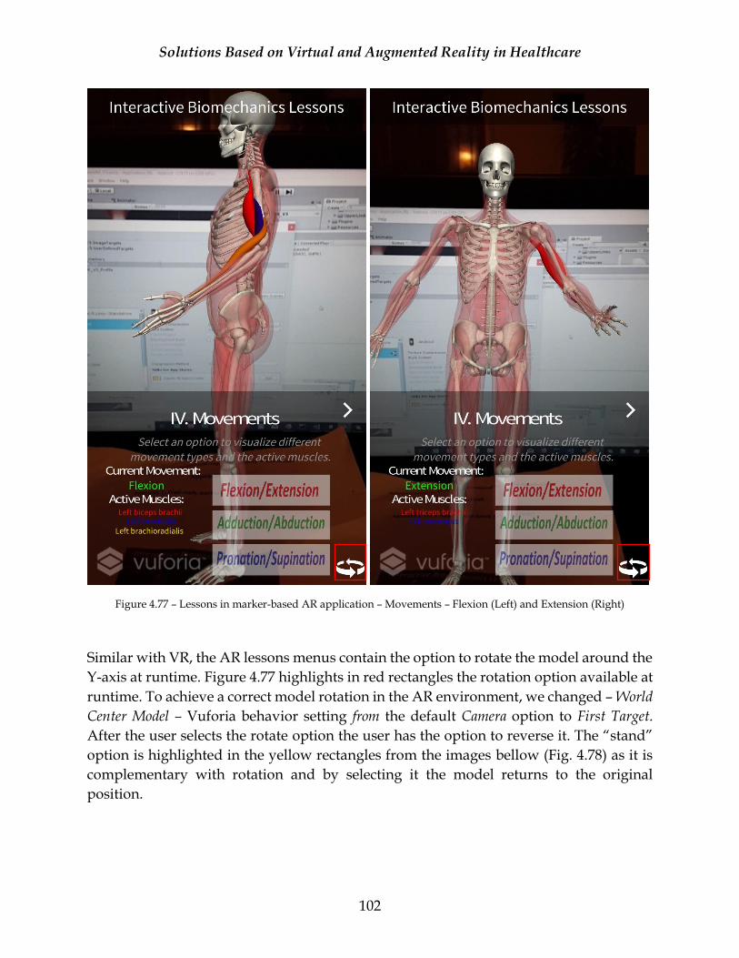
Solutions Based on Virtual and Augmented Reality in Healthcare
102
Figure 4.77 – Lessons in marker-based AR application – Movements – Flexion (Left) and Extension (Right)
Similar with VR, the AR lessons menus contain the option to rotate the model around the
Y-axis at runtime. Figure 4.77 highlights in red rectangles the rotation option available at
runtime. To achieve a correct model rotation in the AR environment, we changed – World
Center Model – Vuforia behavior setting from the default Camera option to First Target.
After the user selects the rotate option the user has the option to reverse it. The “stand”
option is highlighted in the yellow rectangles from the images bellow (Fig. 4.78) as it is
complementary with rotation and by selecting it the model returns to the original
position.
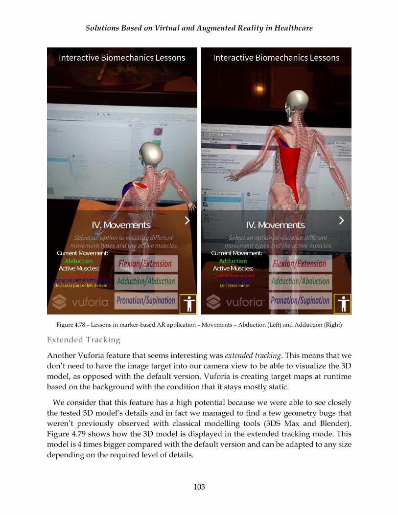
Solutions Based on Virtual and Augmented Reality in Healthcare
103
Figure 4.78 – Lessons in marker-based AR application – Movements – Abduction (Left) and Adduction (Right)
Extended Tracking
Another Vuforia feature that seems interesting was extended tracking. This means that we
don’t need to have the image target into our camera view to be able to visualize the 3D
model, as opposed with the default version. Vuforia is creating target maps at runtime
based on the background with the condition that it stays mostly static.
We consider that this feature has a high potential because we were able to see closely
the tested 3D model’s details and in fact we managed to find a few geometry bugs that
weren’t previously observed with classical modelling tools (3DS Max and Blender).
Figure 4.79 shows how the 3D model is displayed in the extended tracking mode. This
model is 4 times bigger compared with the default version and can be adapted to any size
depending on the required level of details.
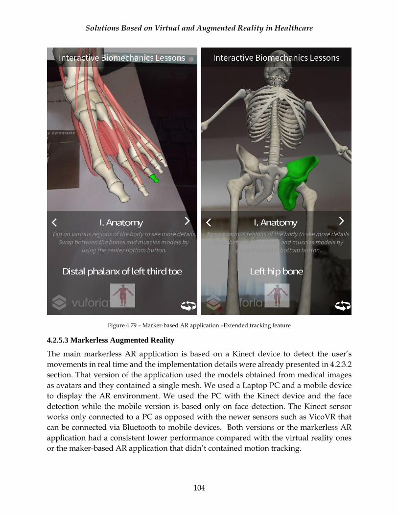
Solutions Based on Virtual and Augmented Reality in Healthcare
104
Figure 4.79 – Marker-based AR application –Extended tracking feature
4.2.5.3 Markerless Augmented Reality
The main markerless AR application is based on a Kinect device to detect the user’s
movements in real time and the implementation details were already presented in 4.2.3.2
section. That version of the application used the models obtained from medical images
as avatars and they contained a single mesh. We used a Laptop PC and a mobile device
to display the AR environment. We used the PC with the Kinect device and the face
detection while the mobile version is based only on face detection. The Kinect sensor
works only connected to a PC as opposed with the newer sensors such as VicoVR that
can be connected via Bluetooth to mobile devices. Both versions or the markerless AR
application had a consistent lower performance compared with the virtual reality ones
or the maker-based AR application that didn’t contained motion tracking.
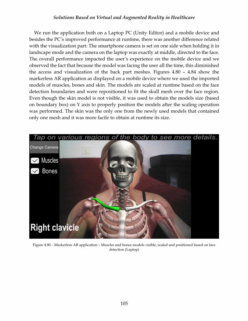
Solutions Based on Virtual and Augmented Reality in Healthcare
105
We run the application both on a Laptop PC (Unity Editor) and a mobile device and
besides the PC’s improved performance at runtime, there was another difference related
with the visualization part: The smartphone camera is set on one side when holding it in
landscape mode and the camera on the laptop was exactly at middle, directed to the face.
The overall performance impacted the user’s experience on the mobile device and we
observed the fact that because the model was facing the user all the time, this diminished
the access and visualization of the back part meshes. Figures 4.80 – 4.84 show the
markerless AR application as displayed on a mobile device where we used the imported
models of muscles, bones and skin. The models are scaled at runtime based on the face
detection boundaries and were repositioned to fit the skull mesh over the face region.
Even though the skin model is not visible, it was used to obtain the models size (based
on boundary box) on Y axis to properly position the models after the scaling operation
was performed. The skin was the only one from the newly used models that contained
only one mesh and it was more facile to obtain at runtime its size.
Figure 4.80 – Markerless AR application – Muscles and bones models visible, scaled and positioned based on face
detection (Laptop)
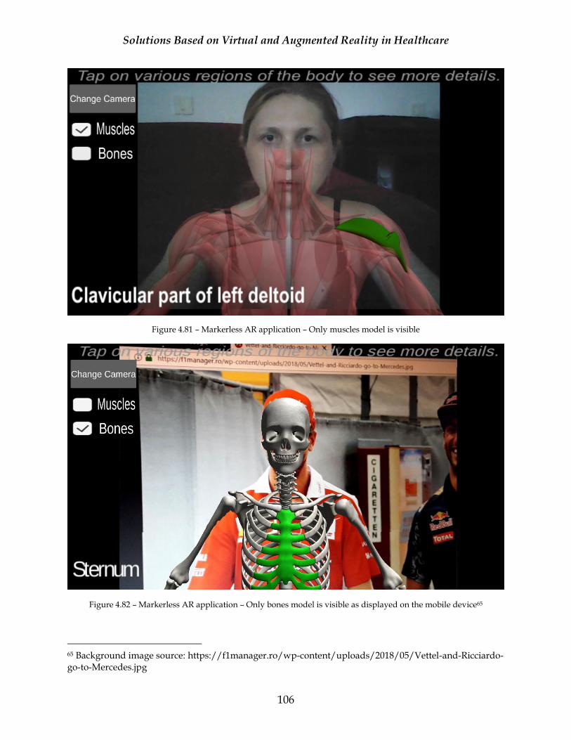
Solutions Based on Virtual and Augmented Reality in Healthcare
106
Figure 4.81 – Markerless AR application – Only muscles model is visible
Figure 4.82 – Markerless AR application – Only bones model is visible as displayed on the mobile device65
65 Background image source: https://f1manager.ro/wp-content/uploads/2018/05/Vettel-and-Ricciardo-go-to-Mercedes.jpg
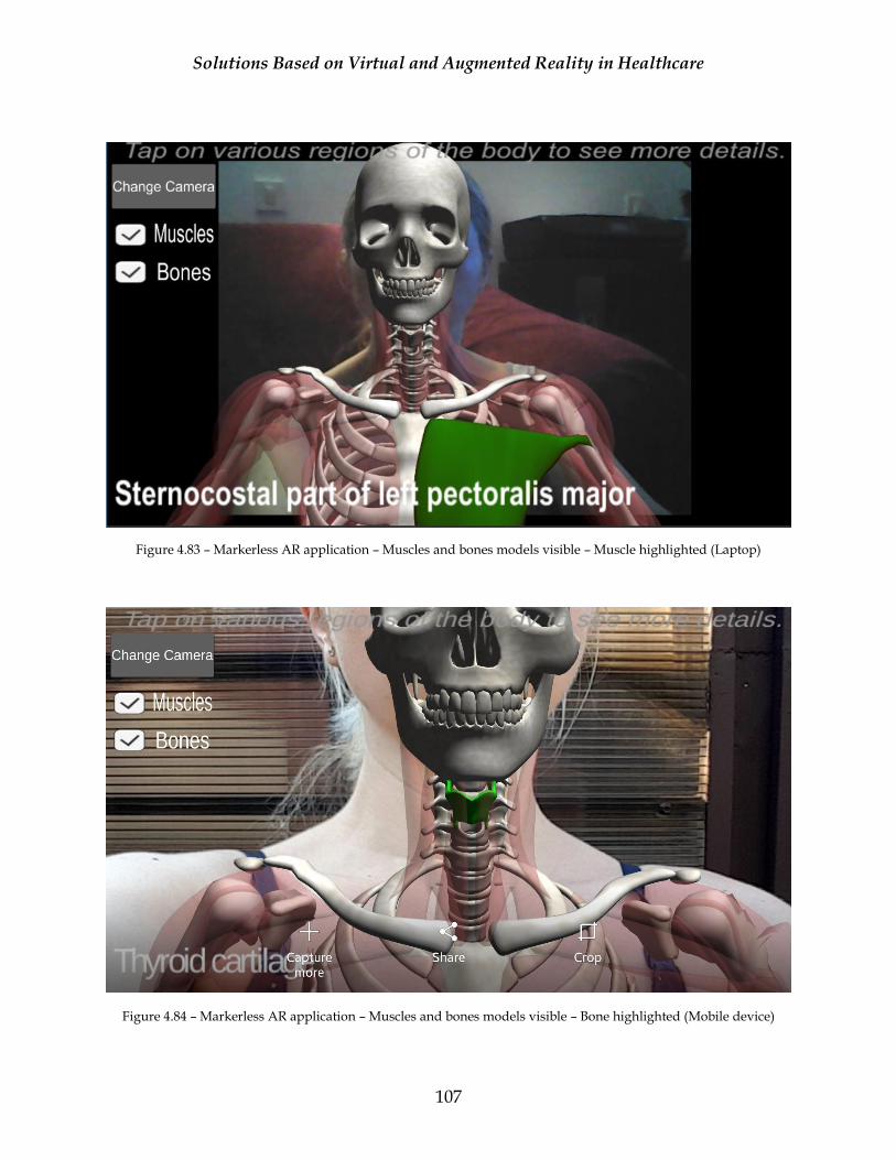
Solutions Based on Virtual and Augmented Reality in Healthcare
107
Figure 4.83 – Markerless AR application – Muscles and bones models visible – Muscle highlighted (Laptop)
Figure 4.84 – Markerless AR application – Muscles and bones models visible – Bone highlighted (Mobile device)
Solutions Based on Virtual and Augmented Reality in Healthcare
108
4.2.6 Performance Analysis
When building an application, either it is for desktop, mobile, VR or AR, the performance
metrics are very important. Usual metrics that are tracked in applications development
are: framerate, memory, application size or the processor heat. The last one is special for
Android devices as it was noticed that during the utilization of heavy computational
applications the mobile devices start to heat. This was observed during author’s extensive
experience in the mobile game development on various titles and Android devices had a
higher occurrence of the heating issues. This issue is obvious usually at extensive usage
of the application and as a result the application’s framerate is impacted due to CPU and
GPU throttling. After the processor reached a certain temperature, it starts to function at
a smaller frequency and in consequence it affects the processing power. Taking into
consideration the fact that the presented applications contain a small number of
interactive lessons and the usual session time is maximum 10 minutes, we didn’t invest
to much in monitoring this metric but considering the complexity of the obtained 3D
models this one should be closely supervised if the solution is extended.
Regarding the other metrics, we gathered data for FPS, memory and rendering usage
using Unity profiler and Android Monitor (from Android Studio). This data was
available only on the development build and we should mention that usually a
development build has lower performance results compared with a release build because
the code has a lower level of optimization. One can add a proprietary tracking method to
gather the performance results with minimal consumption. Due to the available time for
this operation we based our information exclusively on the existing tools.
The AR markerless scenario has two tracking methods: one on the mobile device
(Samsung S6), only with OpenCV face detection and model scaling functionality on, and
one on the PC with all the functionalities enabled. We added the performance data
extracted from the mobile device to be able to make a clear comparison between all 4
scenarios.
Table 4.2 shows the high-level results of the tracked metrics on all 4 scenarios and
Figures 4.85 – 4.88 display the results, as obtained with Unity profiler.
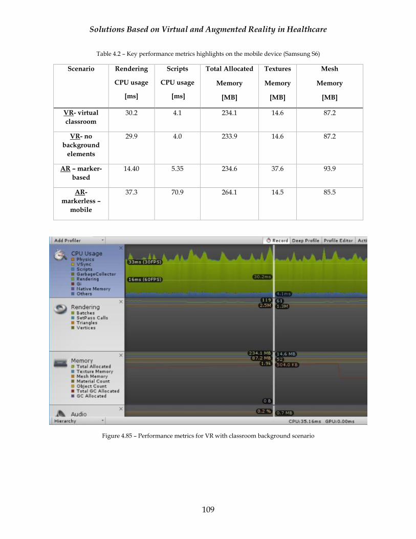
Solutions Based on Virtual and Augmented Reality in Healthcare
109
Table 4.2 – Key performance metrics highlights on the mobile device (Samsung S6)
Scenario Rendering
CPU usage
[ms]
Scripts
CPU usage
[ms]
Total Allocated
Memory
[MB]
Textures
Memory
[MB]
Mesh
Memory
[MB]
VR- virtual
classroom
30.2 4.1 234.1 14.6 87.2
VR- no
background
elements
29.9 4.0 233.9 14.6 87.2
AR – marker-
based
14.40 5.35 234.6 37.6 93.9
AR-
markerless –
mobile
37.3 70.9 264.1 14.5 85.5
Figure 4.85 – Performance metrics for VR with classroom background scenario
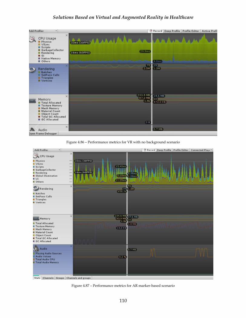
Solutions Based on Virtual and Augmented Reality in Healthcare
110
Figure 4.86 – Performance metrics for VR with no background scenario
Figure 4.87 – Performance metrics for AR marker-based scenario
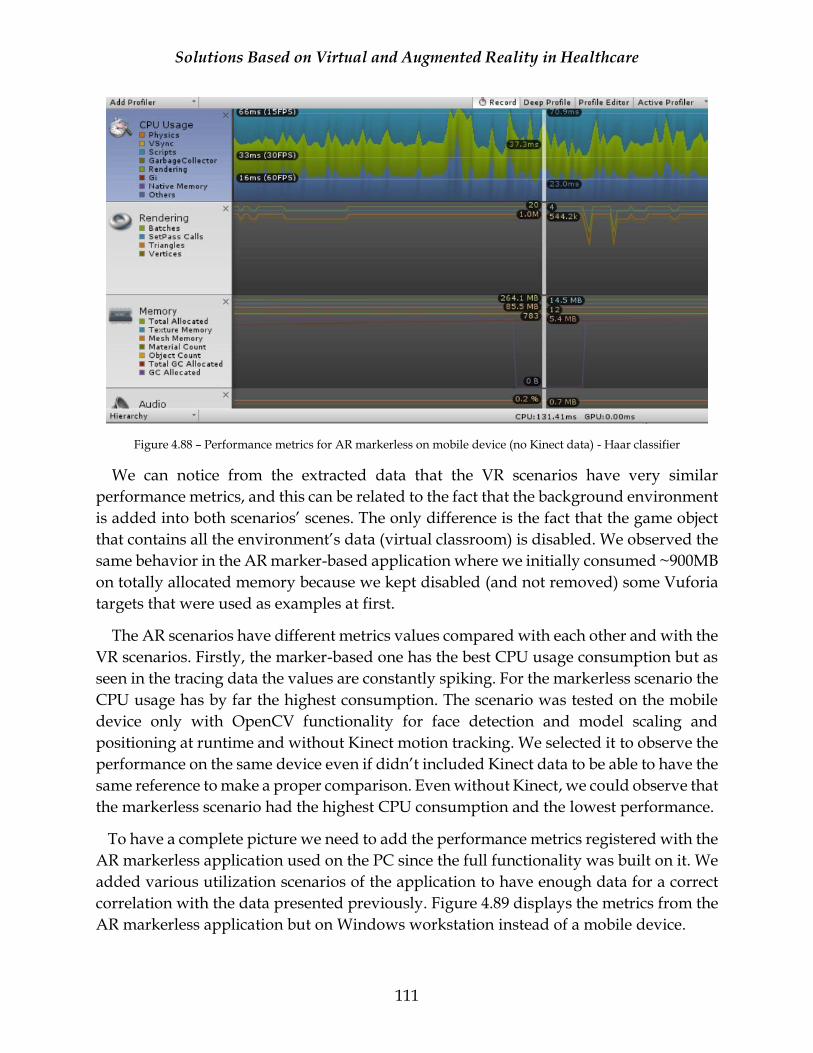
Solutions Based on Virtual and Augmented Reality in Healthcare
111
Figure 4.88 – Performance metrics for AR markerless on mobile device (no Kinect data) - Haar classifier
We can notice from the extracted data that the VR scenarios have very similar
performance metrics, and this can be related to the fact that the background environment
is added into both scenarios’ scenes. The only difference is the fact that the game object
that contains all the environment’s data (virtual classroom) is disabled. We observed the
same behavior in the AR marker-based application where we initially consumed ~900MB
on totally allocated memory because we kept disabled (and not removed) some Vuforia
targets that were used as examples at first.
The AR scenarios have different metrics values compared with each other and with the
VR scenarios. Firstly, the marker-based one has the best CPU usage consumption but as
seen in the tracing data the values are constantly spiking. For the markerless scenario the
CPU usage has by far the highest consumption. The scenario was tested on the mobile
device only with OpenCV functionality for face detection and model scaling and
positioning at runtime and without Kinect motion tracking. We selected it to observe the
performance on the same device even if didn’t included Kinect data to be able to have the
same reference to make a proper comparison. Even without Kinect, we could observe that
the markerless scenario had the highest CPU consumption and the lowest performance.
To have a complete picture we need to add the performance metrics registered with the
AR markerless application used on the PC since the full functionality was built on it. We
added various utilization scenarios of the application to have enough data for a correct
correlation with the data presented previously. Figure 4.89 displays the metrics from the
AR markerless application but on Windows workstation instead of a mobile device.
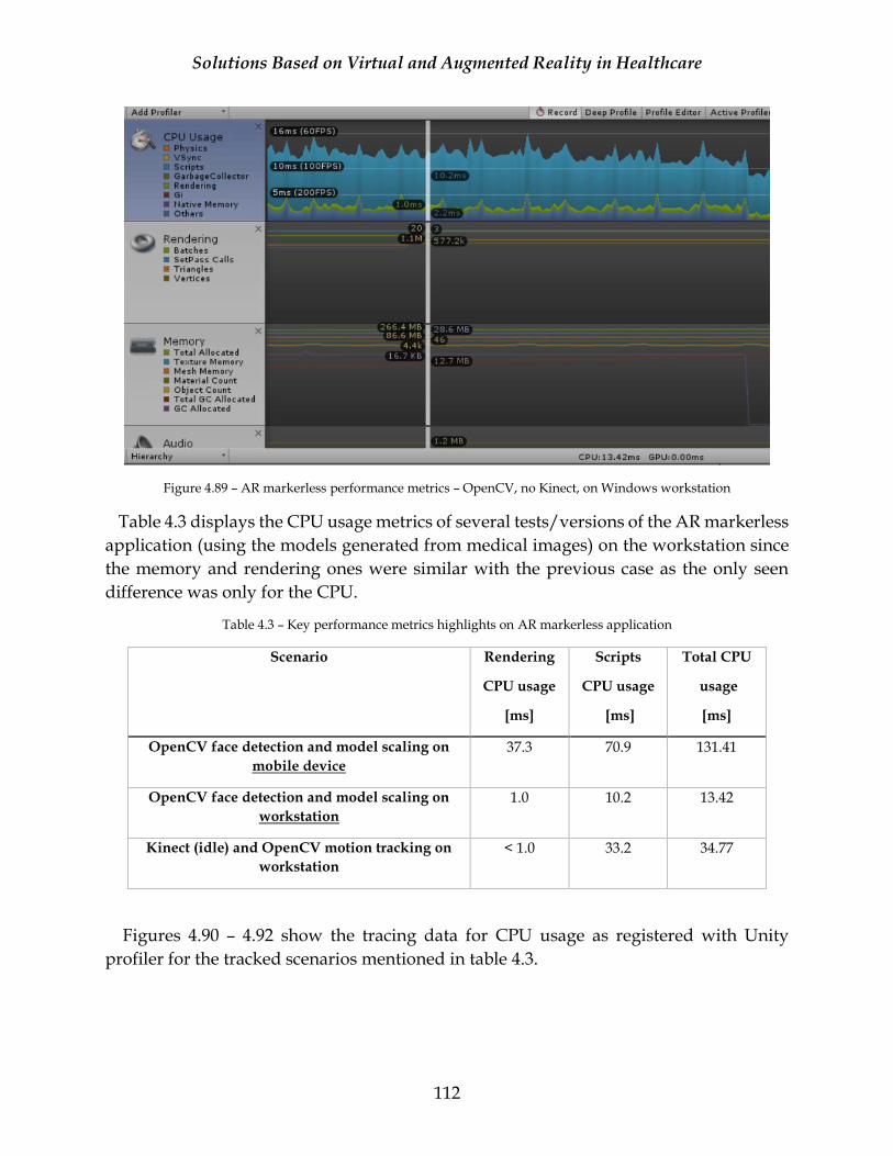
Solutions Based on Virtual and Augmented Reality in Healthcare
112
Figure 4.89 – AR markerless performance metrics – OpenCV, no Kinect, on Windows workstation
Table 4.3 displays the CPU usage metrics of several tests/versions of the AR markerless
application (using the models generated from medical images) on the workstation since
the memory and rendering ones were similar with the previous case as the only seen
difference was only for the CPU.
Table 4.3 – Key performance metrics highlights on AR markerless application
Scenario Rendering
CPU usage
[ms]
Scripts
CPU usage
[ms]
Total CPU
usage
[ms]
OpenCV face detection and model scaling on
mobile device
37.3 70.9 131.41
OpenCV face detection and model scaling on
workstation
1.0 10.2 13.42
Kinect (idle) and OpenCV motion tracking on
workstation
< 1.0 33.2 34.77
Figures 4.90 – 4.92 show the tracing data for CPU usage as registered with Unity
profiler for the tracked scenarios mentioned in table 4.3.
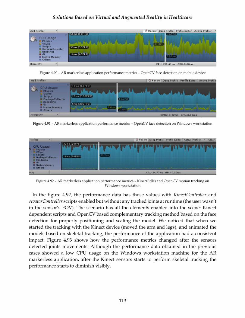
Solutions Based on Virtual and Augmented Reality in Healthcare
113
Figure 4.90 – AR markerless application performance metrics – OpenCV face detection on mobile device
Figure 4.91 – AR markerless application performance metrics – OpenCV face detection on Windows workstation
Figure 4.92 – AR markerless application performance metrics – Kinect(idle) and OpenCV motion tracking on
Windows workstation
In the figure 4.92, the performance data has those values with KinectController and
AvatarController scripts enabled but without any tracked joints at runtime (the user wasn’t
in the sensor’s FOV). The scenario has all the elements enabled into the scene: Kinect
dependent scripts and OpenCV based complementary tracking method based on the face
detection for properly positioning and scaling the model. We noticed that when we
started the tracking with the Kinect device (moved the arm and legs), and animated the
models based on skeletal tracking, the performance of the application had a consistent
impact. Figure 4.93 shows how the performance metrics changed after the sensors
detected joints movements. Although the performance data obtained in the previous
cases showed a low CPU usage on the Windows workstation machine for the AR
markerless application, after the Kinect sensors starts to perform skeletal tracking the
performance starts to diminish visibly.
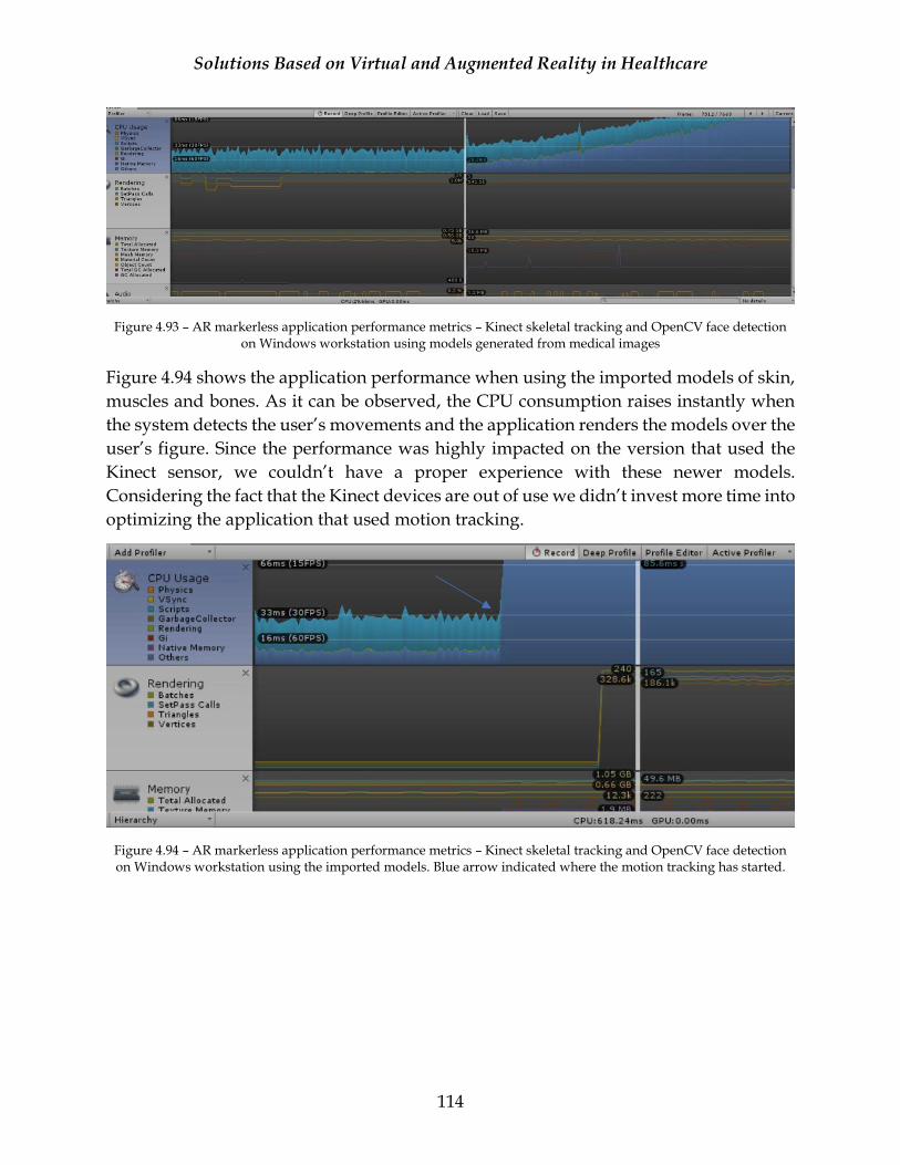
Solutions Based on Virtual and Augmented Reality in Healthcare
114
Figure 4.93 – AR markerless application performance metrics – Kinect skeletal tracking and OpenCV face detection on Windows workstation using models generated from medical images
Figure 4.94 shows the application performance when using the imported models of skin,
muscles and bones. As it can be observed, the CPU consumption raises instantly when
the system detects the user’s movements and the application renders the models over the
user’s figure. Since the performance was highly impacted on the version that used the
Kinect sensor, we couldn’t have a proper experience with these newer models.
Considering the fact that the Kinect devices are out of use we didn’t invest more time into
optimizing the application that used motion tracking.
Figure 4.94 – AR markerless application performance metrics – Kinect skeletal tracking and OpenCV face detection on Windows workstation using the imported models. Blue arrow indicated where the motion tracking has started.
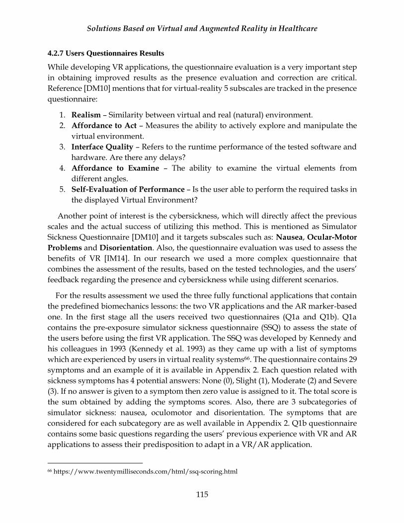
Solutions Based on Virtual and Augmented Reality in Healthcare
115
4.2.7 Users Questionnaires Results
While developing VR applications, the questionnaire evaluation is a very important step
in obtaining improved results as the presence evaluation and correction are critical.
Reference [DM10] mentions that for virtual-reality 5 subscales are tracked in the presence
questionnaire:
1. Realism – Similarity between virtual and real (natural) environment.
2. Affordance to Act – Measures the ability to actively explore and manipulate the
virtual environment.
3. Interface Quality – Refers to the runtime performance of the tested software and
hardware. Are there any delays?
4. Affordance to Examine – The ability to examine the virtual elements from
different angles.
5. Self-Evaluation of Performance – Is the user able to perform the required tasks in
the displayed Virtual Environment?
Another point of interest is the cybersickness, which will directly affect the previous
scales and the actual success of utilizing this method. This is mentioned as Simulator
Sickness Questionnaire [DM10] and it targets subscales such as: Nausea, Ocular-Motor
Problems and Disorientation. Also, the questionnaire evaluation was used to assess the
benefits of VR [IM14]. In our research we used a more complex questionnaire that
combines the assessment of the results, based on the tested technologies, and the users’
feedback regarding the presence and cybersickness while using different scenarios.
For the results assessment we used the three fully functional applications that contain
the predefined biomechanics lessons: the two VR applications and the AR marker-based
one. In the first stage all the users received two questionnaires (Q1a and Q1b). Q1a
contains the pre-exposure simulator sickness questionnaire (SSQ) to assess the state of
the users before using the first VR application. The SSQ was developed by Kennedy and
his colleagues in 1993 (Kennedy et al. 1993) as they came up with a list of symptoms
which are experienced by users in virtual reality systems66. The questionnaire contains 29
symptoms and an example of it is available in Appendix 2. Each question related with
sickness symptoms has 4 potential answers: None (0), Slight (1), Moderate (2) and Severe
(3). If no answer is given to a symptom then zero value is assigned to it. The total score is
the sum obtained by adding the symptoms scores. Also, there are 3 subcategories of
simulator sickness: nausea, oculomotor and disorientation. The symptoms that are
considered for each subcategory are as well available in Appendix 2. Q1b questionnaire
contains some basic questions regarding the users’ previous experience with VR and AR
applications to assess their predisposition to adapt in a VR/AR application.
66 https://www.twentymilliseconds.com/html/ssq-scoring.html
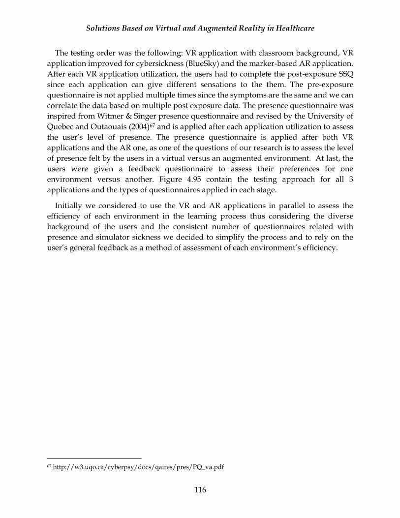
Solutions Based on Virtual and Augmented Reality in Healthcare
116
The testing order was the following: VR application with classroom background, VR
application improved for cybersickness (BlueSky) and the marker-based AR application.
After each VR application utilization, the users had to complete the post-exposure SSQ
since each application can give different sensations to the them. The pre-exposure
questionnaire is not applied multiple times since the symptoms are the same and we can
correlate the data based on multiple post exposure data. The presence questionnaire was
inspired from Witmer & Singer presence questionnaire and revised by the University of
Quebec and Outaouais (2004)67 and is applied after each application utilization to assess
the user’s level of presence. The presence questionnaire is applied after both VR
applications and the AR one, as one of the questions of our research is to assess the level
of presence felt by the users in a virtual versus an augmented environment. At last, the
users were given a feedback questionnaire to assess their preferences for one
environment versus another. Figure 4.95 contain the testing approach for all 3
applications and the types of questionnaires applied in each stage.
Initially we considered to use the VR and AR applications in parallel to assess the
efficiency of each environment in the learning process thus considering the diverse
background of the users and the consistent number of questionnaires related with
presence and simulator sickness we decided to simplify the process and to rely on the
user’s general feedback as a method of assessment of each environment’s efficiency.
67 http://w3.uqo.ca/cyberpsy/docs/qaires/pres/PQ_va.pdf
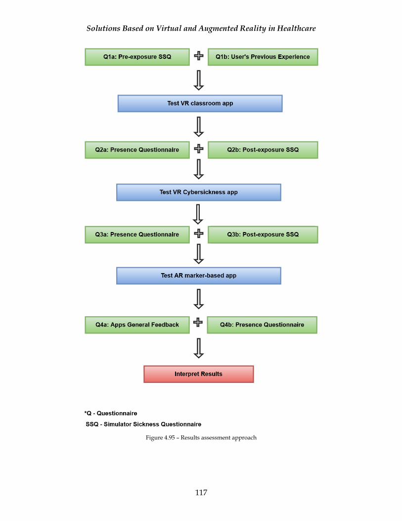
Solutions Based on Virtual and Augmented Reality in Healthcare
117
Figure 4.95 – Results assessment approach
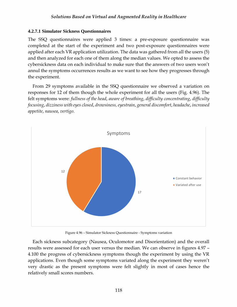
Solutions Based on Virtual and Augmented Reality in Healthcare
118
4.2.7.1 Simulator Sickness Questionnaires
The SSQ questionnaires were applied 3 times: a pre-exposure questionnaire was
completed at the start of the experiment and two post-exposure questionnaires were
applied after each VR application utilization. The data was gathered from all the users (5)
and then analyzed for each one of them along the median values. We opted to assess the
cybersickness data on each individual to make sure that the answers of two users won’t
annul the symptoms occurrences results as we want to see how they progresses through
the experiment.
From 29 symptoms available in the SSQ questionnaire we observed a variation on
responses for 12 of them though the whole experiment for all the users (Fig. 4.96). The
felt symptoms were: fullness of the head, aware of breathing, difficulty concentrating, difficulty
focusing, dizziness with eyes closed, drowsiness, eyestrain, general discomfort, headache, increased
appetite, nausea, vertigo.
Figure 4.96 – Simulator Sickness Questionnaire - Symptoms variation
Each sickness subcategory (Nausea, Oculomotor and Disorientation) and the overall
results were assessed for each user versus the median. We can observe in figures 4.97 –
4.100 the progress of cybersickness symptoms though the experiment by using the VR
applications. Even though some symptoms variated along the experiment they weren’t
very drastic as the present symptoms were felt slightly in most of cases hence the
relatively small scores numbers.
17
12
Symptoms
Constant behavior
Variated after use
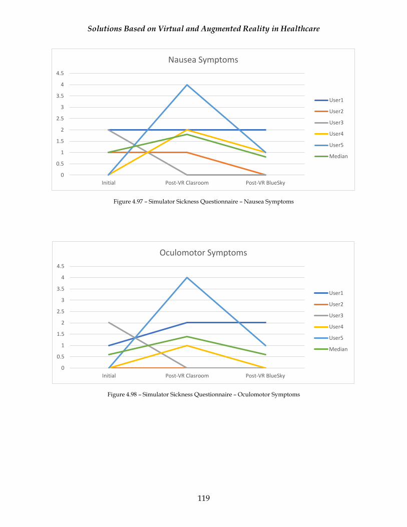
Solutions Based on Virtual and Augmented Reality in Healthcare
119
Figure 4.97 – Simulator Sickness Questionnaire – Nausea Symptoms
Figure 4.98 – Simulator Sickness Questionnaire – Oculomotor Symptoms
0
0.5
1
1.5
2
2.5
3
3.5
4
4.5
Initial Post-VR Clasroom Post-VR BlueSky
Nausea Symptoms
User1
User2
User3
User4
User5
Median
0
0.5
1
1.5
2
2.5
3
3.5
4
4.5
Initial Post-VR Clasroom Post-VR BlueSky
Oculomotor Symptoms
User1
User2
User3
User4
User5
Median
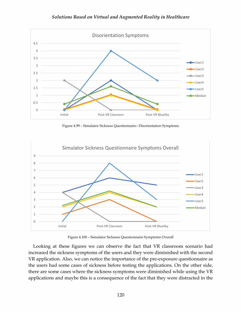
Solutions Based on Virtual and Augmented Reality in Healthcare
120
Figure 4.99 – Simulator Sickness Questionnaire– Disorientation Symptoms
Figure 4.100 – Simulator Sickness Questionnaire Symptoms Overall
Looking at these figures we can observe the fact that VR classroom scenario had
increased the sickness symptoms of the users and they were diminished with the second
VR application. Also, we can notice the importance of the pre-exposure questionnaire as
the users had some cases of sickness before testing the applications. On the other side,
there are some cases where the sickness symptoms were diminished while using the VR
applications and maybe this is a consequence of the fact that they were distracted in the
0
0.5
1
1.5
2
2.5
3
3.5
4
4.5
Initial Post-VR Clasroom Post-VR BlueSky
Disorientation Symptoms
User1
User2
User3
User4
User5
Median
0
1
2
3
4
5
6
7
8
9
Initial Post-VR Clasroom Post-VR BlueSky
Simulator Sickness Questionnaire Symptoms Overall
User1
User2
User3
User4
User5
Median
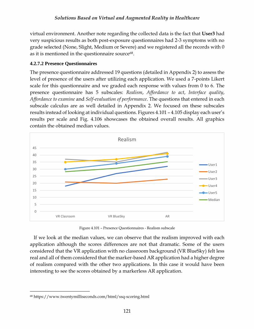
Solutions Based on Virtual and Augmented Reality in Healthcare
121
virtual environment. Another note regarding the collected data is the fact that User3 had
very suspicious results as both post-exposure questionnaires had 2-3 symptoms with no
grade selected (None, Slight, Medium or Severe) and we registered all the records with 0
as it is mentioned in the questionnaire source68.
4.2.7.2 Presence Questionnaires
The presence questionnaire addressed 19 questions (detailed in Appendix 2) to assess the
level of presence of the users after utilizing each application. We used a 7-points Likert
scale for this questionnaire and we graded each response with values from 0 to 6. The
presence questionnaire has 5 subscales: Realism, Affordance to act, Interface quality,
Affordance to examine and Self-evaluation of performance. The questions that entered in each
subscale calculus are as well detailed in Appendix 2. We focused on these subscales
results instead of looking at individual questions. Figures 4.101 – 4.105 display each user’s
results per scale and Fig. 4.106 showcases the obtained overall results. All graphics
contain the obtained median values.
Figure 4.101 – Presence Questionnaires - Realism subscale
If we look at the median values, we can observe that the realism improved with each
application although the scores differences are not that dramatic. Some of the users
considered that the VR application with no classroom background (VR BlueSky) felt less
real and all of them considered that the marker-based AR application had a higher degree
of realism compared with the other two applications. In this case it would have been
interesting to see the scores obtained by a markerless AR application.
68 https://www.twentymilliseconds.com/html/ssq-scoring.html
0
5
10
15
20
25
30
35
40
45
VR Clasroom VR BlueSky AR
Realism
User1
User2
User3
User4
User5
Median
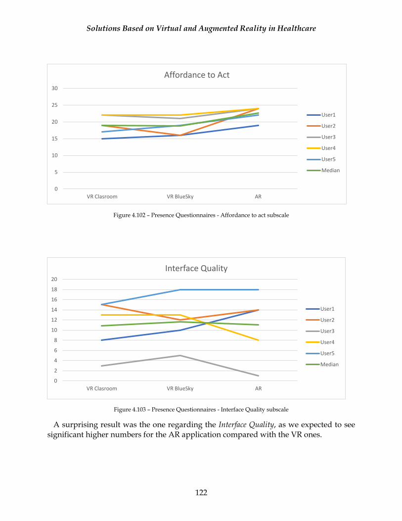
Solutions Based on Virtual and Augmented Reality in Healthcare
122
Figure 4.102 – Presence Questionnaires - Affordance to act subscale
Figure 4.103 – Presence Questionnaires - Interface Quality subscale
A surprising result was the one regarding the Interface Quality, as we expected to see significant higher numbers for the AR application compared with the VR ones.
0
5
10
15
20
25
30
VR Clasroom VR BlueSky AR
Affordance to Act
User1
User2
User3
User4
User5
Median
0
2
4
6
8
10
12
14
16
18
20
VR Clasroom VR BlueSky AR
Interface Quality
User1
User2
User3
User4
User5
Median
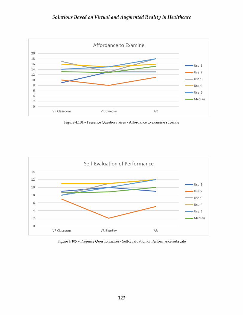
Solutions Based on Virtual and Augmented Reality in Healthcare
123
Figure 4.104 – Presence Questionnaires - Affordance to examine subscale
Figure 4.105 – Presence Questionnaires - Self-Evaluation of Performance subscale
0
2
4
6
8
10
12
14
16
18
20
VR Clasroom VR BlueSky AR
Affordance to Examine
User1
User2
User3
User4
User5
Median
0
2
4
6
8
10
12
14
VR Clasroom VR BlueSky AR
Self-Evaluation of Performance
User1
User2
User3
User4
User5
Median
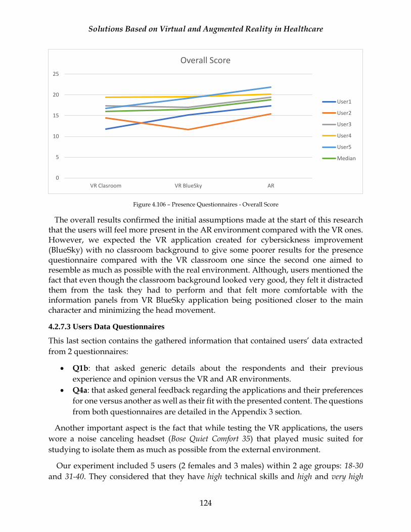
Solutions Based on Virtual and Augmented Reality in Healthcare
124
Figure 4.106 – Presence Questionnaires - Overall Score
The overall results confirmed the initial assumptions made at the start of this research that the users will feel more present in the AR environment compared with the VR ones. However, we expected the VR application created for cybersickness improvement (BlueSky) with no classroom background to give some poorer results for the presence questionnaire compared with the VR classroom one since the second one aimed to resemble as much as possible with the real environment. Although, users mentioned the fact that even though the classroom background looked very good, they felt it distracted them from the task they had to perform and that felt more comfortable with the information panels from VR BlueSky application being positioned closer to the main character and minimizing the head movement.
4.2.7.3 Users Data Questionnaires
This last section contains the gathered information that contained users’ data extracted
from 2 questionnaires:
• Q1b: that asked generic details about the respondents and their previous
experience and opinion versus the VR and AR environments.
• Q4a: that asked general feedback regarding the applications and their preferences
for one versus another as well as their fit with the presented content. The questions
from both questionnaires are detailed in the Appendix 3 section.
Another important aspect is the fact that while testing the VR applications, the users
wore a noise canceling headset (Bose Quiet Comfort 35) that played music suited for
studying to isolate them as much as possible from the external environment.
Our experiment included 5 users (2 females and 3 males) within 2 age groups: 18-30
and 31-40. They considered that they have high technical skills and high and very high
0
5
10
15
20
25
VR Clasroom VR BlueSky AR
Overall Score
User1
User2
User3
User4
User5
Median
Solutions Based on Virtual and Augmented Reality in Healthcare
125
interest in technology. All of them knew what VR is while only one didn’t knew what AR
is. Also, 3 out of 5 respondents said that they have previously tried VR applications and
that they didn’t felt sick after they used them. Two users said that their VR experience
was neither good or bad while one mentioned that it was a very good experience as all 3 of
them felt present in the displayed virtual environment. Regarding the previous AR
applications usage, 3 users responded that they had a good experience while one said that
the experience was neither good or bad. One user considers both AR and VR interesting
while the other 3 said that they prefer the AR applications. For this assessment was
excluded the one user that said that he/she didn’t knew what AR is.
Regarding their feedback for the applications described in this thesis, two users said
that they rate the overall experience good while 3 responded that they considered it very
good.
From the total users, four said that they preferred augmented reality application versus
the virtual reality ones while one said that he/she preferred the virtual reality application
with no background as no one favored the VR application with the classroom
background. During the experiment the users mentioned the fact that the second VR
application was more suited for them because the displayed elements were closer to the
3D model and they could see the information better, along the fact that they felt better in
the virtual environment because there was no background environment (although one
complained about the lighting conditions and the chosen color – blue sky). They
mentioned the fact that it felt that the classroom environment was a disturbance factor
and couldn’t focus as well on the tasks. On the same note, regarding the disturbance
factors, three users agreed the fact that the lack on interruptions within VR applications
testing was beneficial while another one strongly agreed with this. Surprisingly, one user
completely disagreed with this approach (Fig 4.107).
While all of them responded that the app with AR environment is technically more
suited for the learning experience, for VR one we didn’t saw complementary responses
as these questions are opposing themselves in this experiment. Two users said that they
consider VR technically more reliable for the learning experience, other two disagreed
and one was neutral.
Four users agreed and strongly agreed with the fact that the applications are easy to use
and have a low learning curve while another one completely disagreed.
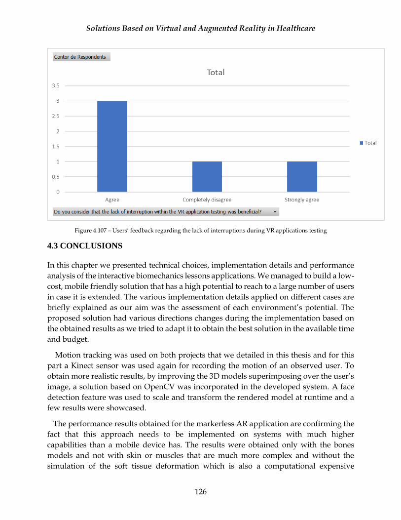
Solutions Based on Virtual and Augmented Reality in Healthcare
126
Figure 4.107 – Users’ feedback regarding the lack of interruptions during VR applications testing
4.3 CONCLUSIONS
In this chapter we presented technical choices, implementation details and performance
analysis of the interactive biomechanics lessons applications. We managed to build a low-
cost, mobile friendly solution that has a high potential to reach to a large number of users
in case it is extended. The various implementation details applied on different cases are
briefly explained as our aim was the assessment of each environment’s potential. The
proposed solution had various directions changes during the implementation based on
the obtained results as we tried to adapt it to obtain the best solution in the available time
and budget.
Motion tracking was used on both projects that we detailed in this thesis and for this
part a Kinect sensor was used again for recording the motion of an observed user. To
obtain more realistic results, by improving the 3D models superimposing over the user’s
image, a solution based on OpenCV was incorporated in the developed system. A face
detection feature was used to scale and transform the rendered model at runtime and a
few results were showcased.
The performance results obtained for the markerless AR application are confirming the
fact that this approach needs to be implemented on systems with much higher
capabilities than a mobile device has. The results were obtained only with the bones
models and not with skin or muscles that are much more complex and without the
simulation of the soft tissue deformation which is also a computational expensive
Solutions Based on Virtual and Augmented Reality in Healthcare
127
operation. We could also observe the fact that during Kinect skeletal tracking the
application’s performance consistently drops. Taking into account the fact that Microsoft
recently announced that moving forward they are offering only support for Kinect
technology, the usage of a Kinect sensor in this type of applications is not the best option
for further development. We consider that new technology such as VicoVR is a much
better fit as it has its own processing source that minimizes the display device overhead
during the skeletal tracking.
The most interesting point from this chapter is the results assessment based on users’
testing and feedback. One of the questions of this research was the fact that in AR
applications users should feel more present while the minimization of external
disturbance factors will aid the learning experience. Both assumptions were confirmed
although we expected a clearer difference. Another interesting point is the fact that while
we tried to create a world that resembles as much as possible with the reality in an VR
scenario (with the classroom background) we actually managed to introduce disturbance
factors as the users felt distracted by its composition. The majority of users said prior the
experiment that they preferred AR applications and the same trend was somehow kept
at the end of our experiment. One user preferred the VR application with no background
created to improve cybersickness. These responses are actually in the same trend with
the market forecasts. If initially it was tough that VR would have a great evolution along
the years, now AR seems to have the lead between these two thus both technological
systems have overall positive feedback.
As a future perspective we can think of algorithms and solution based only on
computer vision for full body skeletal tracking thus the current ones are just at the start
and it required more research to be applied at large scale even though there are a few
approaches that seem to be promising [RD15]. In this direction, one of the newest
approaches is to use human pose estimation implemented with TensorFlow69. We
managed to get the current implementation and to set the appropriate parameters to run
acceptable on our machines. This solution is implemented in JavaScript and offers the
possibility to obtain human poses in real time in a browser and this is available for PC
and mobile devices as well. We added Three.js70 (JavaScript 3D Library) to be able to
render the 3D elements. Even though the visualization and functionality has still a long
way to come this seems to be a viable approach to track the movements of an observed
user in real time without additional sensors. However, in this case the performance has a
tradeoff with the tracking quality as determined by the parameters’ settings (e.g. output
69 https://medium.com/tensorflow/real-time-human-pose-estimation-in-the-browser-with-tensorflow-js-7dd0bc881cd5 70 https://threejs.org/

Solutions Based on Virtual and Augmented Reality in Healthcare
128
stride that affects the height and the width of the layers in the neural network. The lower
the value of the output stride the higher the accuracy but slower the speed71).
71 https://storage.googleapis.com/tfjs-models/demos/posenet/camera.html
Solutions Based on Virtual and Augmented Reality in Healthcare
129
CHAPTER 5
CONCLUSIONS AND FUTURE WORK
5.1 THE ORIGINAL CONTRIBUTIONS OF THIS THESIS
A large part of this thesis’ content is practice based research as it is composed by various
experimental results or novel implementations.
Chapter 3 contains the author’s contribution to TRAVEE – an informatic solution based
on VR for neuromotor rehabilitation. The first part tackled the adaptation of the rendered
3D models based on the patient’s body characteristics such as the age, weight, height,
skin and hair color. This is followed by the initial work at the VR module where the
content was displayed using an Oculus Rift device and the results were published at
CSCS15 conference [AV15b]. Another contribution is the development of the kinematics
module as the system incorporated two tracking devices: Leap Motion and Kinect. The
obtained results were disseminated at EHB2015 conference [AV15a].
Chapter 4 contains the complete details of a solution based on VR and AR in medical
education. It is designed and built completely by the author and the obtained results are
analyzed based on the visual feedback and users’ responses to questionnaires. Not only
the efficiency of the implemented methods is considered but also the responses to the
presence and simulator sickness related questions. The initial proposal of this solution
that aims to improve the learning process in biomechanics study by using VR and AR
was published at ICERI2016 conference and received positive feedback [AV16b].
The author implemented a novel solution for obtaining rigged 3D models for bones,
skin and muscles of the human body based on medical images. One of the most important
aspects is the fact that the 3D model generation was obtained for the whole body not for
smaller regions, as the previously available solutions, bringing new content for advanced
visualization. The results were published at CSCS17 conference [AV17].
The tests section contains various experimental results obtained while testing the
available AR and VR technologies. The results were obtained with Oculus Rift, Google
Cardboard, Gear VR, HoloLens Emulator and Vuforia platform. Another contribution is
related to the real time motion tracking of a human body. This part provides details
regarding the whole-body tracking of an observed user and rendering the 3D models
accordingly with the detected movements. The initial assessment of the viable solutions
was published into an article published at MVAR workshop from ICMI2016 conference
[AV16a].
Solutions Based on Virtual and Augmented Reality in Healthcare
130
The author implemented a solution based on a tracking sensor (Kinect) and computer
vison (OpenCV) to display the 3D model superimposed over the tracked user’s image at
the same scale into an AR environment. The initial results of this approach were
published at CSCS17 conference [AV17].
The biomechanics lessons implementation and results assessment were included into
an article published in the Scientific Bulletin of UPB [AV18]. The most important original
contribution of this thesis is related with the results obtained after testing the developed
applications. The VR application that was placed into a classroom environment generated
an increase of the simulator sickness symptoms of the users and they were diminished
with the second VR application that targeted the cybersickness improvement. In some
cases, the sickness symptoms were diminished while using VR and this might be a
consequence of the fact that they were distracted in the virtual environment.
The results obtained from presence questionnaires confirmed the assumptions of this research that the users will feel more present in the AR environment compared with the VR ones. However, we expected that the VR application created for cybersickness improvement (BlueSky), with no classroom background, to give some poorer results for the presence questionnaire compared with the VR classroom one since the second one aimed to resemble as much as possible with the real environment. The users mentioned the fact that even though the classroom background looked very good, they felt it distracted them from the task they had to perform and that felt more comfortable with the information being positioned closer to the character and minimizing the head movement. Also, they appreciated the fact that the lack of interruptions within VR applications testing was beneficial. Overall, the augmented reality application was preferred in most cases.
5.2 CONCLUSIONS
This thesis is focused on solutions based on virtual and augmented reality in healthcare
and two main subjects were discussed: rehabilitation and medical education. The
solutions covered in this thesis are complex as they contained realistic 3D models and
real-time motion tracking of an observed user in various combinations. We had the
opportunity to add motion tracking to both VR and AR. Even though the functionality
exists, there are future improvements that could be applied to these implementations. We
tried to improve the tracking performance, the encountered issues with some poses and
the visual output displayed in the applications. Unfortunately, due to time restrictions
we couldn’t have applied all the improvements that we wanted, and we aim to do this in
the future.
The original contributions of this thesis were validated by various experimental results
and the proposal of a novel solution that targets both VR and AR systems. Not all the
features were at the desired level of quality as the time was limited or alternative
Solutions Based on Virtual and Augmented Reality in Healthcare
131
solutions would have required additional costs. The focus was to design and implement
a mobile friendly solution since the market forecasts and current indices show a high
interest into this area. Also, the cost of the proposed solution was an important aspect as
we had to develop it with minimal additional costs. We believe that this novel solution is
an excellent candidate for a funded project and in that scenario, we could deliver a better
visual and informational content.
5.3 FUTURE PERSPECTIVES
The future perspectives of the current research are based mainly on the registered
progress in the Interactive Biomechanics Lessons project. A first area of improvement
will be the quality enhancements and skinning adjustments to the 3D models of muscles
and skin obtained from medical images. Moving forward, we are considering the
extension of the existing biomechanics lessons with a more mathematical approach to the
biomechanics applications. The simulation of muscles deformation and its visualization
in the VR and AR scene is the most interesting point of the future perspective. This subject
was already tacked by various researches and it would be a novelty to be applied to
virtual and augmented reality though we are aware of the computational requirements
challenges.
Another point of interest will be the thoroughgoing study of the full body motion
tracking. An interesting point will be the acquisition of novel tracking sensors that are
compatible with the mobile development, but this would require funding. Another
solution with a high potential in the future is to build an efficient skeletal tracking
solution based only on computer vision which will bring the idea of mobility to a whole
new level.

Solutions Based on Virtual and Augmented Reality in Healthcare
132
Acronyms
ADL Activities of Daily Living AR Augmented Reality BCI Brain Computer Interface BMI Body Mass Index CAVE Cave Automatic Virtual Environment CT Computed Tomography DICOM Digital Imaging and Communications in Medicine DK Development Kit DOF Degree of Freedom EMG Electromyography EPPO Electric Powered Prehension Orthosis FBX FilmBox file format FES Functional Electrical Stimulation FOV Field of View HMD Head-Mounted Display HT Head and Torso HTLL Head, Torso and Lower Limbs IBL Interactive Biomechanics Lessons LOD Level of Details LL Lower Limbs MRA Magnetic Resonance Angiography MRI Magnetic Resonance Imagistics NMES Neuromuscular Electrical Stimulation OGRE Object-Oriented Graphics Rendering Engine OHMD Optical Head-Mounted Display OR Operating Room PET Positron Emission Tomography PTM Personalized Training Module PVM Patient Virtual Model RGB Red Green Blue RGS Rehabilitation Game System sEMG Surface Electromyography SPECT Single-photon emission computer tomography SSQ Simulator Sickness Questionnaire STL STereoLitography file format TVM Therapist Virtual Model UI User Interface ULH Upper Limbs Head UWP Universal Windows Platform VR Virtual Reality
Solutions Based on Virtual and Augmented Reality in Healthcare
133
Bibliography
[AG08] A. Gorini and G. Riva, “Virtual Reality in Anxiety Disorders: the past and the future,
in Neurotherapeutics, vol. 8, no. 2, pp. 215-233, 2008.
[AK16] A. Karashanov, A. Manolova and N. Neshov, “Application for Hand
Rehabilitation Using Leap Motion Sensor based on Gamification Approach” in
International Journal of Advance Research in Science and Engineering,
Volume 5, Issue 2, ISSN: 2319-8354, February 2016.
[AV15a] A. Voinea, A. Moldoveanu, F. Moldoveanu and O. Ferche, “Motion Detection
and Rendering for Upper Limb Post-Stroke Rehabilitation” at E-Health and
Bioengineering Conference, Iasi, Romania, November 19-21,
DOI:10.1109/EHB.2015.7391471, ISBN: 978-1-4673-7544-3, pages1-4, 2015.
[AV15b] A. Voinea, A. Moldoveanu and F. Moldoveanu, “3D Visualization in IT Systems
Used for Post Stroke Recovery: Rehabilitation Based on Virtual Reality” at CSCS20:
The 20th International Conference on Control Systems and Computer
Science, 27-29 May, Bucharest, Romania. 10.1109/CSCS.2015.123, p856-862,
ISBN: 978-1-4799-1779-2, 2015.
[AV15c] A. Voinea, A. Moldoveanu, F. Moldoveanu and O. Ferche, “ICT Supported
Learning for Neuromotor Rehabilitation - Achievements, Issues and Trends” at The
International Scientific Conference eLearning and Software for Education,
Bucharest, Romania, Issue 1, p594-601. 8p, 2015.
[AV16a] A. Voinea, A. Moldoveanu and F. Moldoveanu, “Bringing the Augmented Reality
Benefits to Biomechanics Study” in MVAR ’16 Proceedings of the 2016 workshop
on Multimodal Virtual and Augmented Reality, Tokyo, Japan, DOI:
10.1145/3001959.3001969, ISBN: 978-1-4503-4559-0, November 2016.
[AV16b] A. Voinea, A. Moldoveanu and F. Moldoveanu, “Efficient Learning Technique in
Medical Education Based on Virtual and Augmented Reality” in ICERI2016
Proceedings from 9th Annual International Conference of Education,
Research and Innovation, Seville, Spain, DOI: 10.21125/iceri.2016.0975, ISBN:
978-84-617-5895-1, November 2016.
[AV17a] A. Voinea, F. Moldoveanu and A. Moldoveanu, “3D Model Generation and
Rendering of Human Musculoskeletal System Based on Image Processing” at the 21st
Solutions Based on Virtual and Augmented Reality in Healthcare
134
International Conference on Control Systems and Computer Science,
Bucharest, Romania, 2017.
[AV18] Alexandra Voinea and Florica Moldoveanu, “A Novel Solution Based on Virtual and Augmented Reality for Biomechanics Study” in Scientific Bulletin of UPB, Series C. vol. 80, no.2/2018, ISSN 2286-3540, pp.29-40.
[BG09] B. Girard, V. Turcotte, S. Bouchard and Br. Girard, “Crushing Virtual Cigarettes
Reduces Tobacco Addiction and Treatment Discontinuation” in CyberPhychology
& Behavior, vol. 12, no. 5, 2009.
[BK15] B. Khelil and H. Amiri, “Using Kinect System and OpenCV Library for Digits
Recognition” in International Journal of Computer Science and Software
Engineering, Volume 4, Issue 12, ISSN: 2409-4285, pp. 315-322, December 2015.
[BP14] A. Bauer, F. Paclet, V. Cahouet, A. Dicko, O. Palombi, F. Faure and J.Trocaz, “Interactive Visualization of Muscles Activity during Limb Movements” in Eurographics Workshop on Visual Computing for Biology and Medicine, 2014.
[CB13] C. Botella, A. Garcia-Palacios, Y. Vizcaino, R. Herrero, R. M. Banos and Miguel
Angel Belmonte, ”Virtual Reality in the Treatment of Fibromyalgia: A Pilot Study”
in Cyberpsychology, Behavior, and Social Networking, Volume 16, Number
3, DOI: 10.1089/cyber.2012.1572, 2013.
[CK14] C. Kamphuis, E. Barsom, M. Schijven and N. Cristoph, “Augmented Reality in
medical education?”, in Perspectives on Medical Education, vol. 3, 2014, pp.3,
pp. 300-311, 2014.
[DL15] D. Lee, G. Baek, Y. Lim and H. Lim, “Virtual Reality Contents using Oculus Rift
and Kinect”, in Mathematics and Computers in Science and Industry, ISBN:
978-1-61804-327-6, 2015.
[DM10] D. Michaliszyn, A. Marchand, S. Bouchard, MO. Martel and J. Poirier-Bisson,
“A Randomized, Controlled Clinical Trial of In Vitro and In Vivo Exposure for Spider
Phobia” in Cyberpsychology, Behavior and Social Networking, Volume 13,
Number 6, DOI: 10.1089/cyber.2009.0277, 2010.
[ECL11] E. Chen Lu, “Development of an Upper Limb Robotic Device for Stroke
Rehabilitation”, thesis for Master of Health Science Graduate Department of
Institute of Biomaterials and Biomedical Engineering in the University of
Toronto, 2011.

Solutions Based on Virtual and Augmented Reality in Healthcare
135
[ES03] E. Steele, K. Grimmer, B. Thomas, B. Mulley, I. Fulton and H. Hoffman, “Virtual
Reality as a Pediatric Pain Modulation Technique: A Case Study”, in
CyberPsychology & Behavior, vol. 6, no. 6, 2003.
[FC14] F. Cutolo, P.D. Parchi and V.Ferarri, “Video See Trough AR Head-Mounted
Display for Medical Procedures” at IEEE International Symposium on Mixed
and Augmented Reality, 2014.
[FT14] F. Tecchia, G. Avveduto, R. Brondi, M.Carrozzino, M. Bergamasco and L.Alem,
“I’m in VR! Using your own hands in a fully immersive MR system” in Proceedings
of the 20th ACM Symposium on Virtual Reality Software and Technology
(VRST 2014), p.73-76, 2014.
[GS15] G. Sankaranarayanan, B. Li, K. Manser, S. Jones, D. Jones, S. Schwaitzberg, C.
Cao and S. De, “Face and construct validation of a next generation virtual reality
(Gen2-VR) surgical simulator”, in Surgical Endoscopy, June 2015.
[HH14] H. Hua and B. Javidi, “A 3D Integral Imaging Optical See-Trough Head-Mounted
Display” in Optics Express, 22(11), 13484-13491. DOI:10.1364/OE.22.013484,
2014.
[HS16] Ha. Scharffenberger and E. van der Heide, “Multimodal Augmented Reality – The
Norm Rather Than the Exception” in Proceedings of the ACM Workshop on
Multimodal Virtual and Augmented Reality, DOI:10.1145/3001959.3001960,
2016.
[IACG15] I. A. Chicchi Gigloli, F. Pallavicini, E. Pedroli, S. Serino and Giuseppe Riva,
“Augmented Reality: A Brand-New Challenge for the Assessment and Treatment of
Psychological Disorders” in Computational and Mathematical Methods in
Medicine, Hindawi Publishing Corporation, Volume 2015, Article ID 862942,
12p, DOI: http://dx.doi.org/10.1155/2015/862942, 2015.
[IM14] I. Mahalil, M. E. Rusli, A. M. Yusof, M. Z. M. Yusof and A. R. R. Zainudin,
“Virtual Reality-based Technique for Stress Therapy” at the 4th International
Conference on Engineering Technology and Technopreneurship (ICE2T),
Kuala Lumpur, Malaysia, 2014.
[JL02] J. Lee, J. Ku, D. Jang, D. Kim, Y. Choi, I. Kim and S. Kim, “Virtual Reality Systems
for Treatment of the Fear of Public Speaking Using Image-Based Rendering and
Moving Pictures”, in CyberPhychology & Behavior, vol. 5, no. 3, 2002.
Solutions Based on Virtual and Augmented Reality in Healthcare
136
[JT05] J. Teran, E.Sifakis, S. S. Blemker, V. Ng-Thow-Hing, C. Lau and R. Fedkiw,
“Creating and Simulating Skeletal Muscle from the Visible Human Data Set” in IEEE
Transactions on Visualization and Computer Graphics, Volume 11, Issue 3,
DOI: 10.1109/TVCG.2005.42, May-June 2005.
[JVWR14] Overview: Virtual Reality in Medicine, Journal of Virtual Worlds Research,
ISSN:1941-8477, Lantern Part ½, Volume 7, No. 1, January 2014.
[KK14] K. Karolczak and A. Klepaczk, “A stereoscopic viewer of the results of vessel
segmentation in 3D magnetic resonance angiography images”, in Proceedings of the
International Conference on Signal Processing: Algorithms Architectures
Arrangements and Applications, Sept. 2014.
[LDLL14] L.D. Lledo, S. Ezquerro, F.J. Badesa, R. Morales, N. Garcia -Aracil and J.M.
Sabater, ”Implementation of 3D visualization applications based on physical-haptics
principles to perform rehabilitation tasks” at 5th IEEE RAS & EMBS International
Conference on Biomedical Robotics and Biomechatronics (BioRob), Sao
Paulo, Brazil, August 2014.
[LDP11] L. De Paolis, G.Aloisio and M. Pulimeno, “An Augmented Reality Application
for the Enhancement of Surgical Decisions” at ACHI 2011 – The Fourth
International Conference on Advances in Computer-Human Interactions,
ISBN:978-1-61208-117-5, pg.192-196, 2011.
[MCJ06] M. C. Juan, R. Banos, C. Botella, D. Perez, M. Alcaniz and C. Monserrat, ”An
augmented reality system for the treatment of acrophobia: The sense of presence using
immersive photography” in Presence: Teleoperators and Virtual Environments,
vol.15, no.4, pp.393-402, 2006.
[MCJ10] M. C. Juan and D.Prez, “Using Augmented and Virtual Reality for the development
of acrophobic scenarios. Comparison of the levels of presence and anxiety” in
Computers and Graphics, vol.34, no.6, pp.756-766, 2010.
[MM14] M. Mekni and A. Lemieux, “Augmented Reality: Applications, Challenges and
Future Trends” in Applied Computational Science, ISBN: 978-960-474-368-1,
2014.
[MCFM14a] M.C.F. Macedo and A. L. Apolinario Jr., “A Semi-Automatic Markerless
Augmented Reality Approach for On-Patient Volumetric Medical Data Visualization”
Solutions Based on Virtual and Augmented Reality in Healthcare
137
at XVI Symposium on Virtual and Augmented Reality, 978-1-4799-4261-9/14,
DOI: 10.1109/SVR.2014.29, 2014.
[MCFM14b] M. Macedo, A. Apolinario, A. Souza and G. Giraldi, “High-Quality On-
Patient Medical Data Visualization in a Markerless Augmented Reality
Environment”, in SBC Journal in Interactive Systems, vol. 5, no. 3, 2014.
[MCFM15] M. C.F. Macedo, C. S. de B. Almeida, A.C.S. Souza, J. P/ Silva, A. L. Apolinario
Jr. and G.A. Giraldi, “A Markerless Augmented Reality Environment for Medical
Data Visualization” in: Proceedings of Conference on Computer Graphics,
Patterns and Images (SIBGRAPI 2015), Salvador, Brazil, 2015.
[MK14] M. Khademi, H. Mousavi Hondori, A. McKenzie, L. Dodakian, C. Videira
Lopes and S. C. Cramer, “Free-hand Interaction with Leap Motion Controller for
Stroke Rehabilitation” in Proceedings of CHI EA ’14 – Extended Abstracts of
Human Factors in Computing Systems, ISBN: 978-1-4503-2474-8, DOI:
10.1145/2559206.2581203, 2014.
[MLA97] ML. Aisen, HI. Krebs, F. McDowell and BT. Volpe, “The effect of robot-assisted
therapy and rehabilitative training on motor recovery following stroke” in Archives
of Neurology, 54(4):443-6, April 1997.
[MSC10] M.S. Cameirao, S. Bermudez i Badia, E. Duarte Oller and P. FMJ Vershure,
“Neurorehabilitation using the virtual reality based Rehabilitation Gaming System:
methodology, design, psychometrics, usability and validation” in Journal of
NeuroEngineering and Rehabilitation, 2010.
[NF14] N. Friedman, V. Chan, AN. Reinkensmeyer, A. Beroukhim, GJ. Zambrano, M.
Bachman and DJ. Reinkensmeyer, “Retraining and Assessing Hand Movement
After Stroke Using the MusicGlove: comparison with conventional hand therapy and
isometric grip training” in Journal of NeuroEngineering and Rehabilitation,
DOI: 10.1186/1743-0003-11-76, 2014.
[OB05] O. Bimber, R. Raskar and M. Inami, “Spatial Augmented Reality”, AK Peters
Wellesley, 2005.
[OF15] O. Ferche, A. Moldoveanu, D. Cinteza, C. Toader, F. Moldoveanu, A.
Voinea, C. Taslitchi, “From Neuromotor Command to Feedback: A survey of
techniques for rehabilitation through altered perception” at E-Health and
Bioengineering Conference (EHB), Iasi, Romania, November 2015.
Solutions Based on Virtual and Augmented Reality in Healthcare
138
[OMF15] O. M. Ferche, A. Moldoveanu, F. Moldoveanu, A. Voinea, V. Asavei and I.
Negoi, “Challenges and issues for successfully applying virtual reality in medical
rehabilitation” at The International Scientific Conference eLearning and
Software for Education, Bucharest, Romania, Issue 1, p494-501, 8p, April 2015.
[PM14] P. Maciejasz, J. Eschweiler, K. Gerlash-Hahn, A. Jansen-Troy and S. Leonhardt,
“A Survey on Robotic Devices for Upper Limb Rehabilitation” in Journal of
NeuroEngineering and Rehabilitation, DOI: 10.1186/1743-0003-11-3, 2014.
[RD15] R. Damle, A. Gurjar, A. Joshi and K. Nagre, “Human Body Skeleton Detection and
Tracking” in International Journal of Technical Research and Applications,
Volume 3, Issue 6 (November-December 2015), pp.222-225, e-ISSN:2320-8163,
2015.
[RK01] R. Kuo, F. Delvecchio, and G. Preminger, “Virtual Reality: Current Urologic
Applications and Future Developments”, in Journal of Endourology, vol. 15, no.
1, Feb. 2001.
[RM00] R. Myers and T. Bierig, “Virtual Reality and Left Hemineglect: A Technology for
Assessment and Therapy”, in CyberPshychology & Behavior, vol. 3. No. 3, 2000.
[RTA97] R. T. Azuma et al, “A survey of Augmented Reality” in Presence, vol.6, no. 4,
pp.355-385, 1997.
[SD14] S. Davis, K. Nesbitt and E.Nalivaiko, “A Systematic Review of Cybersickness”, at
Interactive Entertainment Conference, Newcastle, Australia, 2014.
[SJH09] SJ. Housman, KM. Scott and DJ. Reinkensmeyer, “A randomized controlled trial
of gravity-supported, computer-enhanced arm exercise for individuals with severe
hemiparesis” in Neurorehabilitation and Neural Repair, February 2009.
[SG15] S. Guennouni, A. Ahaitouf and A. Mansouri, “A Comparative Study of Multiple
Object Detection Using Haar-Like Feature Selection and Local Binary Patterns in
Several Platforms”, in Modelling and Simulation in Engineering, Article ID
948960, 2015.
[SH05] S. Hesse, C. Werner, M. Pohl, S. Rueckriem, J. Mehrholz and ML. Lingnau,
“Computerized arm training improves the motor control of the severely affected arm
after stroke: a single-blinded randomized trial in two centers” in Stroke, DOI:
10.1161/01.STR.0000177865. 37334.ce, August 2005.
Solutions Based on Virtual and Augmented Reality in Healthcare
139
[SL15] S.Livatino, L. De Paolis, M. D’Agostino, A. Zocco, A. Agrimi, A. De Santis, L.
Bruno and M. Lapressa, “Sterepscopic Visualization and 3-D Technologies in
Medical Endoscopic Teleoperation”, in IEEE Transactions on Industrial
Electronics, vol. 62, no. 1, Jan. 2015.
[SS12] Sanni Siltanen, in book: Theory and applications of marker-based augmented
reality, VTT Science 3, ISBN: 978-951-38-7449-0, 2012.
[SY14] S. Yoshida, K. Kihara, H. Takeshita and Y. Fuji, “Instructive head-mounted display
system: pointing device using a vision-based finger tracking technique applied to
surgical education”, in Videosurgery Mininv, vol. 9, no. 3, 2014.
[TB12] T. Blum, V. Kleeberger, C. Bichlmeier and N. Navab, “Miracle: An Augmented
Reality magic mirror system for anatomy education” in IEEE Virtual Reality
Workshops (VRW), Costa Mesa, CA. DOI: 10.1109/VR.2012.6180909, 2012.
[TM14] T. Mota, M. Mello, L. Nedel, A. Maciel and F. Faria, “Mobile Simulator for Risk
Analysis”, at XVI Symposium of Virtual and Augmented Reality, DOI:
10.1109/SVR.2014.52, 2014.
[TO96] T. Ojala, M. Pietikäinen, and D. Harwood, “A comparative study of texture
measures with classification based on feature distributions” in Pattern Recognition,
29, 1996.
[YC01] Y. Choi, D. Jang, J. Ku, M. Shin and S. Kim, “Short-Term Treatment of Acrophobia
with Virtual Reality Therapy (VRT): A Case Report” in CyberPsychology &
Behavior, vol. 4, no. 3, 2001.
[YL14] Y. Liu, “Virtual Neurosurgical Education for Image-guided Deep Brain Stimulation
Neurosurgery”, in 2014 International Conference on Audio, Language and
Image Processing, July 2014.
[YR06] Yann Rodriguez, “Face Detection and Verification using Local Binary Patterns”,
PHD Thesis at École Polytechnique Fédérale De Lausanne, 2006.
[ZG15] Z. Geyao, “Walking in Virtual Reality”, Design & Technology, Parsons the New
School for Design, 2015.

Solutions Based on Virtual and Augmented Reality in Healthcare
140
[ZY15] Ziv Yaniv and Cristian Linte, “Applications of Augmented Reality in Operating
Room”, in book: Fundamentals of Wearable Computers and Augmented
Reality, second Edition, pp.485-518, DOI: 10.1201/b18703-23, 2015.
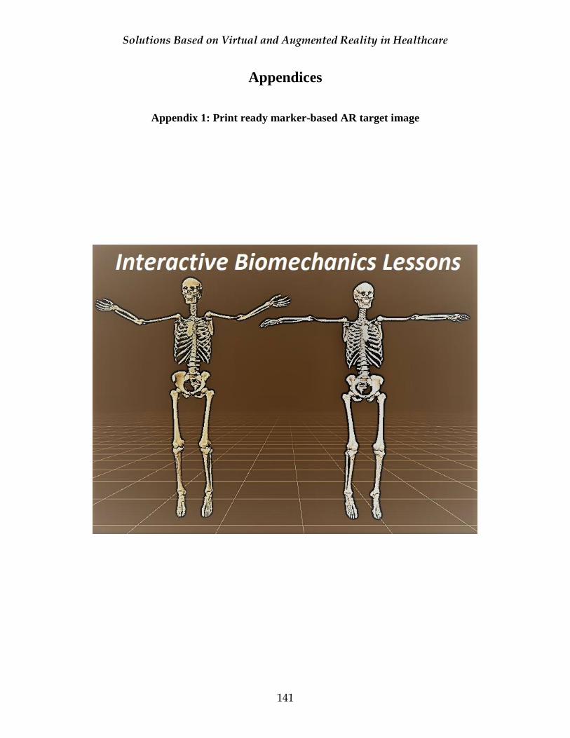
Solutions Based on Virtual and Augmented Reality in Healthcare
141
Appendices
Appendix 1: Print ready marker-based AR target image
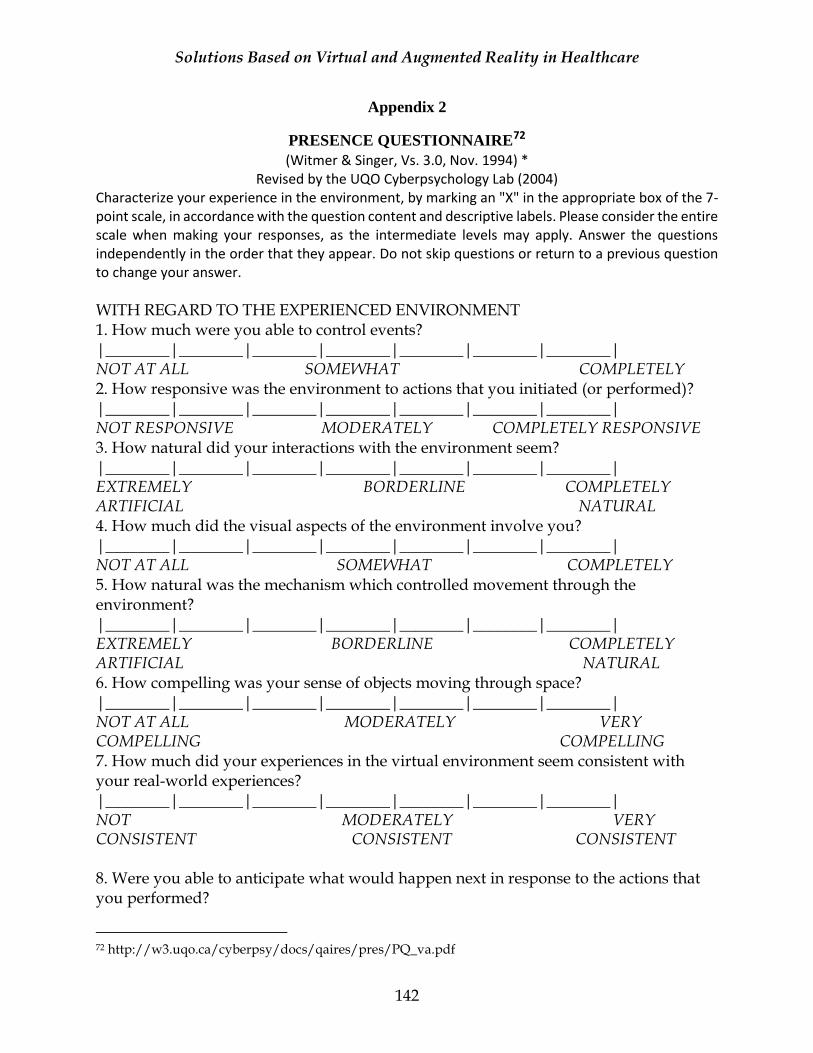
Solutions Based on Virtual and Augmented Reality in Healthcare
142
Appendix 2
PRESENCE QUESTIONNAIRE72 (Witmer & Singer, Vs. 3.0, Nov. 1994) *
Revised by the UQO Cyberpsychology Lab (2004) Characterize your experience in the environment, by marking an "X" in the appropriate box of the 7-point scale, in accordance with the question content and descriptive labels. Please consider the entire scale when making your responses, as the intermediate levels may apply. Answer the questions independently in the order that they appear. Do not skip questions or return to a previous question to change your answer.
WITH REGARD TO THE EXPERIENCED ENVIRONMENT 1. How much were you able to control events? |________|________|________|________|________|________|________| NOT AT ALL SOMEWHAT COMPLETELY 2. How responsive was the environment to actions that you initiated (or performed)? |________|________|________|________|________|________|________| NOT RESPONSIVE MODERATELY COMPLETELY RESPONSIVE 3. How natural did your interactions with the environment seem? |________|________|________|________|________|________|________| EXTREMELY BORDERLINE COMPLETELY ARTIFICIAL NATURAL 4. How much did the visual aspects of the environment involve you? |________|________|________|________|________|________|________| NOT AT ALL SOMEWHAT COMPLETELY 5. How natural was the mechanism which controlled movement through the environment? |________|________|________|________|________|________|________| EXTREMELY BORDERLINE COMPLETELY ARTIFICIAL NATURAL 6. How compelling was your sense of objects moving through space? |________|________|________|________|________|________|________| NOT AT ALL MODERATELY VERY COMPELLING COMPELLING 7. How much did your experiences in the virtual environment seem consistent with your real-world experiences? |________|________|________|________|________|________|________| NOT MODERATELY VERY CONSISTENT CONSISTENT CONSISTENT 8. Were you able to anticipate what would happen next in response to the actions that you performed?
72 http://w3.uqo.ca/cyberpsy/docs/qaires/pres/PQ_va.pdf
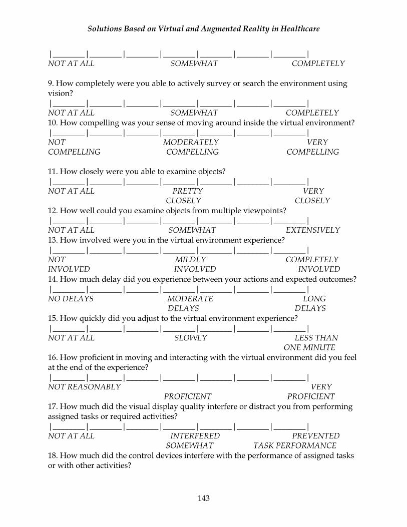
Solutions Based on Virtual and Augmented Reality in Healthcare
143
|________|________|________|________|________|________|________| NOT AT ALL SOMEWHAT COMPLETELY 9. How completely were you able to actively survey or search the environment using vision? |________|________|________|________|________|________|________| NOT AT ALL SOMEWHAT COMPLETELY 10. How compelling was your sense of moving around inside the virtual environment? |________|________|________|________|________|________|________| NOT MODERATELY VERY COMPELLING COMPELLING COMPELLING 11. How closely were you able to examine objects? |________|________|________|________|________|________|________| NOT AT ALL PRETTY VERY CLOSELY CLOSELY 12. How well could you examine objects from multiple viewpoints? |________|________|________|________|________|________|________| NOT AT ALL SOMEWHAT EXTENSIVELY 13. How involved were you in the virtual environment experience? |________|________|________|________|________|________|________| NOT MILDLY COMPLETELY INVOLVED INVOLVED INVOLVED 14. How much delay did you experience between your actions and expected outcomes? |________|________|________|________|________|________|________| NO DELAYS MODERATE LONG DELAYS DELAYS 15. How quickly did you adjust to the virtual environment experience? |________|________|________|________|________|________|________| NOT AT ALL SLOWLY LESS THAN ONE MINUTE 16. How proficient in moving and interacting with the virtual environment did you feel at the end of the experience? |________|________|________|________|________|________|________| NOT REASONABLY VERY PROFICIENT PROFICIENT 17. How much did the visual display quality interfere or distract you from performing assigned tasks or required activities? |________|________|________|________|________|________|________| NOT AT ALL INTERFERED PREVENTED SOMEWHAT TASK PERFORMANCE 18. How much did the control devices interfere with the performance of assigned tasks or with other activities?
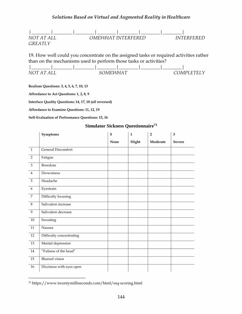
Solutions Based on Virtual and Augmented Reality in Healthcare
144
|________|________|________|________|________|________|________| NOT AT ALL OMEWHAT INTERFERED INTERFERED GREATLY 19. How well could you concentrate on the assigned tasks or required activities rather than on the mechanisms used to perform those tasks or activities? |________|________|________|________|________|________|________| NOT AT ALL SOMEWHAT COMPLETELY
Realism Questions: 3, 4, 5, 6, 7, 10, 13
Affordance to Act Questions: 1, 2, 8, 9
Interface Quality Questions: 14, 17, 18 (all reversed)
Affordance to Examine Questions: 11, 12, 19
Self-Evaluation of Performance Questions: 15, 16
Simulator Sickness Questionnaire73
Symptoms 0
None
1
Slight
2
Moderate
3
Severe
1 General Discomfort
2 Fatigue
3 Boredom
4 Drowsiness
5 Headache
6 Eyestrain
7 Difficulty focusing
8 Salivation increase
9 Salivation decrease
10 Sweating
11 Nausea
12 Difficulty concentrating
13 Mental depression
14 “Fulness of the head”
15 Blurred vision
16 Dizziness with eyes open
73 https://www.twentymilliseconds.com/html/ssq-scoring.html
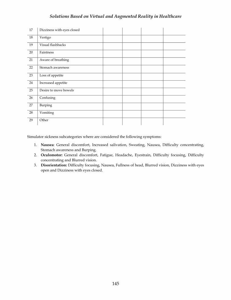
Solutions Based on Virtual and Augmented Reality in Healthcare
145
17 Dizziness with eyes closed
18 Vertigo
19 Visual flashbacks
20 Faintness
21 Aware of breathing
22 Stomach awareness
23 Loss of appetite
24 Increased appetite
25 Desire to move bowels
26 Confusing
27 Burping
28 Vomiting
29 Other
Simulator sickness subcategories where are considered the following symptoms:
1. Nausea: General discomfort, Increased salivation, Sweating, Nausea, Difficulty concentrating,
Stomach awareness and Burping.
2. Oculomotor: General discomfort, Fatigue, Headache, Eyestrain, Difficulty focusing, Difficulty
concentrating and Blurred vision.
3. Disorientation: Difficulty focusing, Nausea, Fullness of head, Blurred vision, Dizziness with eyes
open and Dizziness with eyes closed.
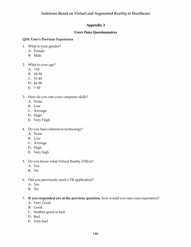
Solutions Based on Virtual and Augmented Reality in Healthcare
146
Appendix 3
Users Data Questionnaires
Q1b: User’s Previous Experience
1. What is your gender?
A. Female
B. Male
2. What is your age?
A. <18
B. 18-30
C. 31-40
D. 41-50
E. > 50
3. How do you rate your computer skills?
A. None
B. Low
C. Average
D. High
E. Very High
4. Do you have interest in technology?
A. None
B. Low
C. Average
D. High
E. Very high
5. Do you know what Virtual Reality (VR) is?
A. Yes
B. No
6. Did you previously used a VR application?
A. Yes
B. No
7. If you responded yes at the previous question, how would you rate your experience?
A. Very Good
B. Good
C. Neither good or bad
D. Bad
E. Very bad
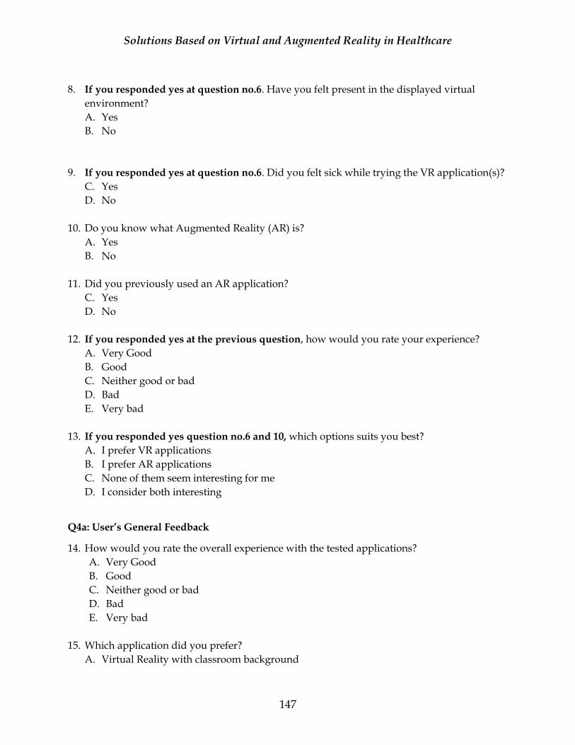
Solutions Based on Virtual and Augmented Reality in Healthcare
147
8. If you responded yes at question no.6. Have you felt present in the displayed virtual
environment?
A. Yes
B. No
9. If you responded yes at question no.6. Did you felt sick while trying the VR application(s)?
C. Yes
D. No
10. Do you know what Augmented Reality (AR) is?
A. Yes
B. No
11. Did you previously used an AR application?
C. Yes
D. No
12. If you responded yes at the previous question, how would you rate your experience?
A. Very Good
B. Good
C. Neither good or bad
D. Bad
E. Very bad
13. If you responded yes question no.6 and 10, which options suits you best?
A. I prefer VR applications
B. I prefer AR applications
C. None of them seem interesting for me
D. I consider both interesting
Q4a: User’s General Feedback
14. How would you rate the overall experience with the tested applications?
A. Very Good
B. Good
C. Neither good or bad
D. Bad
E. Very bad
15. Which application did you prefer?
A. Virtual Reality with classroom background
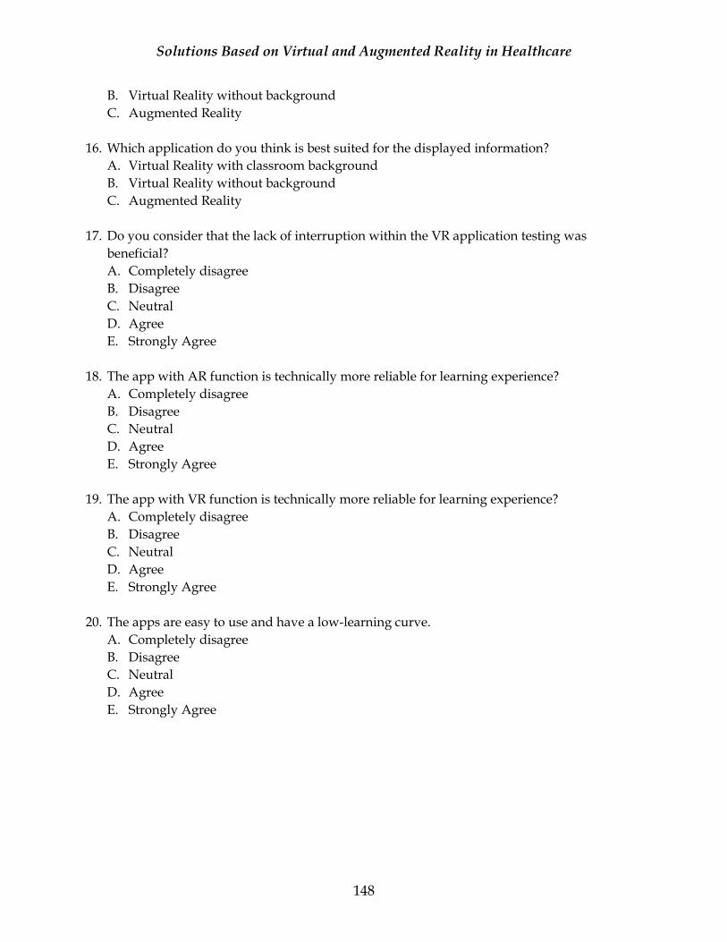
Solutions Based on Virtual and Augmented Reality in Healthcare
148
B. Virtual Reality without background
C. Augmented Reality
16. Which application do you think is best suited for the displayed information?
A. Virtual Reality with classroom background
B. Virtual Reality without background
C. Augmented Reality
17. Do you consider that the lack of interruption within the VR application testing was
beneficial?
A. Completely disagree
B. Disagree
C. Neutral
D. Agree
E. Strongly Agree
18. The app with AR function is technically more reliable for learning experience?
A. Completely disagree
B. Disagree
C. Neutral
D. Agree
E. Strongly Agree
19. The app with VR function is technically more reliable for learning experience?
A. Completely disagree
B. Disagree
C. Neutral
D. Agree
E. Strongly Agree
20. The apps are easy to use and have a low-learning curve.
A. Completely disagree
B. Disagree
C. Neutral
D. Agree
E. Strongly Agree