113264016 Anatomia Omului
299
BODY ORGANIZATION Anatomical position and terms of direction KAPLAJ 4) MEDICAL ....... a A ` I b
-
Upload
biologie-profesor -
Category
Documents
-
view
241 -
download
0
Transcript of 113264016 Anatomia Omului

8/13/2019 113264016 Anatomia Omului
http://slidepdf.com/reader/full/113264016-anatomia-omului 1/298
B O D Y
O R G A N I Z A T I O N
Anatomical position and terms of direction
aA
Ib
8/13/2019 113264016 Anatomia Omului
http://slidepdf.com/reader/full/113264016-anatomia-omului 2/298
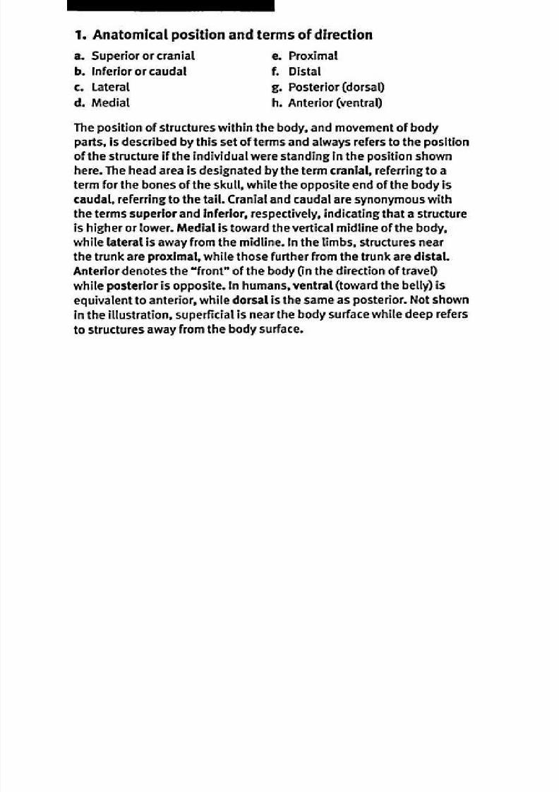
1. Anatomical position and terms of direction
a. Sup erior or cranial . Proximal
b. Inferior or caud al . Distal
c . Lateral . Poste rior (do rsal)
d . M edial . An terior (ventral)
The p osit ion of structures within the body, and mo vemen t of body
parts, is described by th is set of term s and always refers to the p osition
of the structure if the individual were standing in the p osit ion shown
here. The head area is designated by th e term cranial, referring to aterm for the bon es of the skull, while the opp osite en d o f the bod y is
caudal, referring to th e tail. Cranial and caud al are synonym ous w ith
the term s superior and Inferior, respectively, indicating th at a structure
is higher or lower. M edial is toward th e vertical midline o f the bod y,
wh ile lateral is away from the m idline. In the limbs, structures near
the tru nk are proximal, while those further from the tru nk are d istal.An terior den otes the front of the body (in the direction of travel)
wh ile po sterior is op posite. In hum ans, ven tral (tow ard th e belly) is
equ ivalent t o anter ior, wh ile do rsal is the same as posterior. Not show n
in the il lustration, super ficial is near the bo dy surface w hile deep refers
to structures away from the bod y surface.

8/13/2019 113264016 Anatomia Omului
http://slidepdf.com/reader/full/113264016-anatomia-omului 3/298
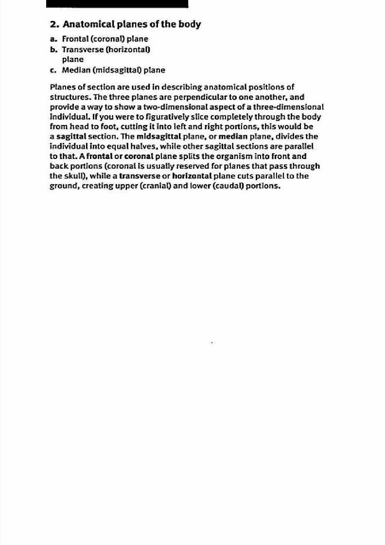
2natomical planes of the body
KAPLAN) MEDICAL".....
8/13/2019 113264016 Anatomia Omului
http://slidepdf.com/reader/full/113264016-anatomia-omului 4/298
8/13/2019 113264016 Anatomia Omului
http://slidepdf.com/reader/full/113264016-anatomia-omului 5/298
8/13/2019 113264016 Anatomia Omului
http://slidepdf.com/reader/full/113264016-anatomia-omului 6/298
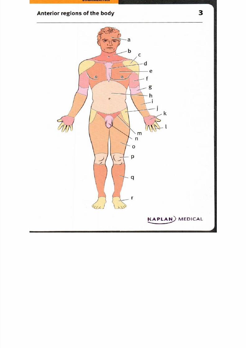
3. Anterior regions of the body
a. Head
b. Neck (cervical)
c. Deltoid
d . Sternal
e. Pecto ral (chest)
1. Brachial (arm)
g. Cubital
h. Abdominal
I. An tebrachial (forearm)
j. Trochanteric
k. Palmar
L D igital (fingers)
m . Inguinal and p ubic
n . Pen is (genital)
o. F emoral (thigh)
p . Knee
q. Lig
r. Do rsal foot
The head is connected to the trun k through th e cervical or neck region.
Th e trunk includ es the chest and sternal regions, the abdom en, and the
inguin al/pu bic and gen ital region s (the p en is, of cou rse, is an or gan
that is only foun d in the m ale). The u pp er lim bs may be divided into
the d eltoid (shou lder), brachial (up per arm ), cubital (fron t of elbow),antebrach ial (lower arm ), palmar (hand ) and d igital (fingers) regions,
wh ile the lower limbs include trochanteric (hip), femoral (upp er leg),
knee, leg and foot. It may be helpful to rem em ber that some region s
are correlated w ith the nam es of underlying structures: the d eltoid and
pecto ral regions are nam ed for the m uscles in th at area, while sternal
and troc hanteric regions refer to skeletal structur es under neath.

8/13/2019 113264016 Anatomia Omului
http://slidepdf.com/reader/full/113264016-anatomia-omului 7/298
O R G A N I Z A T I O N
Po sterior regions o f the b ody
II
m
KAPLAN) MEDICAL

8/13/2019 113264016 Anatomia Omului
http://slidepdf.com/reader/full/113264016-anatomia-omului 8/298
4. Posterior regions of the body
a. Head
b. Neck cervical)
c. Scapular (shoulder blade)
d. Brachial arm)
e. Vertebral
f. Olecranon elbow)
g. Lumbar
h. Antebrachial forearm)
I. Gluteal
j. Femoral thigh)
k. Popliteal
I. Surat calf)
m. Calcaneal
From the posterior aspect, one can see areas not visible from the
anterior view, such as scapular (shoulder blade), vertebral, lumbar
(lower back) and gluteal (buttocks) regions in the trunk. The upper
limbs include the olecranon or elbow region, while the lower limbs
include popliteal (back of knee), sural (calf) and calcaneal (heel)
regions. The olecranon and calcaneus are bone structures in their
respective regions.
8/13/2019 113264016 Anatomia Omului
http://slidepdf.com/reader/full/113264016-anatomia-omului 9/298
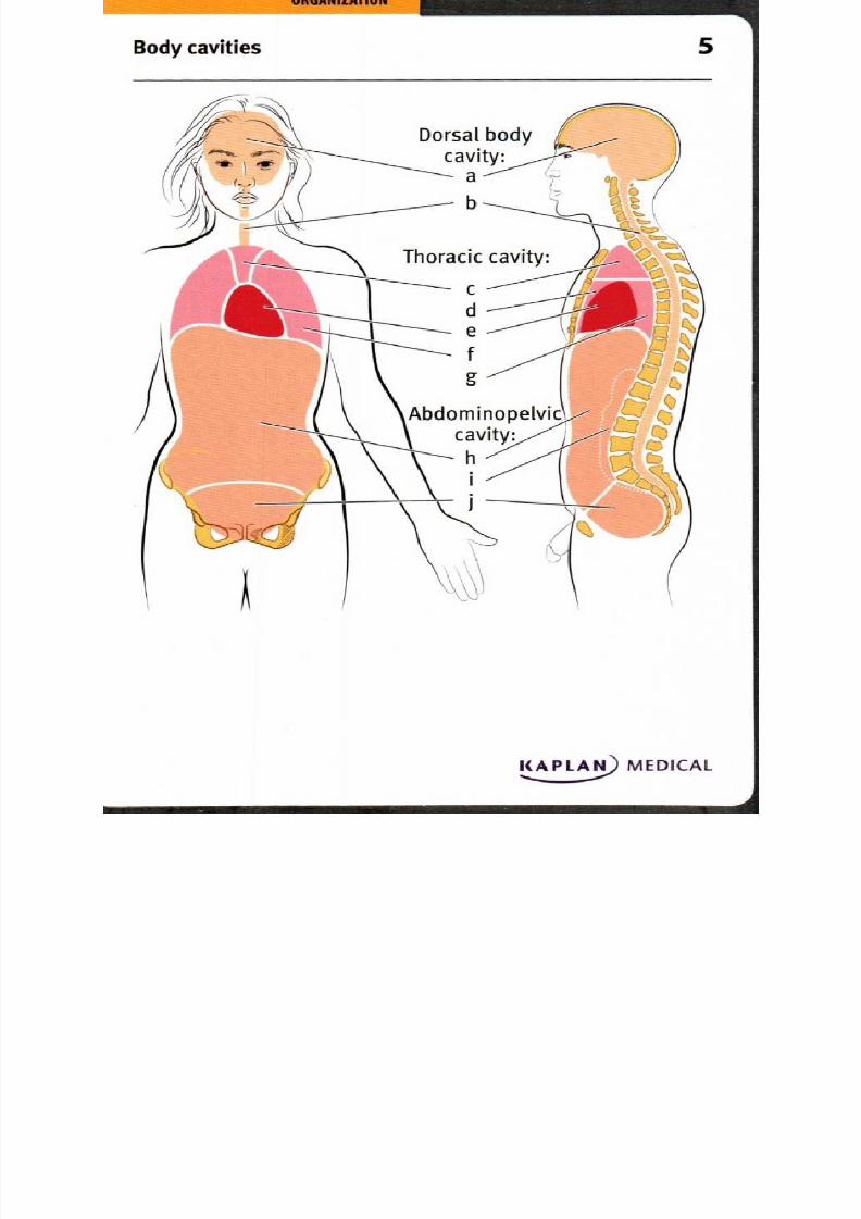
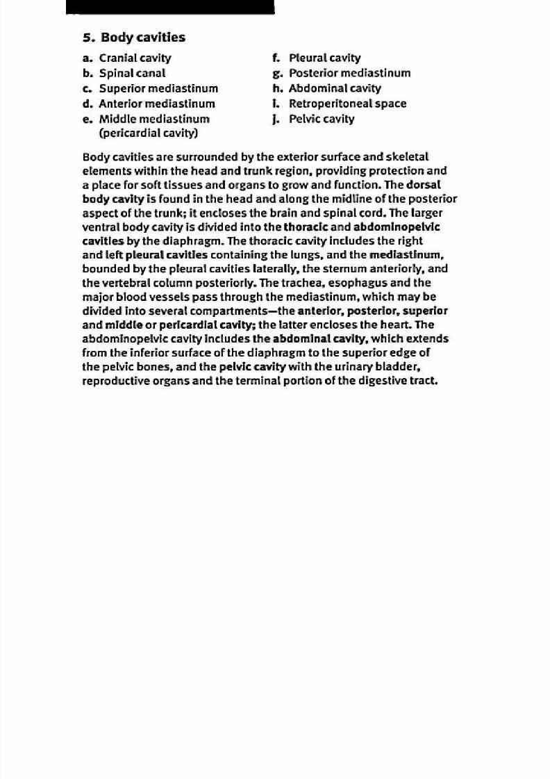
5ody cavities
Dorsal bodycavity:
a
b
1 1 4 7 — T— d
e
f
g
Abdominopelviccavity:
Thoracic cavity:
V
KAPLAN) MEDICAL
•
8/13/2019 113264016 Anatomia Omului
http://slidepdf.com/reader/full/113264016-anatomia-omului 10/298

8/13/2019 113264016 Anatomia Omului
http://slidepdf.com/reader/full/113264016-anatomia-omului 11/298
O R G N I Z TIO N
keletal and visceral structures of the head and neck
KAPLAN MEDIC L
8/13/2019 113264016 Anatomia Omului
http://slidepdf.com/reader/full/113264016-anatomia-omului 12/298
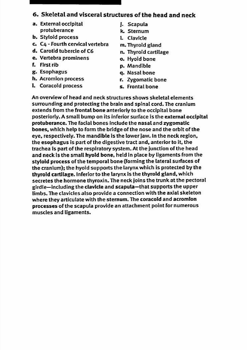
6. Sk eletal and visceral structures o f the hea d an d n eck
a. Extern al occipital
protuberance
b. Styloid pro cessc. C4 - Fou rth cervical vertebra
d . Carotid tubercle of C6
e. Vertebra prom inens
f. First rib
g. Esophagus
h. Acro mion process
I. Coracoid process
j. Scapula
k. Sternum
I. Clavicle
m . Thyroid gland
n . Thyroid cartilage
o . H yoid bone
p . Mandible
q. Nasal bon e
r. Zygom atic bone
s. F rontal bone
A n ove rview of head and n eck structures shows skeletal elemen ts
surroun ding and p rotecting the brain and spinal cord. The cranium
exten ds from th e frontal bone ante riorly to the occipital bone
posteriorly. A small bum p o n its inferior surface is the ex ternal occipital
protu berance. The facial bones includ e the nasal and zygomatic
bones, which help to form th e bridge of the nose and the orbit of the
eye, respectively. The m andible is the lower jaw. In the neck region,
the e sophagus is part of th e d igestive tract and , anterior to i t , the
trachea is part of the respiratory system . A t the junction o f the head
and n eck is the sm all hyoid bone, held in place by ligamen ts from the
styloid p rocess of the tem poral bone (form ing the lateral surfaces of
the cranium ); the hyo id suppor ts the larynx wh ich is protec ted by the
thyroid cartilage. Inferior to the larynx is the thyroid gland , which
secretes the hor mo ne thyrox in. The n eck joins the trunk at the pectoral
girdle— including the clavicle and scapula— that supp orts the upper
l im bs. The clavicles also prov ide a con nect ion w ith the axial skeleton
whe re they articulate with the sternu m. The coracoid and acrom ion
proc esses of the scapula prov ide an attachm ent po int for num erou s
m uscles and l igamen ts.
8/13/2019 113264016 Anatomia Omului
http://slidepdf.com/reader/full/113264016-anatomia-omului 13/298
ab
Thoracic, abdominal and pelvic viscera, anterior view
KAPLAN) MEDICAL

8/13/2019 113264016 Anatomia Omului
http://slidepdf.com/reader/full/113264016-anatomia-omului 14/298
7. Thoracic, abdominal and pelvic viscera, anterior view
a. Thyroid cartilage
b. Thyroid gland
c. Clavicle
d. Arch of aorta
(behind sternum)
e. Third rib
f. Outline of heart
g. Left lung
h. Spleen
1 Stomach
j. Transverse colon
k. Small intestine
1 Outline of descending colon
(behind small intestine)
m . Sigmoid colonn. Ou tline of rectum
o. Urinary bladder
p. Ascending colon
q. Gall bladder
r. Liver
s. Right lung
t. Superior vena cava
(behind sternum)
u. Hyo id bone
Organs of several body systems share the space within the cavities
of the trunk. The superior part of these cavities is protected by the
ribs, sternum and vertebral column of the axial skeleton, while the
inferior portion is supported by the pelvic girdle. The thoracic cavity
contains the lungs which surround the pericardial cavity containing
the heart. Venous blood enters the heart through the vena cava and
is pumped from the heart to the body through the aorta; this critical
area is well protected behind the sternum. In the abdominal cavity, the
digestive tract includes the stomach, small intestine, colon (ascending,
transverse, descending and sigmoid), and rectum. Accessory glands of
the digestive system include the liver, gall bladder and pancreas. The
spleen is an organ of the circulatory, lymphatic and immune systems.
The urinary bladder is located in the lower part of the abdominal cavity.
8/13/2019 113264016 Anatomia Omului
http://slidepdf.com/reader/full/113264016-anatomia-omului 15/298
Thoracic, abdominal and pelvic viscera, posterior view
KAPLAN) MEDICAL

8/13/2019 113264016 Anatomia Omului
http://slidepdf.com/reader/full/113264016-anatomia-omului 16/298
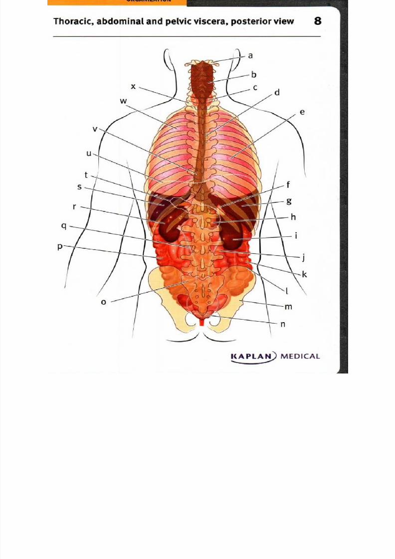
8. Thoracic, abdominal and pelvic viscera, posterior view
a. Atlas
b. Pharynx
c. Thyroid gland
d. Trachea
e. Right lung
1 . Right adrenal gland
g. Liver
h. Pancreas
I . Right kidney
j. Small intestine
k. Ascending colon
L Iliac crest
m . Pelvic girdle
n. Seminal vesicle
o. Sacrum
p. Descending colon
q. Left ureter
r. Left kidney
s. Spleen
t. Outline of pancreas
u. Diaphragm
v. Esophagus
w. Left lung
x. First thoracic vertebra
The posterior view clearly shows the position of the vertebral column,
extending from the atlas that articulates with the cranium to the fused
vertebrae that form the sacrum, which articulates with the pelvic girdle.
The wide pharynx at the rear of the nose and mouth divides into two
passageways—the posterior esophagus leading to the stomach and
the anterior trachea or windpipe. The rear of the thoracic cav ity is filled
with the lungs. The diaphragm is a thin sheet of muscle that marks the
boundary between the thoracic and abdominal cavities, and functionsin breathing. Accessory digestive organs include a large liver and the
deep, mostly hidden pancreas, while the small intestine and parts of
the colon are also visible from this view. The excretory system includes
the dorsal, paired kidneys that form urine, and the ureters that carry
the urine to the bladder. Superior to the kidney lie paired adrenal
gland s, part o f the endocrine system. The only reproductive structures
in view are the sem inal vesicles, fou nd on ly in th e m ale.
8/13/2019 113264016 Anatomia Omului
http://slidepdf.com/reader/full/113264016-anatomia-omului 17/298
O R G A N I Z A T I O N
Thoracic, abdominal and pelvic viscera,
right lateral view
KAPLAN) MEDICAL
8/13/2019 113264016 Anatomia Omului
http://slidepdf.com/reader/full/113264016-anatomia-omului 18/298
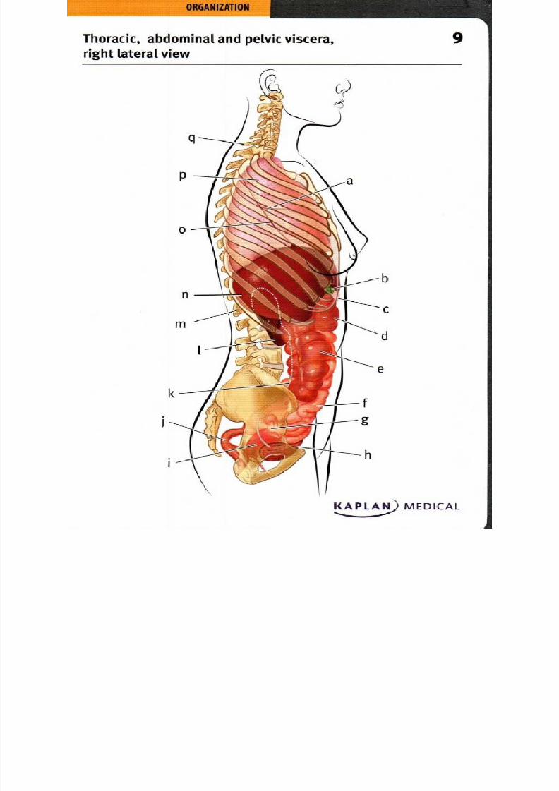
9 . Thoracic, abdominal and pelvic viscera, right lateral view
a. Horizontal fissure of lung . Rectum
b. Gall bladd er . Ureter
c. Stomach Right kidney
d . Transverse colon . L i vertebra
e. Ascending colon . Liver
1. Small intestine . Oblique fissure of lung
g. Ovary . Right lung
h. Urinary bladd er . C7 vertebraI. Uterus
The right lung is divided into three lobes by the horizontal and oblique
fissures. In the abdominal cavity, the digestive tract occupies the largest
part of the space, including the stomach, small intestine, colon and
rectum. The small intestine terminates on the right side, leading into
the ascending colon and then to the transverse colon. The gall bladder,
an accessory organ of the digestive system, is found on the posterior
surface of the right lobe of the liver. The kidneys lie near the posterior
wall of the abdominal cavity, with the right kidney being positioned
slightly inferior compared with the left. The kidneys and ascending
colon are retroperitoneal, while the small intestine and transverse colon
are peritoneal. Ureters lead from the kidneys down to the inferior and
anterior position of the bladder. The female reproductive structures, the
ovary and uterus may be found between the bones that form the pelvis.

8/13/2019 113264016 Anatomia Omului
http://slidepdf.com/reader/full/113264016-anatomia-omului 19/298
=Thoracic, abdominal and pelvic viscera, 0
left lateral view
KAPLAN) MEDICAL
8/13/2019 113264016 Anatomia Omului
http://slidepdf.com/reader/full/113264016-anatomia-omului 20/298
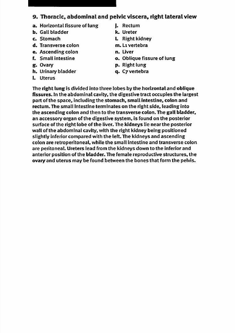
10. Thoracic, abdominal and pelvic viscera, left lateral view
a. C7 v ertebra
b. O blique fissure of lung
c. L eft dome of d iaphragm
d. Spleen
e. L 1 vertebra
1. Prostate gland
j. Urinary bladde r
k. D escending colon
1. Small intestine
m. Transverse colon
1. Left kidney . Stomach
g. Ureter . Liver
h. Rectum . Left lung
The left lung is divided in to two lobes by an oblique fissure. The
diaphragm is located betwee n the thoracic and abdom inal cavit ies,
and forms a d om e shape w hen relaxed. Usually lying left and ventral
to the lobes of the l iver is the stomach , which th en leads to the small
intest ine, colon, and rectu m . O n the right side, the transverse colon
leads to the d escending colon, before und igested m aterials em pty into
the rectum for com paction and el imination. Posterior to the stom ach
is the left kidney, with the u reter fun neling urine to th e bladd er.
A lthough many m ale reprod uctive structures are located outside the
abdom inope lvic cavity, the pr ostate gland is found near the bladde r.

8/13/2019 113264016 Anatomia Omului
http://slidepdf.com/reader/full/113264016-anatomia-omului 21/298
I N T E G U M E N T A R Y
S Y S T E M
Layers o f the sk in and ass ociated structures 1
8/13/2019 113264016 Anatomia Omului
http://slidepdf.com/reader/full/113264016-anatomia-omului 22/298
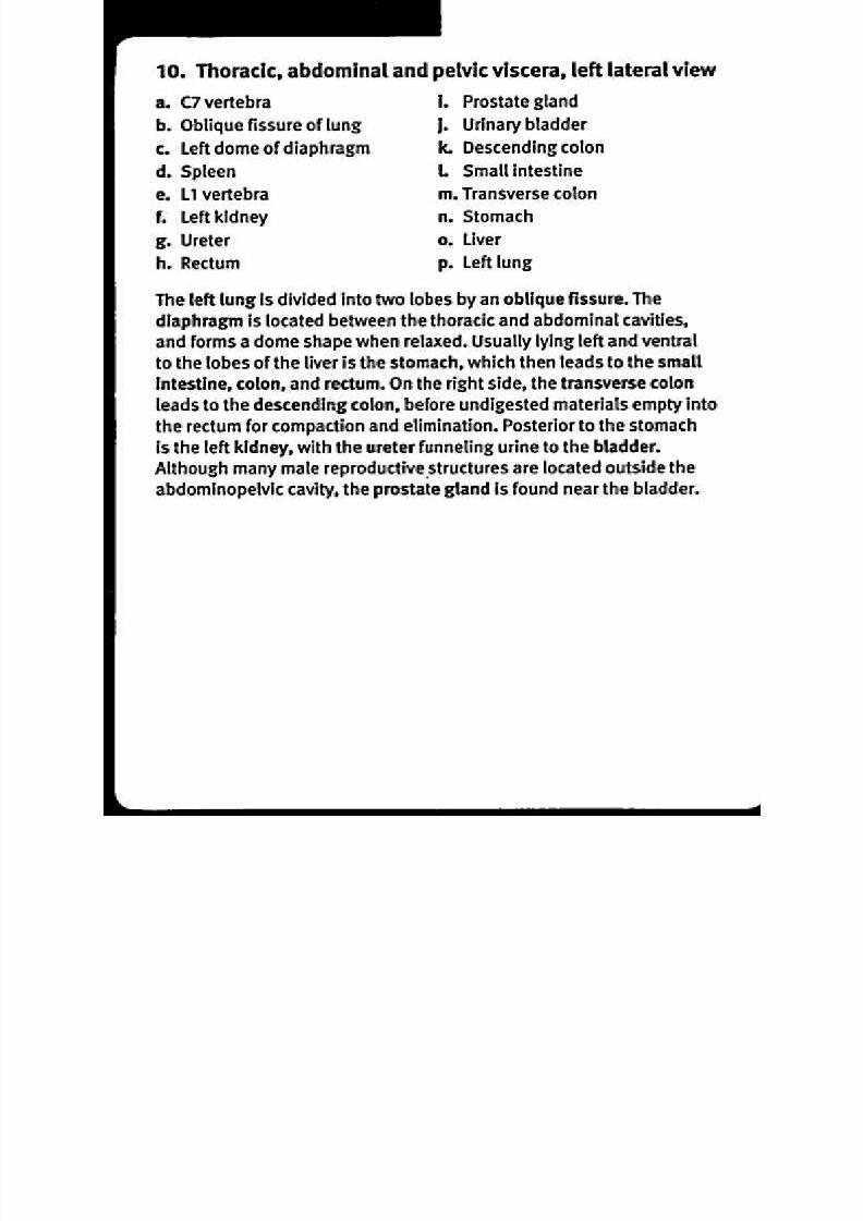
11. L ayers of the skin
a. Sweat gland
b. Meissner's corpuscle
c. Haird. Epidermis
e. Dermis
1 . Hypodermis
and associated structu res
g. Papillary layer
h. Reticular layer
I . Hair follicle
j. Sebaceous gland
k. Pacinian corpuscle
I . Arrector pili m uscle
The integumentary system includes the largest organ of the body—
the skin. It functions to protect underlying body parts from water loss,
chemical insult, and physical harm. Specialized structures detect
pressure, pain or temperature stimuli; Meissner's corpuscles sense
light touch while Pacinian corpuscles sense deep pressure. Sebaceous
glands secrete lipids that inhibit bacteria and lubricate the hair shaft.
Sweat glands secrete water, waste products and electrolytes, in part to
cool the skin and reduce body temperature. A cross-section of humanskin reveals layers of the skin; the interface between the layers is often
indistinct as one layer merges into the next. The epidermis consists of
stratified squamous epithelium that provides mechanical protection
against invasion by microorganisms. The dermis has a superficial
papillary layer of areolar tissue, with capillaries and sensory neurons
that supply the epidermis; and a deeper, thicker reticular layer withdense, irregular connective tissue and networks of blood vessels,
lymph vessels, and nerve fibers. Many of the accessory organs such as
hair follicles and sweat glands are embedded in the reticular layer. The
subcutaneous layer, or hypodermis, consists of areolar and adipose
tissue; distribution of body fat in this layer varies between the sexes or
at different times in life (such as the presence of "baby fat").

8/13/2019 113264016 Anatomia Omului
http://slidepdf.com/reader/full/113264016-anatomia-omului 23/298
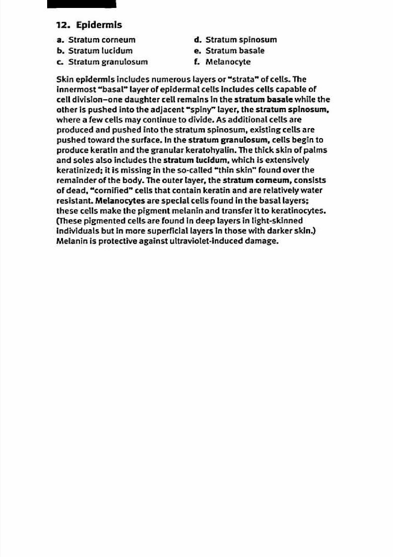
Epidermis
—a
:,......,..44...,
— b. ., .1 .. ...
• 0 ••° "••.. . •'• •
. .- •
• ao 0 0
0
0 ,.., 00 •0, NZ a0 d..
--a
a 0 ••••..., a6
O• • 0 90•
• .' •• •
or •
-o0s 0•
0
—
• • •
•
•
KAPLAN MEDICAL
8/13/2019 113264016 Anatomia Omului
http://slidepdf.com/reader/full/113264016-anatomia-omului 24/298

8/13/2019 113264016 Anatomia Omului
http://slidepdf.com/reader/full/113264016-anatomia-omului 25/298
S Y S T E M
1 .APLA) MEDICAL
8/13/2019 113264016 Anatomia Omului
http://slidepdf.com/reader/full/113264016-anatomia-omului 26/298
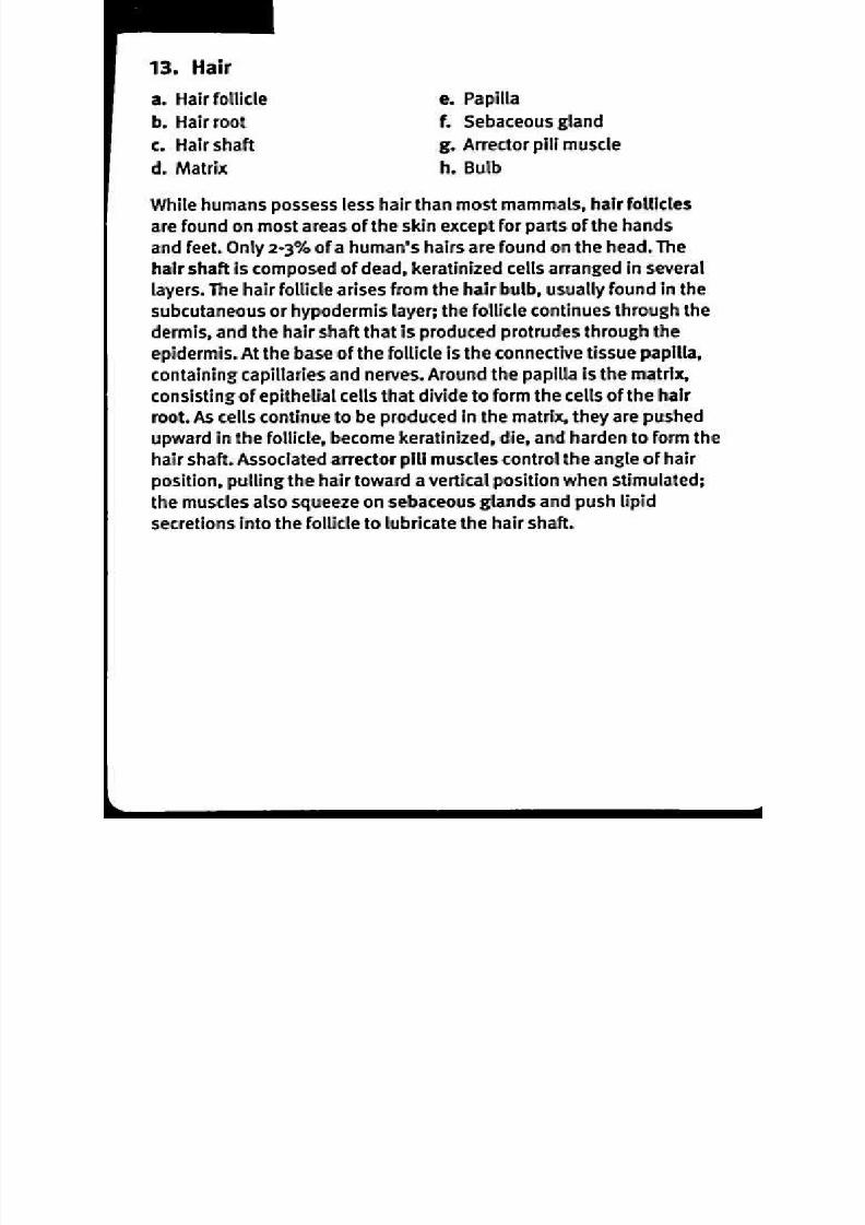
13. Hair
a. H air follicle
b. Hair root
c . H air shaft
d . Matrix
e. Papilla
1 . Sebaceous gland
g. Arrector pili muscle
h. Bulb
W hile hum ans possess less hair than m ost m amm als, hair fol lic les
are found on m ost areas of the skin excep t for parts of the hand s
and feet. Only 2-3% of a hum an's hairs are found on th e head. The
hair shaft is com po sed of dead , keratinized c ells arranged in sever allayers. The hair follicle arises from the h air bulb, usually foun d in th e
subcutaneous or h ypod erm is layer; the fol lic le continu es through the
derm is, and the hair shaft that is prod uced p rotrud es through the
epid erm is. A t the base of the fo llicle is the co nn ective tissue papilla,
con taining capillaries and n erves. Arou nd the p apilla is the m atrix,
con sisting of epith elial cells that d ivide to form the ce lls of the hair
root. As cells continu e to be prod uced in the matrix, they are pushed
up ward in the foll icle, becom e keratinized, die, and hard en to form the
hair shaft. A ssociated arr ecto r pill m uscles contr ol the angle of hair
position, pulling the hair toward a ver tical position whe n stimulated;
the muscles also squeeze on sebaceous glands and push lipid
secretion s into the fo llicle to lubricate the h air shaft.

8/13/2019 113264016 Anatomia Omului
http://slidepdf.com/reader/full/113264016-anatomia-omului 27/298
I N T E G U M E N T A R Y
S Y S T E M
Fingernail
\_
8/13/2019 113264016 Anatomia Omului
http://slidepdf.com/reader/full/113264016-anatomia-omului 28/298
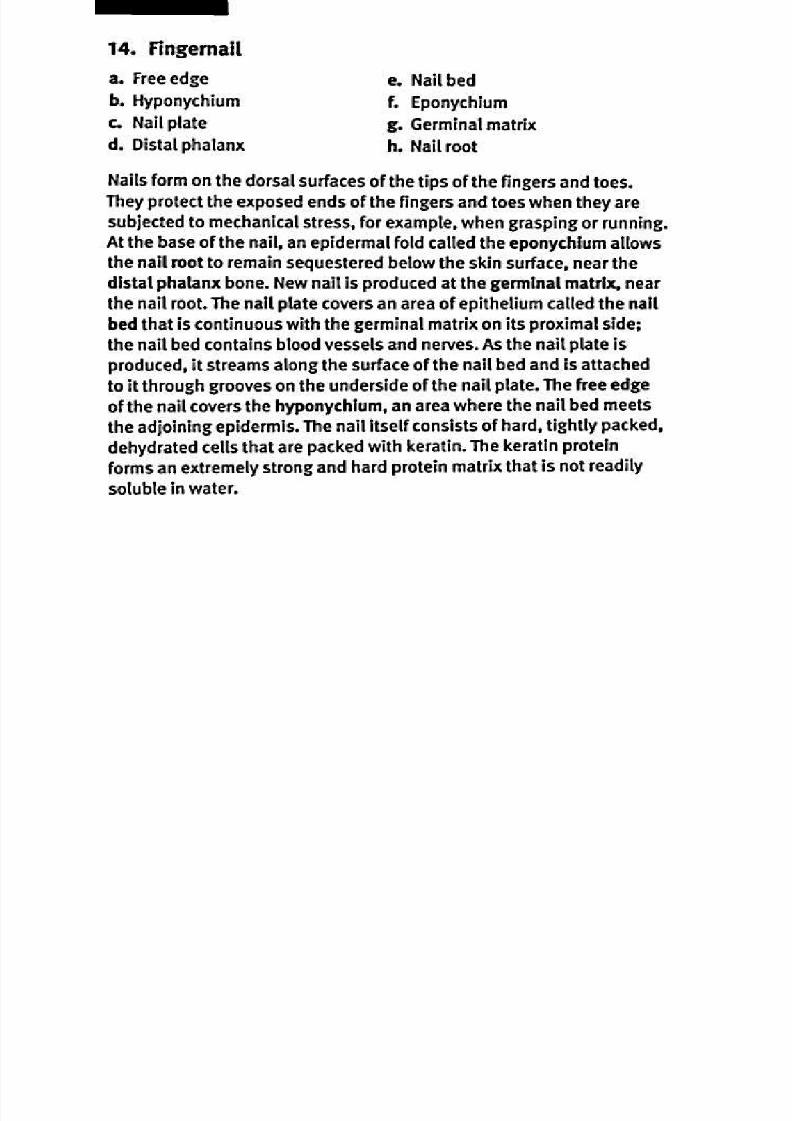
14. Fingernail
a. F ree edge
b. Hyponychium
c . Nail plated . D istal ph alanx
e. Nail bed
1. Ep onychium
g. Germinal m atrix
h . Nail roo t
Nails form on the d orsal surfaces of the tips of th e fingers and to es.
They p rotect the expo sed ends of the f ingers and toes when they are
subjected to m echan ical stress, for exam ple, wh en grasping or run ning.
A t the base of th e nail, an ep idermal fold called the epon ychium allowsthe n ail root to rem ain sequestered below the skin surface, near the
distal ph alanx bon e. New nail is pro du ced at the germ inal m atrix, near
the n ail root. The nail plate covers an area of epithelium called t he n ail
bed that is continuou s with the germ inal matrix on its proximal side;
the n ail bed c on tains blood vessels and ne rves. As the n ail plate is
prod uced, i t streams along the surface of th e nail bed and is attachedto i t through grooves on the un derside o f the nail plate. The free edge
of the n ail covers the hypo nychium , an area where th e nail bed m eets
the ad joining ep iderm is. The n ail itself consists of hard , t ightly packed ,
dehyd rated cells that are packed w ith keratin. The keratin protein
forms an extrem ely strong and hard p rotein m atrix that is not readily
soluble in w ater.
8/13/2019 113264016 Anatomia Omului
http://slidepdf.com/reader/full/113264016-anatomia-omului 29/298
Skeleton, anterior view 15
KAPLAN) MEDICAL
S Y S T E M
8/13/2019 113264016 Anatomia Omului
http://slidepdf.com/reader/full/113264016-anatomia-omului 30/298
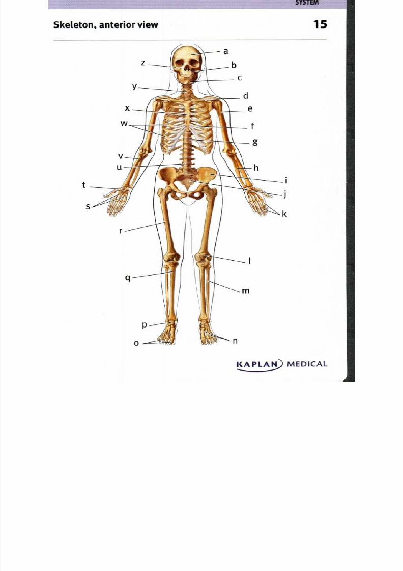
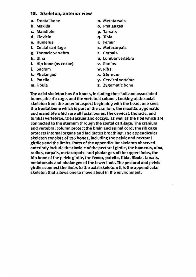
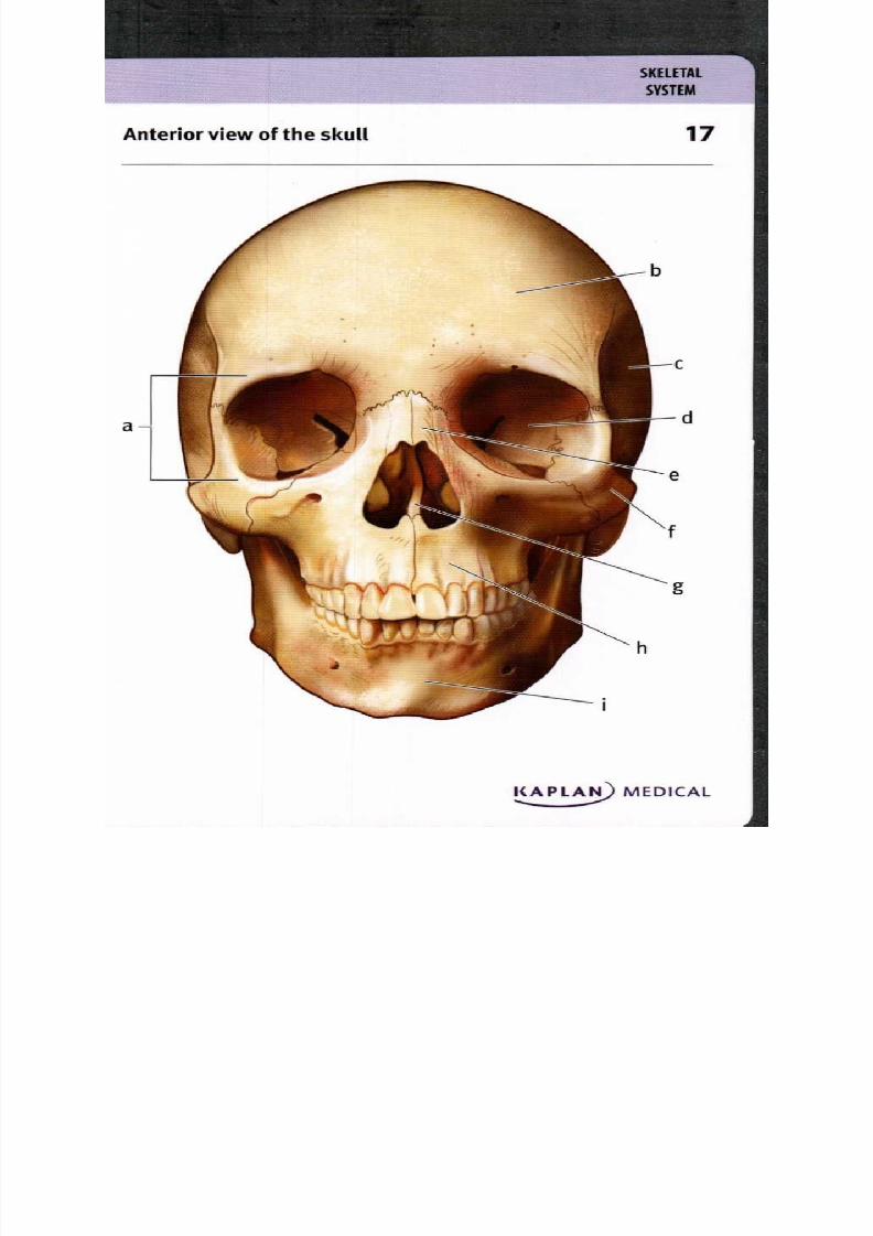
15. Skeleton, anterior view
a. F rontal bone
b. Maxilla
c. Mandible
d . Clavicle
e. Humerus
1. Co stal carti lage
g. Tho racic vertebra
h . Ulna
i. H ip bone (os coxae)
j. Sacrum
k. Phalanges
I. Patella
m. Fibula
n . Metatarsals
o. Phalanges
p . Tarsals
q. Tibia
r. Femur
s. Metacarpals
t . Carpals
u . L umbar vertebra
v. Radius
w . Ribs
x. Sternum
y. Cerv ical vertebra
z. Zygom atic bone
The axial skeleton h as 8o bones, includ ing the skull and associated
bones, the rib cage, and the vertebral column. L ooking at the axial
skeleton from the anterior aspect beginning w ith the head , one sees
the fron tal bone wh ich is part of the cranium , the m axilla, zygomatic
and m andible wh ich are all facial bones, the cerv ical, thoracic, and
lumbar vertebrae, the sacrum and co ccyx, as well as the ribs which are
conn ected to th e sternum th rough th e costal cartilage. The craniumand vertebral colum n p rotect the brain and spinal cord; the rib cage
protec ts internal organs and facilitates breathing. The append icular
skeleton con sists of 126 bon es, includ ing the pe lvic and pect oral
girdles and the limbs. Parts of the append icular skeleton o bserved
anteriorly include the c lavicle of the p ectoral girdle, the hu mer us, ulna,
radius, carpals, m etacarpals, and ph alanges of the up per lim bs, the
hip bo ne o f the p elvic gird le, the fem ur , patella, tibia, fibula, tarsals,
metatarsals and phalanges of the low er limb. The p ectoral and p elvic
girdles conn ect the l imbs to th e axial skeleton; it is the app end icular
skeleton that al lows on e to m ove about in the en vironmen t.

8/13/2019 113264016 Anatomia Omului
http://slidepdf.com/reader/full/113264016-anatomia-omului 31/298
Ske leton , po sterior view
KAPLAN MEDICAL
S Y S T E M

8/13/2019 113264016 Anatomia Omului
http://slidepdf.com/reader/full/113264016-anatomia-omului 32/298
8/13/2019 113264016 Anatomia Omului
http://slidepdf.com/reader/full/113264016-anatomia-omului 33/298
S K E L E T A L
S Y S T E M
17Anterior view of the skull
1
8/13/2019 113264016 Anatomia Omului
http://slidepdf.com/reader/full/113264016-anatomia-omului 34/298

8/13/2019 113264016 Anatomia Omului
http://slidepdf.com/reader/full/113264016-anatomia-omului 35/298
S Y S T E M
Skull, lateral view 8
KAPLAN) MEDICAL.....

8/13/2019 113264016 Anatomia Omului
http://slidepdf.com/reader/full/113264016-anatomia-omului 36/298
18. Sku ll, lateral view
a. Coronal suture . Zygomatic bone
b. Parietal bone . Nasal bone
c. Zygomatic process . Lacrimal bone
d . Temporal bone . Sphenoid bone
e. Squamous suture . Frontal bone
1 . Lambdoid suture . Coronoid process
g. External occ ipital . Mandibular foramen
protuberance . Mandibular notch
h . Occipital bone . Mandibular condyle
I. Mastoid process . Ramus of mandible
j. External acoustic meatus . Angle of mandible
k. Styloid process . Body of mandible
L Mandible . Mental foramen
m . Maxilla
Observed from the lateral aspect, one can identify the large
braincase formed by the cranial bones, including a single frontal,
two parietal , two temp oral, one occipi tal, one sphen oid and one
ethm oid wh ich is not seen in this view. The tem poral bone has
several pro cesses that articulate with bones or pro vide attachm ent
sites for mu scles and l igamen ts, and t he exte rnal acoustic meatu sor extern al ear. The tem po ral bone articulates with the parietal
bone at an imm ovable joint, the squamous suture. The m andible
form s the lower jaw; it articulates with the te m po ral bon e at the
mand ibular co ndyle. The strong temp oralis mu scle that closes the
jaw attaches at the corono id pro cess. Op enings in th e m andible
include the m ental foramen for n erves and the mand ibular foramenfor blood vessels and ne rves. Oth er facial bones visible from this
aspect includ e th e m axilla, zygom atic, nasal and lacrim al bones.
8/13/2019 113264016 Anatomia Omului
http://slidepdf.com/reader/full/113264016-anatomia-omului 37/298
S Y S T E M
Skull, superior view 9
KAPLA) MEDICAL....
8/13/2019 113264016 Anatomia Omului
http://slidepdf.com/reader/full/113264016-anatomia-omului 38/298
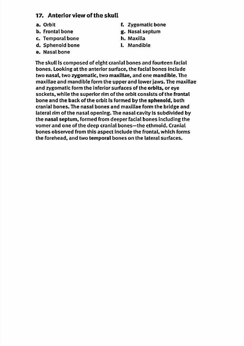
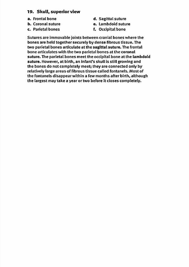
19. Sku ll, sup erior view
a. Frontal bone . Sagittal suture
b. Corona( suture . Lambdoid suture
c. Parietal bones . Occipital bone
Sutures are immovable joints between cranial bones where the
bones are held together securely by dense fibrous tissue. The
two parietal bones articulate at the sagittal suture. The frontal
bone articulates with the two parietal bones at the corona
suture. The parietal bones meet the occipital bone at the lambdoid
suture. However, at birth, an infant's skull is still growing and
the bones do not completely meet; they are connected only by
relatively large areas of fibrous tissue called fontanels. Most of
the fontanels disappear within a few months after birth, although
the largest may take a year or two before it closes completely.
8/13/2019 113264016 Anatomia Omului
http://slidepdf.com/reader/full/113264016-anatomia-omului 39/298
External surface o f the base of the sku ll 20
KAPLA) MEDICAL
8/13/2019 113264016 Anatomia Omului
http://slidepdf.com/reader/full/113264016-anatomia-omului 40/298
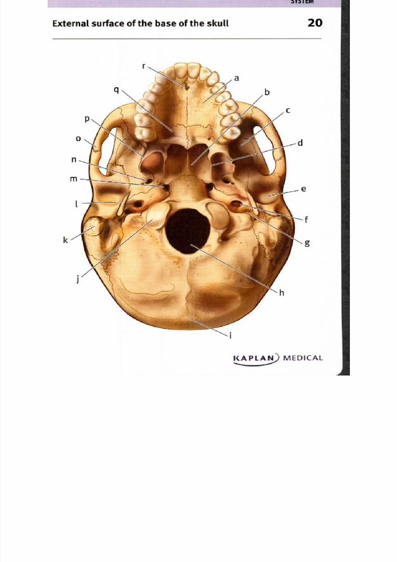
20. External su rface of the b ase of the sk ull
a. Palatine p rocess of m axilla
b. Vomer
c. Greater wing of
sphenoid bone
d . M edial pterygoid plate
of sphenoid bone
e. M and ibular fossa
f. Carotid canal
g. Jugular foramen
h. F oramen magnum
i. External occipital
protuberance
j. Occipital condyle
k. M astoid process
1 Styloid process
m . F oramen lacerum
n . F oramen ovate
o . Zygomatic arch
p . L ateral plate of
sphenoid bone
q. Palatine bone
r. Inc isive fo ssa
The h ard palate is form ed by th e palatine p rocess of the m axillae
anteriorly and th e palatine bo nes po steriorly; the incisive fossa forms
a passageway for nerv es and arteries. The v om er form s the bony
part of the n asal septu m . The p terygoid plates are extensions of the
sphenoid bone that form attachm ent s i tes for muscles that mo ve the
lower jaw. The foram en m agnum is the large hole in the o ccipital bone
throu gh wh ich the spinal cord p asses; on either side , the occ ipital
condyles articulate w ith the first vertebra of the n eck. Between th e
foramen m agnum and th e external occipital protuberance, a bonycrest m arks attachm ent sites for ligam ents stabilizing the ve rtebrae
of the neck. The m astoid pr ocess of the tem poral bone provides an
attachmen t site for m uscles rotating the head ; muscles attached t o
the styloid p rocess contro l the hyoid, the p harynx and th e tongu e.
H oles for passage of blood vessels and nerv es includ e the carotid
canal and foramen lacerum in the tem poral bone, the foramenovate in the sphenoid bon e, and the jugular foramen form ed at the
junction of the t emp oral and occipital bones. A d epression in the
tem poral bone, the m andibular fossa, articulates with th e m andible.
8/13/2019 113264016 Anatomia Omului
http://slidepdf.com/reader/full/113264016-anatomia-omului 41/298

8/13/2019 113264016 Anatomia Omului
http://slidepdf.com/reader/full/113264016-anatomia-omului 42/298

8/13/2019 113264016 Anatomia Omului
http://slidepdf.com/reader/full/113264016-anatomia-omului 43/298
S Y S T E M
Right tem poral and sph enoid bon es 2
Right temporal bone,
lateral view
Sphenoid bone,
superior view
Ki. . 3 . . . . . N MEDICAL
8/13/2019 113264016 Anatomia Omului
http://slidepdf.com/reader/full/113264016-anatomia-omului 44/298
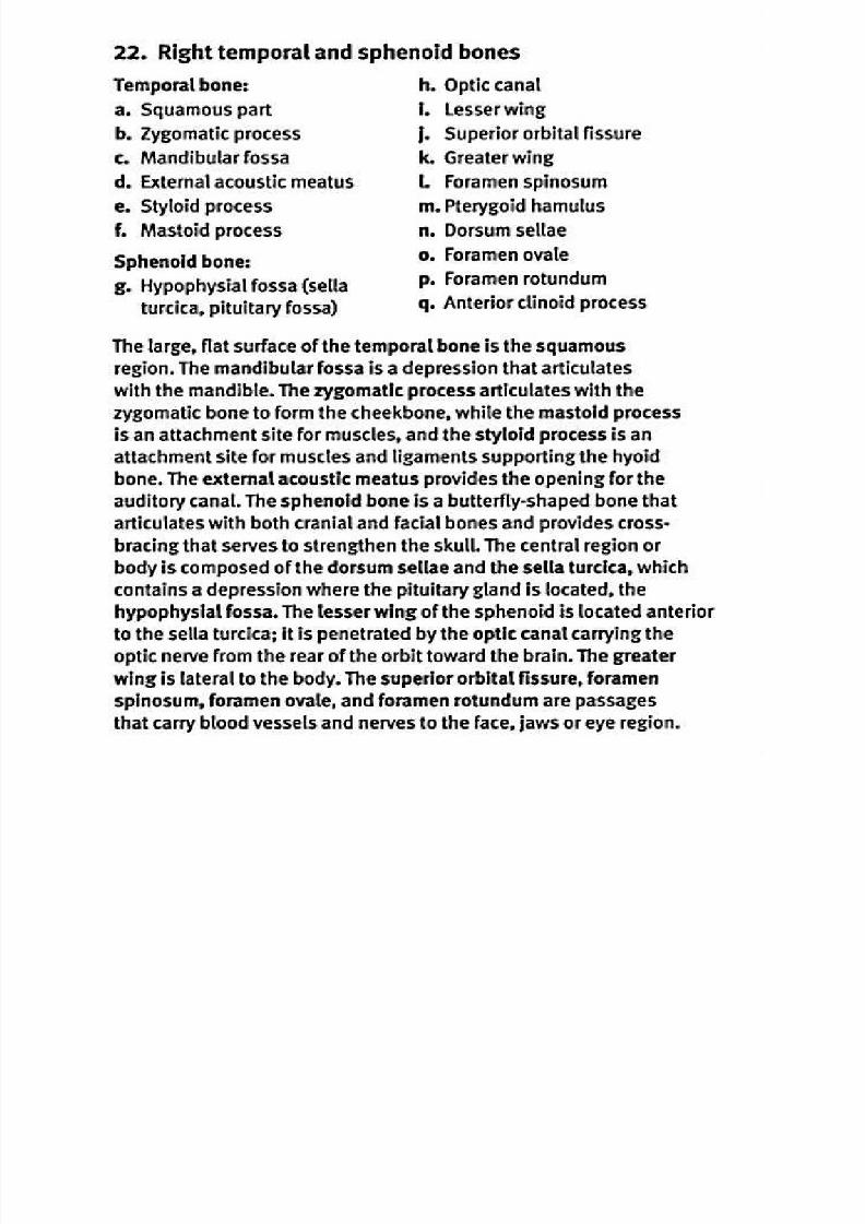
22. Right temporal and sphenoid bones
Temporal bone:
a. Squamous part
b. Zygomatic process
c. Mandibular fossa
d . External acoustic meatus
e. Styloid process
f. Mastoid process
Sphenoid bone:g. Hypophysial fossa sella
turcica, pituitary fossa
h. O ptic canal
i. Lesser wing
I . Superior orbital fissurek. Greater wing
I Foramen spinosum
m . Pterygoid hamulus
n . Dorsum sellae
o. F oramen ovate
P. Foramen rotundum
q• Anterior clinoid process
The large, flat surface of the temporal bone is the squamous
region. The mandibular fossa is a depression that articulates
with the mandible. The zygomatic process articulates with the
zygomatic bone to form the cheekbone, while the mastoid process
is an attachment site for muscles, and the styloid process is an
attachment site for muscles and ligaments supporting the hyoid
bone. The external acoustic meatus provides the opening for the
auditory canal. The sphenoid bone is a butterfly-shaped bone that
articulates with both cranial and facial bones and provides cross-
bracing that serves to strengthen the skull. The central region or
body is composed of the dorsum setae and the sella turcica, which
contains a depression where the pituitary gland is located, the
hypophysial fossa. The lesser wing of the sphenoid is located anterior
to the sella turcica; it is penetrated by the optic canal carrying the
optic nerve from the rear of the orbit toward the brain. The greater
wing is lateral to the body. The superior orbital fissure, foramen
spinosum, foramen ovate, and foramen rotundum are passages
that carry blood vessels and nerves to the face, jaws or eye region.
8/13/2019 113264016 Anatomia Omului
http://slidepdf.com/reader/full/113264016-anatomia-omului 45/298
S Y S T E M
Hyoid bone 3
KAPLAN MEDICAL
8/13/2019 113264016 Anatomia Omului
http://slidepdf.com/reader/full/113264016-anatomia-omului 46/298
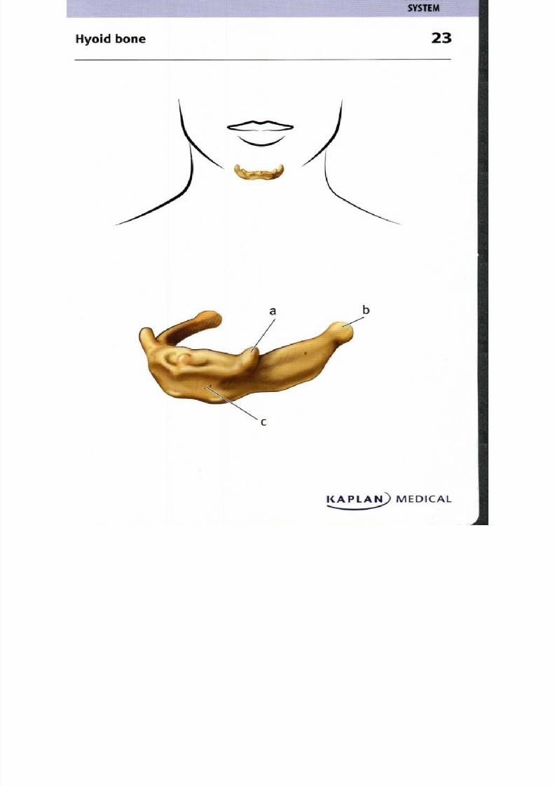
23. Hyoid bone
a. L esser horn
b. Greater horn
c. Body
The small hyoid bone is located at th e base of the to ngue
and im med iately superior to the larynx. It is crucial to hu man
speech as i t braces the ton gue and larynx to allow a wide range
of mo vem ents. The bod y of the hyoid is an attachm ent s i te for
mu scles of the ph arynx, larynx and tongue. The greater horn s
support the larynx and provide attachmen t s ites for mu scles
mo ving the tongue. The lesser horns are suspended from the
styloid p roce sses of the tem po ral bon es via l igame nts.
8/13/2019 113264016 Anatomia Omului
http://slidepdf.com/reader/full/113264016-anatomia-omului 47/298
Vertebral column lateral view 4
KAPLAN MEDICALS
8/13/2019 113264016 Anatomia Omului
http://slidepdf.com/reader/full/113264016-anatomia-omului 48/298
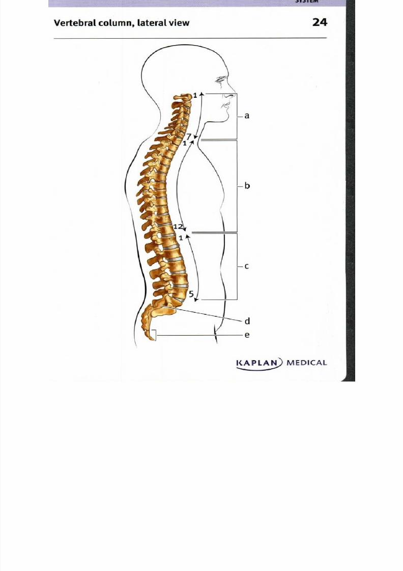
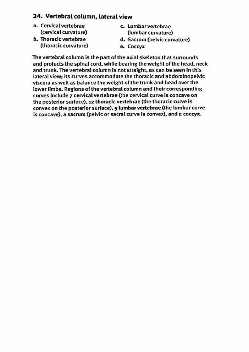
24. V ertebral colum n, lateral view
a. Cervical vertebrae . Lumbar vertebrae
cervical curvature) lumbar curvature)
b. Thoracic vertebrae Sacrum pelvic curvature)
thoracic curvature) . Coccyx
The vertebral column is the part of the axial skeleton that surrounds
and protects the spinal cord, while bearing the weight of the head, neck
and trunk. The vertebral column is not straight, as can be seen in this
lateral view; its curves accommodate the thoracic and abdominopelvicviscera as well as balance the weight of the trunk and head over the
lower limbs. Regions of the vertebral column and their corresponding
curves include 7 cervical vertebrae the cervical curve is concave on
the posterior surface), 12 thoracic vertebrae (the thoracic curve is
convex on the posterior surface), 5 lumbar vertebrae (the lumbar curve
is concave), a sacrum pelvic or sacral curve is convex ), and a coccyx.
8/13/2019 113264016 Anatomia Omului
http://slidepdf.com/reader/full/113264016-anatomia-omului 49/298
S Y S T E M
Po sterior view o f the vertebrae 5
KAPLA5) MEDICAL

8/13/2019 113264016 Anatomia Omului
http://slidepdf.com/reader/full/113264016-anatomia-omului 50/298
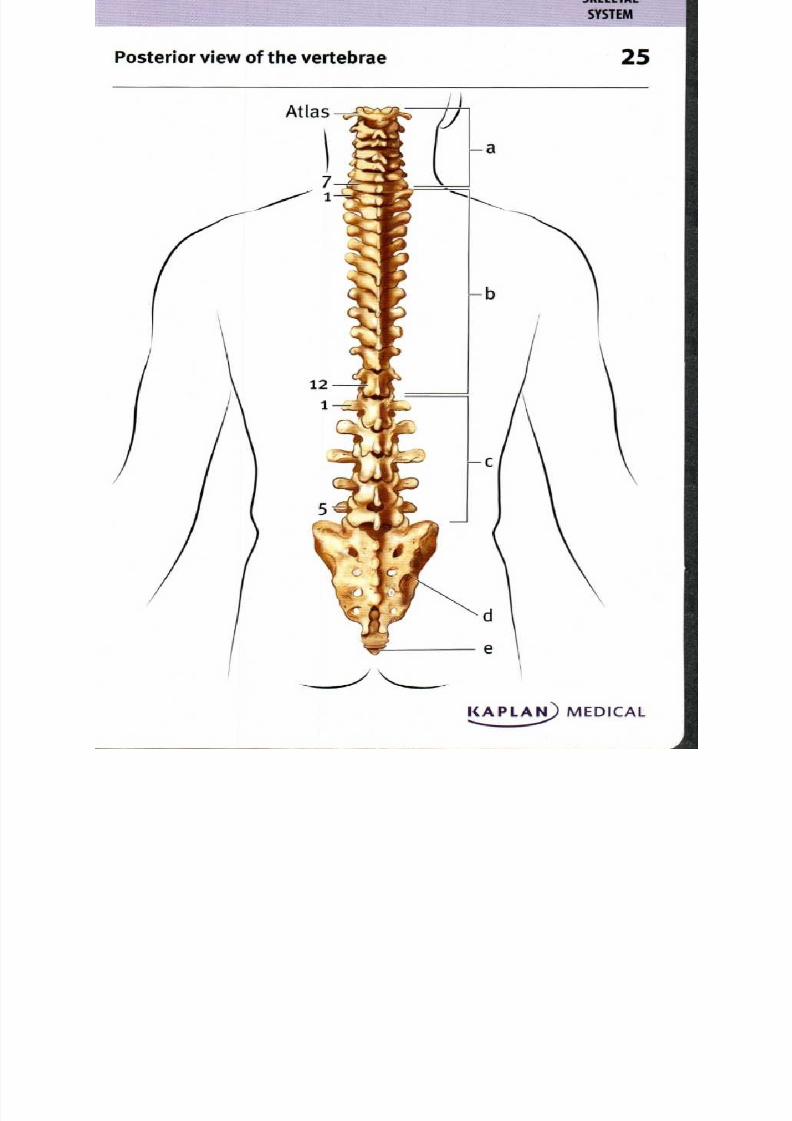
25. Posterior view of the vertebrae
a. Cervical vertebrae . Sacrum
b. Thoracic vertebrae . Coccyx
c. Lumbar vertebrae
The adult human vertebral column consists of 26 bones-7 cervical
vertebrae C1•C7, including the atlas[Ci] and axis[C2D form the
neck, 12 thoracic vertebrae (Ti-T12) support the upper back and
articulate with ribs, 5 lumbar vertebrae (11-15) sup port the lower
back, a sacrum consisting of 5 fused vertebrae articulates with
the pelvis, and a coccyx resulting from the fusion of the final 4-6
vertebrae. Generally, one spinal nerve emerges at each vertebra;
however, although there are only 7 cervical vertebrae, there are
8 cervical nerves. Each individual vertebra consists of a vertebral
body or centrum that transfers weight to the next lower vertebra, a
vertebral arch forming the posterior margin of the vertebral canal,
and variable types of processes that either provide attachment
points for muscles or articulate with ribs. The 5 sacral vertebrae
begin fusing after puberty and are usually completely fused by age
25-30. The coccyx is not completely fused until late in adulthood.
8/13/2019 113264016 Anatomia Omului
http://slidepdf.com/reader/full/113264016-anatomia-omului 51/298
Atlas (Ci) and axis (C2), superior view 6
a
Atlasi
e
Atlas and Axis
KAPLAN) MEDICAL
8/13/2019 113264016 Anatomia Omului
http://slidepdf.com/reader/full/113264016-anatomia-omului 52/298
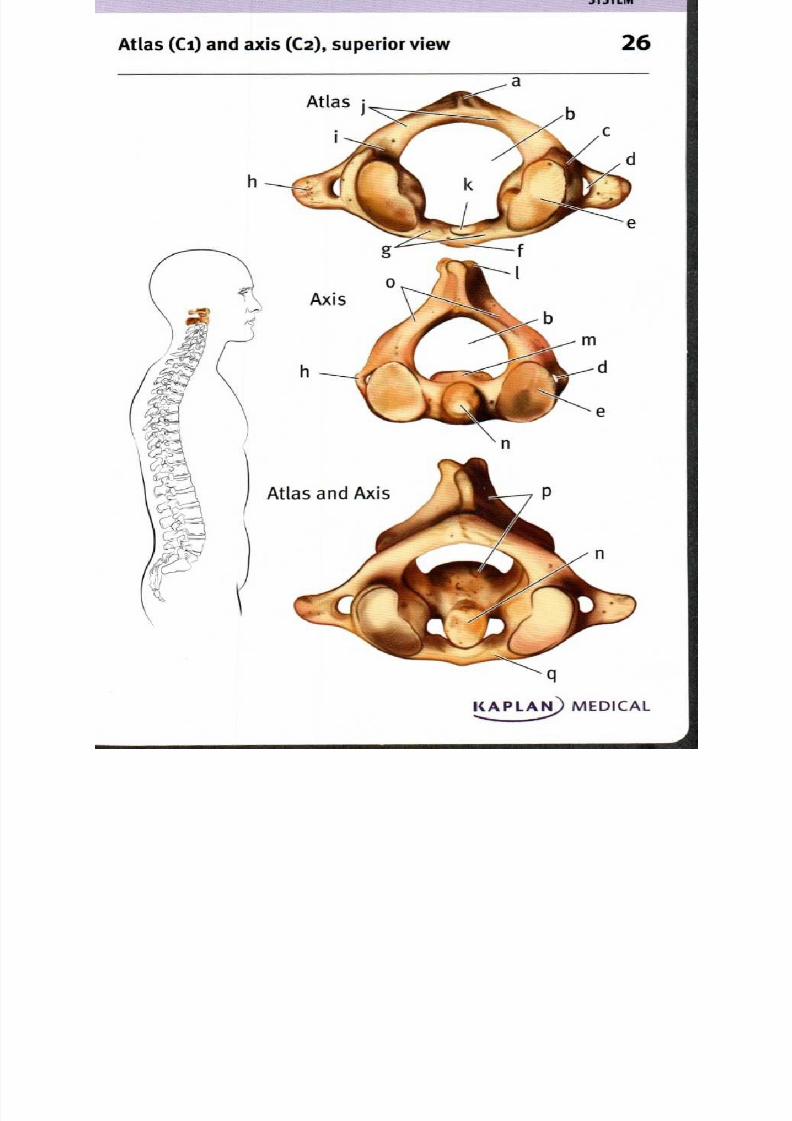
26. Atlas Ci) and axis C2), superior view
a. Posterior tubercle . Posterior arch
b. Vertebral foramen . Facet for odontoid
c. Lateral mass rocess of axis
d. Transverse foramen . Bifid spinous process
e. Superior articular . Body of axis
process (facet) . Odontoid process (dens)
f. Anterior tubercle . Arch of axis lamina)
g. Anterior arch . Axis
h. Transverse process . Atlas
I. Groove for vertebral artery
The first cervical vertebra is called the atlas; its superior articular
processes have facets that articulate with the occipital condyles of
the skull in a type of joint that permits forward•backward motion of
the head. The body of the second cervical vertebra, the axis, has aprominent odontoid process that extends superiorly and articulates
with a facet on the atlas, providing a pivot point to allow rotational
movement of the head. The facet of the superior articular process of
the axis articulates with a similar flat surface on an inferior articular
process of the atlas. Like other individual vertebrae, the axis has
a prominent dorsal spinous process, which is notched as it is in
cervical vertebrae 3-6 and is referred to as bind ; the atlas has a
smaller dorsal process know n as the posterior tu bercle. L aterally, a
transverse process provides attachm ent sites for mu scles, wh ile the
tran sverse foram en allows passage of verte bral arteries and v eins.

8/13/2019 113264016 Anatomia Omului
http://slidepdf.com/reader/full/113264016-anatomia-omului 53/298
Superior view
S Y S T E M
Cervical vertebra, superior and lateral views 7
I(APLA) MEDICAL

8/13/2019 113264016 Anatomia Omului
http://slidepdf.com/reader/full/113264016-anatomia-omului 54/298
27. Cervical vertebra, superior and lateral views
a. Bifid spinous process . Transverse process
b. Vertebral foramen . Body
c. Lam ina of vertebral arch . Uncus of vertebral bodyd. Pedicle of vertebral arch . Transverse foramen
e. Superior articular process . Inferior articular process
Cervical vertebrae have a relatively large vertebral foramen, since
the spinal cord still includes most of the axons that exit the brain,
and the vertebral bod y only needs to support the weight of thehead. The vertebral foramen is bounded by the body anteriorly, the
pedicles laterally, and the laminae posteriorly. Where the lamina
meet is a posteriorly projecting protrusion called the spinous process,
which is notched, or bifid, for C2-C6. Articular processes lie at the
junction between the pedicles and laminae; each has a relatively
flat surface, the facet, which articulates with the articular process of
the neighboring vertebra. The superior articular process articulates
with the vertebra above, and the inferior articular process articulates
with the vertebra below. The uncus is a ridge of bone around the
superior edge of the body in cervical vertebrae, increasing the
stability of the joint with the vertebra above it. Laterally, transverse
processes provide attachment sites for neck muscles; a hole, the
transverse foramen, allows passage of vertebral arteries and veins.

8/13/2019 113264016 Anatomia Omului
http://slidepdf.com/reader/full/113264016-anatomia-omului 55/298
Superior view
h
Lateral view
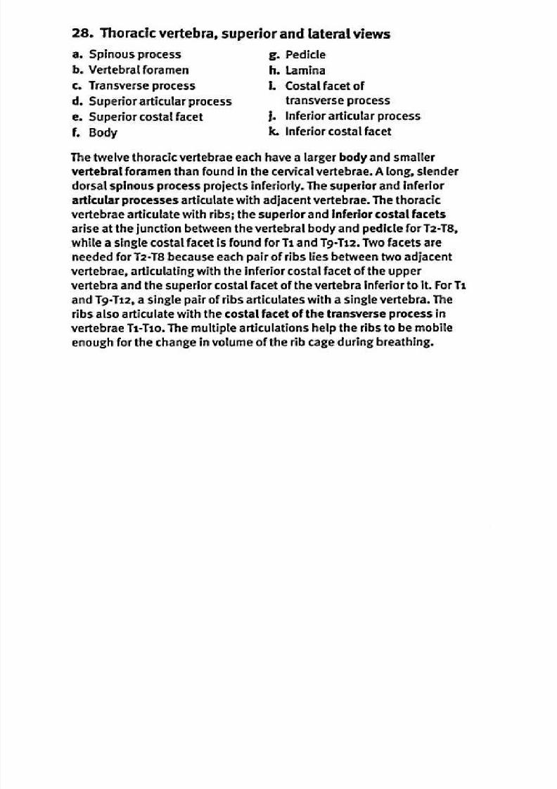
Tho racic ve rtebra, superior and lateral views 8
KAPLAN) MEDICAL
8/13/2019 113264016 Anatomia Omului
http://slidepdf.com/reader/full/113264016-anatomia-omului 56/298
8/13/2019 113264016 Anatomia Omului
http://slidepdf.com/reader/full/113264016-anatomia-omului 57/298
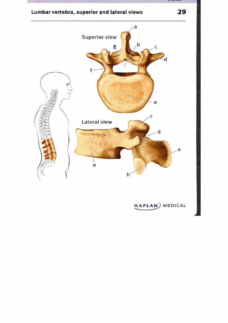
Lum bar vertebra, supe rior an d lateral views 9
Superior view r /
f
Lateral view
e
I(APLA) MEDICAL,••
8/13/2019 113264016 Anatomia Omului
http://slidepdf.com/reader/full/113264016-anatomia-omului 58/298
8/13/2019 113264016 Anatomia Omului
http://slidepdf.com/reader/full/113264016-anatomia-omului 59/298

8/13/2019 113264016 Anatomia Omului
http://slidepdf.com/reader/full/113264016-anatomia-omului 60/298
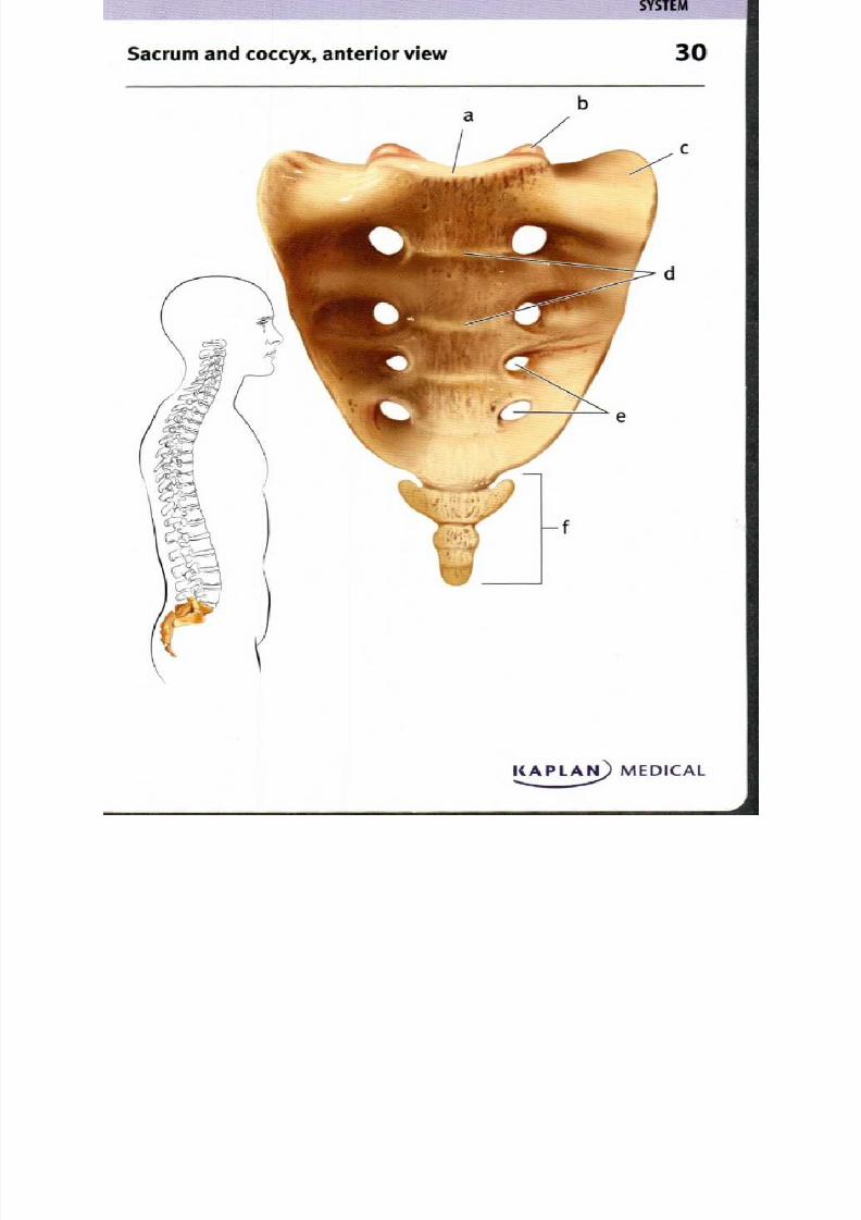
30. Sacrum and coccyx anterior v iew
a. Sacral pro m ontory Transverse l ines
b. Sup erior a rticular p rocess . A nte rior sacral foramina
c. L ateral m ass (ala) Coccyx
F ive sacral vertebrae fuse to form the sacrum , wh ile 3.5 coccygeal
vertebrae fuse to form the co ccyx. These vertebrae begin fusing after
pu berty; the sacrum is usually comp letely fused by the m id•twen ties,
wh ile the coccyx is not co m pletely fused until late in adu lthood . The
coccyx is a vestigial rem nant of the tail of evo lutionary ancestors,
but in hum ans, has no vertebral foramen and does not surroun d
a part of the spinal cord. The regions of the sacrum include the
sacral pro mon tory that articulates with the last lumbar vertebra
(L 5), the tw o broad lateral masses (ala) on either side, and the
central sacral bod y, correspond ing to the fu sed vertebral bodies;
the transverse l ines mark the p osition of the interv ertebral discs
between the bod ies of the fused vertebrae. The anterior sacral
foram ina pro vide p assageways for sacral nerves as well as arteries.

8/13/2019 113264016 Anatomia Omului
http://slidepdf.com/reader/full/113264016-anatomia-omului 61/298
8/13/2019 113264016 Anatomia Omului
http://slidepdf.com/reader/full/113264016-anatomia-omului 62/298
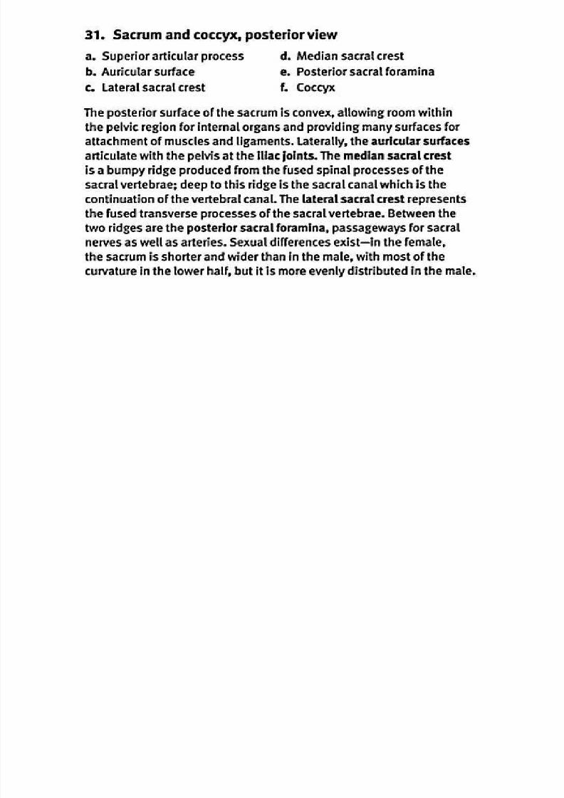
31. Sacrum and coccyx, posterior view
a. Superior articular process . Median sacral crest
b. Auricular surface . Posterior sacral foramina
c. Lateral sacral crest . Coccyx
The posterior surface of the sacrum is convex, allowing room within
the pelvic region for internal organs and providing many surfaces for
attachment of muscles and ligaments. Laterally, the auricular surfaces
articulate with the pelvis at the iliac joints. The median sacral crest
is a bumpy ridge produced from the fused spinal processes of the
sacral vertebrae; deep to this ridge is the sacral canal which is the
continuation of the vertebral canal. The lateral sacral crest represents
the fused transverse processes of the sacral vertebrae. Between the
two ridges are the posterior sacral foramina, passageways for sacral
nerves as well as arteries. Sexual differences exist—in the female,
the sacrum is shorter and wider than in the male, with most of the
curvature in the lower half, but it is more evenly distributed in the male.

8/13/2019 113264016 Anatomia Omului
http://slidepdf.com/reader/full/113264016-anatomia-omului 63/298
8/13/2019 113264016 Anatomia Omului
http://slidepdf.com/reader/full/113264016-anatomia-omului 64/298

8/13/2019 113264016 Anatomia Omului
http://slidepdf.com/reader/full/113264016-anatomia-omului 65/298
S K E L E T A L
S Y S T E M
Sternum , anterior view 3
MEDICAL

8/13/2019 113264016 Anatomia Omului
http://slidepdf.com/reader/full/113264016-anatomia-omului 66/298
33. Sternum anterior view
a. Sup rasternal notch
b. Clavicular notch
c. Manubriumd . Stern al angle
e. Co stal notches
1. Body
g. Xiphoid process
The sternum , part of the axial skeleton, functions to p rotect and
support the internal organs of the th oracic cavity, and to form an
attachmen t point for ribs. It has three main po rtions—the superior
section is the triangular manu brium that articulates with the clavicles,
the m ain bod y, and th e small xipho id proc ess located inferior to
the body. The superior edge of the m anubrium has two points of
attachmen t for the clavicles, located laterally on either side of th e
med ial depression kn own as the suprastemal notch. At the po int of
attachmen t between the manubrium and body of the sternum is the
sternal angle, a conven ient m arker located at the level of the secon d
rib. The anterior en ds of ribs 1-7 articulate with the sternum ; the first
rib articulates with the m anubrium at sites inferior to the clavicular
notch es, and ribs 2.7 h ave cart ilage conn ect ions to the bod y of the
sternum at the co stal notch es. R ibs 8.10 are attached by carti lage
to the carti lage conn ections of rib 7. The x iphoid p rocess forms an
attachmen t po int for som e m uscles, including th e d iaphragm.
8/13/2019 113264016 Anatomia Omului
http://slidepdf.com/reader/full/113264016-anatomia-omului 67/298
34ib and vertebra, articulated, superior view;
rib, posterior view
S K E L E T A L
S Y S T E M

8/13/2019 113264016 Anatomia Omului
http://slidepdf.com/reader/full/113264016-anatomia-omului 68/298
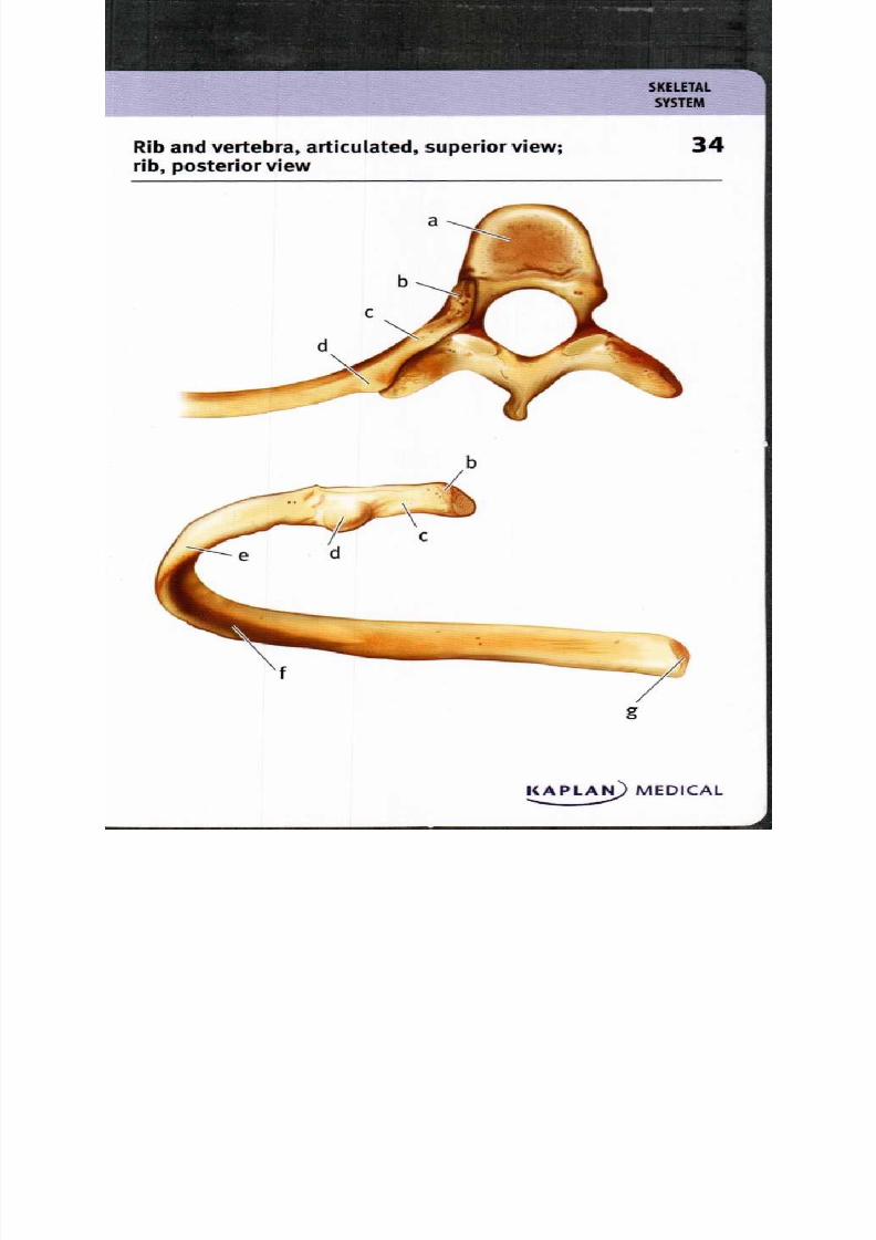
34. R ib and vertebra, articulated, sup erior view;rib, po sterior view
a. Tho racic vertebra . Angle of rib
b. Head of rib . Costal groovec. Neck of rib . Sternal extremity
d. Tubercle of rib
The ribs function to protect the organs of the thoracic cavity and
to provide a flexible cavity for breathing. The thoracic vertebrae
articulate with the ribs; the head of the rib attaches at the costal
facets near the body of the vertebrae while the tubercle of the rib
is positioned at the costal facet of the transverse process. The
shaft of the rib curves anteriorly at the angle of the rib. Along
the inferior border of the internal surface of the rib lies a costal
groove which marks the site where nerves and blood vessels
pass. Cartilage connected to ribs 17 at the sternal extremity
articulate with the sternum at the costal notches. Cartilage
attached to ribs 8•io, in turn, attaches to the cartilage from rib 7.
8/13/2019 113264016 Anatomia Omului
http://slidepdf.com/reader/full/113264016-anatomia-omului 69/298
S Y S T M
Rib cage anterior view 5
KAPLA MEDICAL

8/13/2019 113264016 Anatomia Omului
http://slidepdf.com/reader/full/113264016-anatomia-omului 70/298
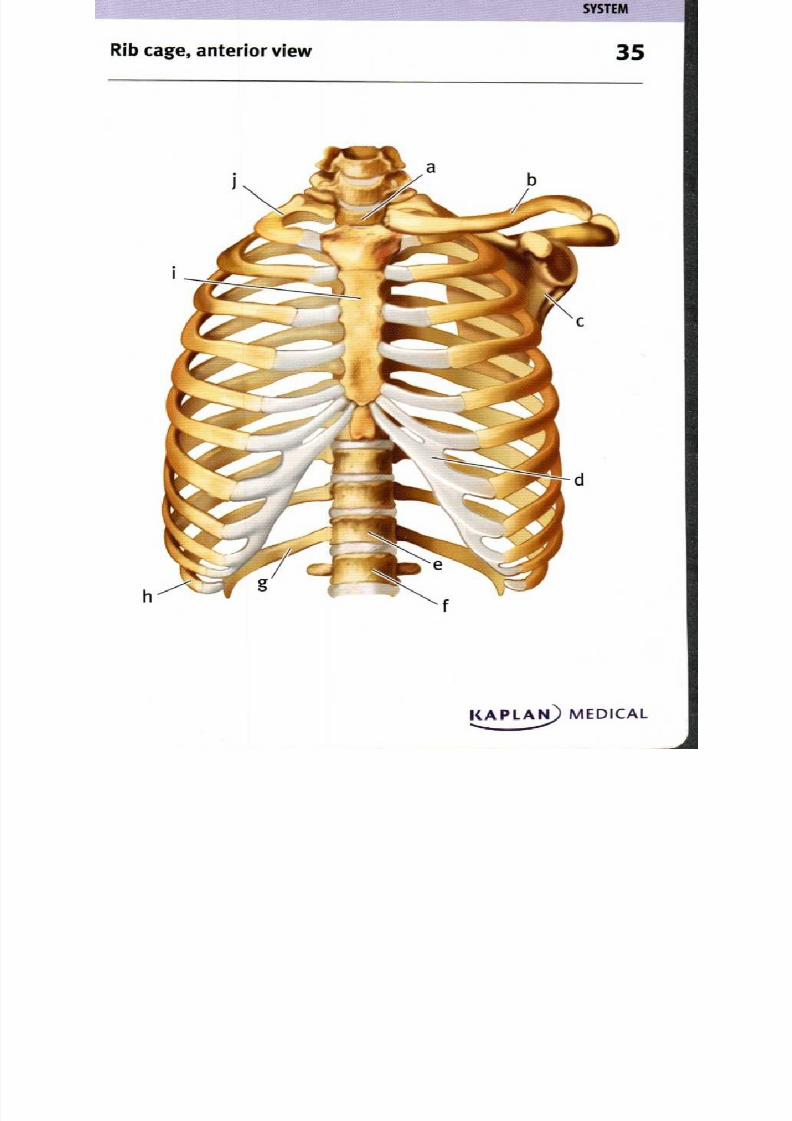
35. R ib cage, anterior view
a. 1st thoracic vert ebra
b. Clavicle
c . Scapula
d . Co stal cartilage
e . 12th th oracic vertebra
f. 1st lum bar vertebra
g. 12th rib
h. nth rib
I. Sternum
j. 1st rib
The rib cage consists of the sternum and the 12 p airs of ribs, wh ich
are attached po steriorly to the thoracic vertebrae. R ibs 1-7 articulate
with th e sternu m thro ugh the co stal cartilage; for ribs 8.io the costalcartilage articulates only indirectly with t he sternum since it fuses
to th e cartilage of rib 7 for sup po rt. Ribs 11-12 do n ot attach to the
sternum at all; they are conn ected w ith other skeletal elem ents only
at the v ertebral end. The articulation between the axial skeleton and
the pector al girdle occurs where th e clavicle, or collarbone, attaches
to the sternum at the m anubrium; in turn , the clavicle articulateswith the scapu la or shoulder blade. The fun ction of the rib cage is
to p rotect the heart, lun gs and other th oracic organs as well as to
serve as an attachm ent po int for mu scles involved in m ovem ents
of the p ectoral girdle and u pp er lim bs, adjustmen ts to the po sition
of the vertebral colum n, and m ost impo rtantly, breathing.

8/13/2019 113264016 Anatomia Omului
http://slidepdf.com/reader/full/113264016-anatomia-omului 71/298

8/13/2019 113264016 Anatomia Omului
http://slidepdf.com/reader/full/113264016-anatomia-omului 72/298
36. Pectoral girdle and upper limb, anterior view
a. Clavicle
b. Acromion process
c. Coracoid process
d. Humerus
e. Radius
1 . Sternum
g. Scapula
h. Ulna
i. Carpals
j. Metacarpals
k. Phalanges
The pectoral girdle is composed of four bones, two clavicles and two
scapulae. The acromion and coracoid processes of the scapulaeare points of attachment for numerous ligaments and muscles. The
clavicle articulates with the sternum of the axial skeleton medially,
and with the scapula laterally. The primary function of the pectoral
girdle is to provide an anchor for movements of the arm. Each upper
limb consists of a humerus in the (upper) arm, an ulna and a radius
in the forearm, eight carpal bones in the wrist, five metacarpal
bones in the hand, and u; phalanges or finger bones. At the
shoulder, the humerus articulates with the scapula to produce a
wide range of arm movements; at the elbow, the humerus articulates
with the radius and ulna to flex the forearm, while articulation
between the radius and ulna allows pronation of the forearm. The
complex wrist joint provides for a wide range of movements while
the finger joints allow flexion and extension of the fingers.
8/13/2019 113264016 Anatomia Omului
http://slidepdf.com/reader/full/113264016-anatomia-omului 73/298
S Y S T E M
Scapula, anterior and lateral views 7
e
d
f
Anterior view
b
Lateral view
MEDICAL
8/13/2019 113264016 Anatomia Omului
http://slidepdf.com/reader/full/113264016-anatomia-omului 74/298
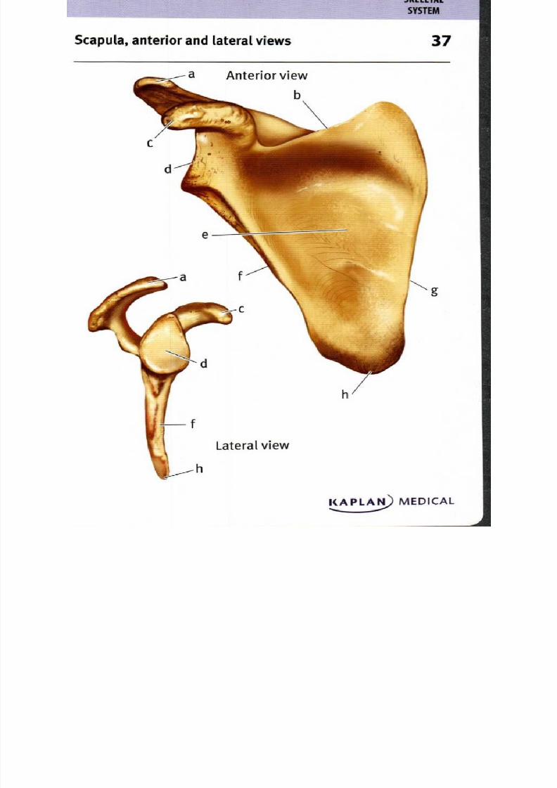
37. Scapula, anterior and lateral views
a. Acrom ion process . Subscapular fossa
b. Sup erior border . L ateral border
c. Coracoid process . M edial borde rd . Gleno id fossa . In ferior angle
Viewed from t he anterior per spective, the scapula has an obvious
large, triangular surface that is roughly con cave, forming th e
subscapu lar fossa; its ed ges are, observ ed in a clockw ise direc tion
from this aspect , the sup erior border, medial border, inferior angle,
and lateral bord er. Betw een th e superior and lateral bord ers, the
scapula articulates with the h um erus at the shou lder joint. The
glenoid fossa is the con cave 'socket ' within which th e rou nded head
of the h um erus rotates. Tw o pro cesses also originate in this area and
extend superiorly; the m ore anterior is the coracoid p rocess which
is an attachme nt p oint for l igamen ts and tend ons; posterior to this
is the larger acrom ion p rocess, which articulates with the clavicle
as well as being the attachm ent p oint for add ition al l igamen ts and
tend ons of th e shoulde r joint. The lateral view clearly shows that
these two processes project from the thin p lane o f the scapula; the
roun ded nature of the glenoid fossa also becom es more o bvious.
8/13/2019 113264016 Anatomia Omului
http://slidepdf.com/reader/full/113264016-anatomia-omului 75/298
Scapula, posterior view 8
KAPLAI9 MEDICAL

8/13/2019 113264016 Anatomia Omului
http://slidepdf.com/reader/full/113264016-anatomia-omului 76/298

8/13/2019 113264016 Anatomia Omului
http://slidepdf.com/reader/full/113264016-anatomia-omului 77/298
Clavicle and related bones, superior view;
clavicle, inferior view
Superior view
8/13/2019 113264016 Anatomia Omului
http://slidepdf.com/reader/full/113264016-anatomia-omului 78/298
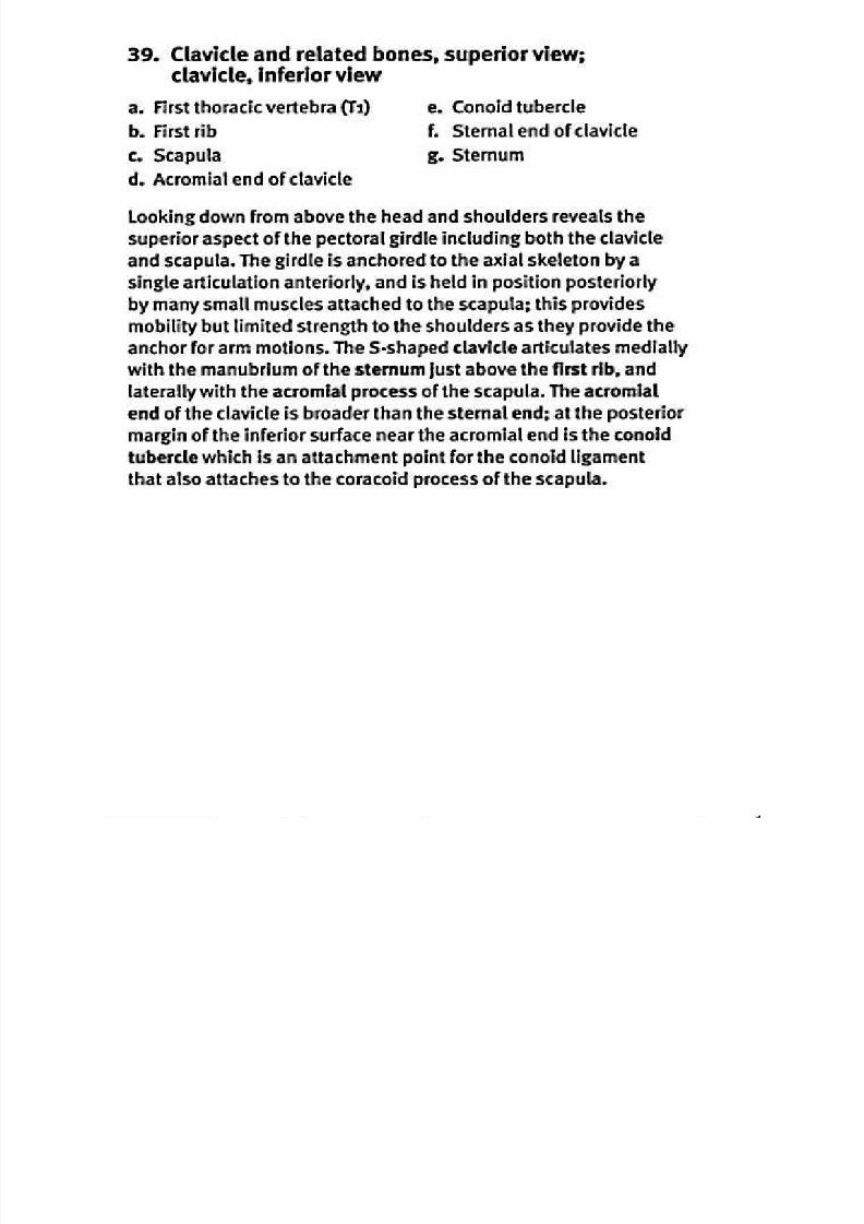
39. Clavicle and related bones, superior view;
clavicle, inferior view
a. First thoracic vertebra (Ti) . Conoid tubercle
b. First rib . Sternal end of claviclec. Scapula . Sternum
d. Acrom ial end of clavicle
Looking down from above the head and shoulders reveals the
superior aspect of the pectoral girdle including both the clavicle
and scapula. The girdle is anchored to the axial skeleton by a
single articulation anteriorly, and is held in position posteriorly
by many small muscles attached to the scapula; this provides
mobility but limited strength to the shoulders as they provide the
anchor for arm motions. The S-shaped clavicle articulates medially
with the manubrium of the sternu m just above the first rib, and
laterally with the acrom ial proc ess of the scapula. The acrom ial
end of the clavicle is broader th an the sternal end; at the po sterior
margin of the inferior surface near the acrom ial end is the cono id
tubercle which is an attachmen t point for the co noid l igame nt
that also attaches to the co racoid p rocess of the scapula.

8/13/2019 113264016 Anatomia Omului
http://slidepdf.com/reader/full/113264016-anatomia-omului 79/298
40um erus, anterior and posterior view s
Anterior view osterior view
S Y S T E M
KAPLA :1) MEDICAL
8/13/2019 113264016 Anatomia Omului
http://slidepdf.com/reader/full/113264016-anatomia-omului 80/298
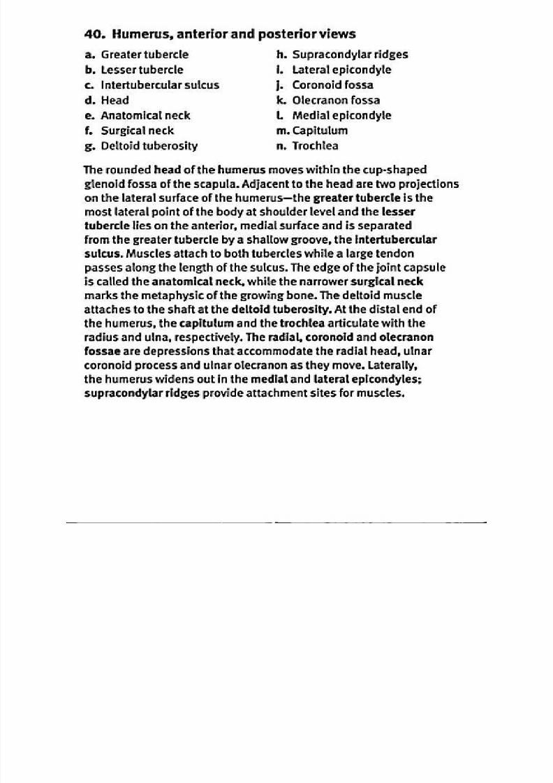
40. H umerus anterior and posterior views
a. Greater tubercle
b. L esser tubercle
c . Intertu bercular sulcusd . Head
e. An atomical neck
1 Surgical neck
g. Deltoid tu berosity
h . Su pracond ylar ridges
i. L ateral epicond yle
j. Co rono id fossak. O lecranon fossa
1 M edial epicondyle
m . Capitulum
n . Trochlea
The roun ded h ead of the hum erus moves wi thin the cup-shapedglenoid fossa of the scapula. A djacent to the head are two project ions
on th e lateral surface of the hum erus— the greater tubercle is the
mo st lateral point of th e bod y at shoulder level and the lesser
tuberc le lies on th e anterior, me dial surface and is separated
from the greater tubercle by a shallow groove, the In tertubercular
sulcus. M uscles attach to both tu bercles while a large tend on
passes along the length o f the sulcus. Th e edge o f the joint cap sule
is called the anatom ical neck, while the n arrower surgical neck
m arks the m etaphysic of the growing bone. The deltoid m uscle
attaches to the shaft at the de ltoid tubero sity. A t the d istal end of
the hu me rus, the capitulum and th e trochlea articulate with the
radius and ulna, respectively. The radial , corono id and olecranon
fossae are depressions that accom m od ate the radial head, ulnar
coron oid pro cess and ulnar olecranon as they move . L aterally,
the h um erus widens o ut in the m edial and lateral epicondyles;
supracond ylar ridges pro vide attachmen t si tes for m uscles.
8/13/2019 113264016 Anatomia Omului
http://slidepdf.com/reader/full/113264016-anatomia-omului 81/298
S Y S T E M
Ulna and radius, lateral and anterior views 41
b
d
e
g
k.
Ulna, lateral view lna and radius, anterior view
KAPLAN MEDICAL
8/13/2019 113264016 Anatomia Omului
http://slidepdf.com/reader/full/113264016-anatomia-omului 82/298
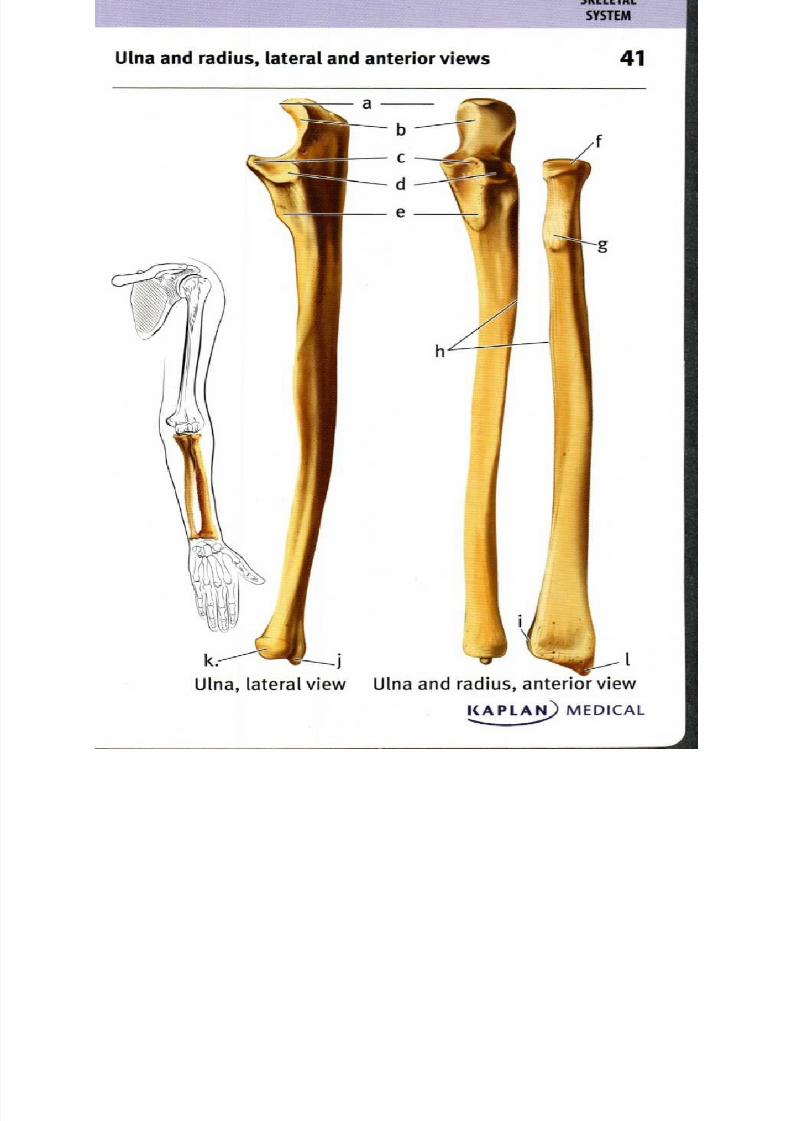
41. Ulna and radius, lateral and anterior views
a. Olecranon process
b. Trochlear notch
c. Coronoid process
d. Rad ial notch
e. Tuberosity of the ulna
f. Head of radius
g. Radial tuberosity
h. Interosseous margin
I . U lnar notch
j. Styloid process of ulna
k. Head of ulna
I. Styloid process of radius
The bones of the forearm are the ulna and radius. The more medial and
longer ulna articulates with the trochlea of the humerus at the trochlearnotch. The superior edge of the trochlear notch is the olecranon process
which fits into the olecranon fossa of the humerus when the forearm is
extended, and the inferior edge of the trochlear notch is the coronoid
process which fits into the coronoid fossa of the humerus when the
forearm is flexed. Lateral to the coronoid process, the radial notch of
the ulna articulates with the head of the radius. Distal to the radialhead, the radial tuberosity forms an attachment site for muscles. A
fibrous sheet called the interosseous membrane connects the radius
and ulna along the interosseous margins, and serves as a site for
muscle attachment. At their distal ends, the ulna and radius articulate
with each other, and the radius articulates with bones of the wrist. The
lateral surface of the ulnar head articulates with the ulnar notch of
the radius. A stytoid process extends distally from each of the bones,
providing many attachment sites for ligaments and muscles of the wrist.

8/13/2019 113264016 Anatomia Omului
http://slidepdf.com/reader/full/113264016-anatomia-omului 83/298
S Y S T E M
Han d, pos terior (dorsal) view 2
KAPLAN) MEDICAL......

8/13/2019 113264016 Anatomia Omului
http://slidepdf.com/reader/full/113264016-anatomia-omului 84/298
42. H and, posterior (dorsal) view
a. Phalanges . Triquetrum
b. Head of m etacarpal . Lunate
c. Shaft of m etacarpal Metacarpal
d. Base of m etacarpal . T rapezoid
e. Hamate . Trapezium
f. Capitate Scaphoid
Eight carpal bones m ake u p the flexible wrist, articulating at
individual joints that allow lim ited, gliding m otion between the
bone surfaces. The proxim al row of carpals includes the scaphoid
bone, lun ate bone, triquetrum , and p isiform bone; the distal row
consists of the trapezium , trapezoid bon e, capitate bone, and the
ham ate bone. Articulating w ith the distal carpal bones are the five
m etacarpal bones, form ing the hand . T he m etacarpals are identified
by rom an nu m erals; m etacarpal I is m ost lateral, form ing the base ofthe thum b, and articulates with the trapezium . The proxim al base of
each m etacarpal articulates with the carpals. T he m etacarpal heads
articulate distally with phalanges, or finger bones. The thum b has
two p halanges; each of the other fingers has three, m aking a total of
14 phalanges on each hand. The joint between m etacarpal I and the
trapezium at the base of the thum b is a saddle joint, allow ing m orerange of m otion than foun d w ith the other m etacarpals, and leading
to the abilities associated w ith having an opp osable thum b.
8/13/2019 113264016 Anatomia Omului
http://slidepdf.com/reader/full/113264016-anatomia-omului 85/298
S Y S T E M
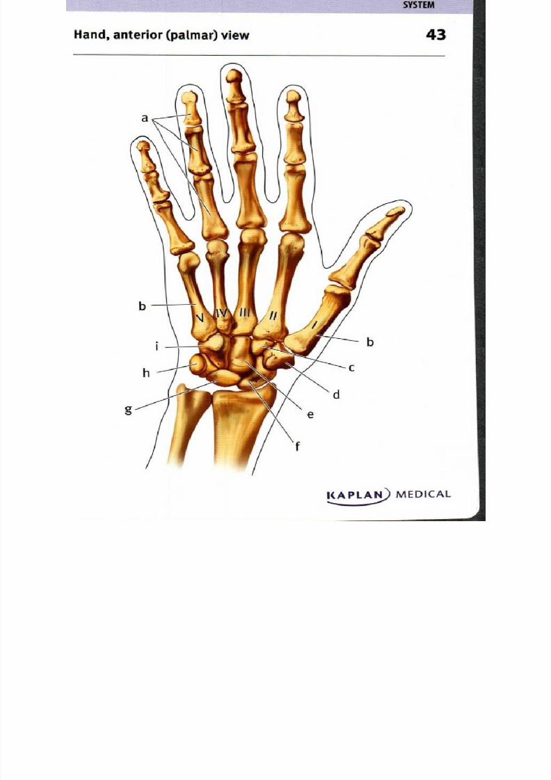
Hand, anterior (palmar) view 3
KAI. . . . . _21 AN MEDICAL

8/13/2019 113264016 Anatomia Omului
http://slidepdf.com/reader/full/113264016-anatomia-omului 86/298
8/13/2019 113264016 Anatomia Omului
http://slidepdf.com/reader/full/113264016-anatomia-omului 87/298
S Y S T E M
44ip bone, lateral view
Adult hip bone
pHip bone of a child
KAPLAN) MEDICAL
•

8/13/2019 113264016 Anatomia Omului
http://slidepdf.com/reader/full/113264016-anatomia-omului 88/298
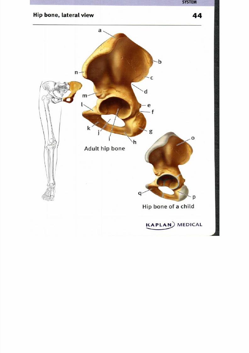
44. H ip bone, lateral view
a. Iliac crest
b. Posterior superior iliac spine
c. Posterior inferior iliac spine
d. G reater sciatic notch
e. Ischia' spine
f. L esser sciatic notch
g. Ischia' tuberosity
h. Ischial ram us
i. Acetabulum
I.
k.
I.
m .
n.
o.
p.
q .
O bturator foramen
Inferior pubic ramu s
Superior pubic ram us
Anterior inferior iliac spine
Anterior superior iliac spine
Ilium
Ischium
Pubis
T he hip bone, or os coxae, is form ed from the fusion of three
bones— the ilium , ischium , and p ubis. T he fusion lines are visible
in the child's hip bone im age, showing how the three bones m eet
to form the acetabulum which is seen clearly in the lateral view and
articulates with the head of the fem ur. An terior to the acetabulumare the superior and inferior ram i of the pubis; posterior to the
acetabulum is the ischium , extending from the Ischia( spine on the
superior edge to the ischial ram us w hich m eets the inferior pu bic
ram us. The Ischia' tuberosity is the rounded p rotrusion that bears
one's weight when seated. The space im m ediately inferior to the
acetabulum is the obdura tor foram en w hich is filled by a sheet ofcollagen fibers that provide sites for attachm ent of m uscles. Superior
to the acetabulum is the large broad ilium , which suppo rts the weight
of the internal organs of the trunk; m uscles, tendons and ligam ents
attach at sites includ ing the iliac crest and variou s iliac spines. The
greater sciatic notch allow s passage of the sciatic nerve to the low er
8/13/2019 113264016 Anatomia Omului
http://slidepdf.com/reader/full/113264016-anatomia-omului 89/298
8/13/2019 113264016 Anatomia Omului
http://slidepdf.com/reader/full/113264016-anatomia-omului 90/298
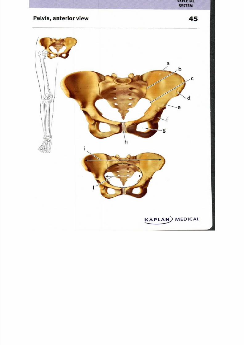
45. Pelvis, anterior view
a. Iliac crest
b. Sacroiliac joint
c. Greater sciatic notch
d. Anterior superior iliac spine
e. An terior inferior iliac spine
1 Acetabulum
g. Obturator foramen
h. Symphysis pubis
I False pelvis
j. True pelvis
The pelvis is formed from the two ossa coxae of the appendicular
skeleton and the sacrum and coccyx of the axial skeleton. Because
it supports the weight of the upper body and mediates the stresses
of locomotion, the bones are larger and heavier than those of the
pectoral girdle. The ilium of the ox coxae articulates with the sacrum
at the sturdy sacroiliac joint. T he iliac crest form s the sup erior,
posterior edge of the pelvis, w hile the anterior su perior iliac spines
m ark the lateral edges. The anterior an d inferior lim it of the pelvis
is comp osed of the pubis bones, med ial to the obdurator foram en;
the pubis bones a re conn ected by fibrocartilage at the sym physis
pu bis. T he true pelvis (or lesser pe lvis) is the cavity po sterior to
the pubic sym physis, anterior to the sacrum and coccyx, and
boun ded by the m edial surfaces of the ilia near the greater sciatic
notch. The fa lse pelvis (or greater pelvis) is the larger, mo re sup erior
cavity bou nded latera lly by the anterior sup erior iliac spines.
8/13/2019 113264016 Anatomia Omului
http://slidepdf.com/reader/full/113264016-anatomia-omului 91/298
8/13/2019 113264016 Anatomia Omului
http://slidepdf.com/reader/full/113264016-anatomia-omului 92/298
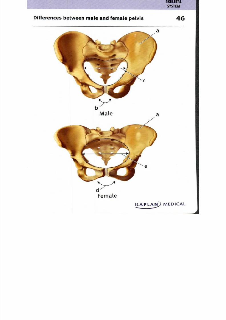
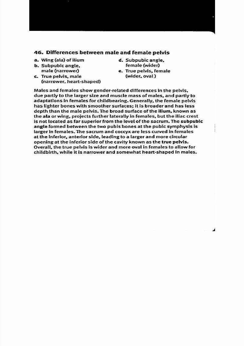
46 Differences between male and female pelvis
a. W ing ala) of ilium . Subpubic angle,
b. Subpubic angle, emale wider)
male narrower) . True pelvis female
c. True pelvis, m ale wider, oval)
(narrower, heart-shaped)
Males and females show gender-related differences in the pelvis,
due partly to the larger size and muscle mass of males, and partly to
adaptations in females for childbearing. Generally, the female pelvis
has lighter bones with smoother surfaces; it is broader and has less
depth than the male pelvis. The broad surface of the ilium, known as
the ala or wing, projects further laterally in females, but the iliac crest
is not located as far superior from the level of the sacrum. The subpubic
angle formed between the two pubis bones at the pubic symphysis islarger in females. The sacrum and coccyx are less curved in females
at the inferior, anterior side, leading to a larger and more circular
opening at the inferior side of the cavity known as the true pelvis.
Overall the true pelvis is wider and more oval in females to allow for
childbirth, while it is narrower and somewhat heart-shaped in males.
8/13/2019 113264016 Anatomia Omului
http://slidepdf.com/reader/full/113264016-anatomia-omului 93/298
ower limb anterior view 7
pl AN MEDICAL
8/13/2019 113264016 Anatomia Omului
http://slidepdf.com/reader/full/113264016-anatomia-omului 94/298
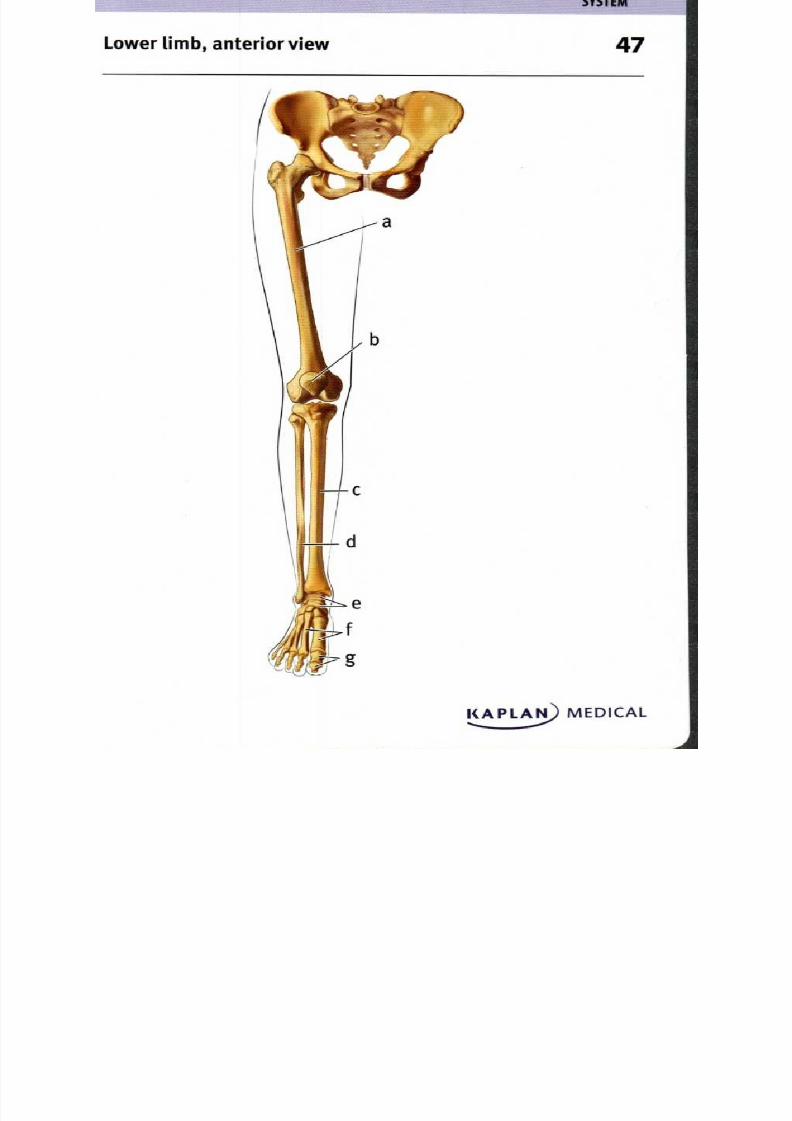
47. Lower limb, anterior view
a. Femur . Tarsals
b. Patella Metatarsals
c. Tibia . Phalanges
d. Fibula
T he lower lim b m ust withstand the stresses of locom otion and bearing
the body's weight; for this reason, the bones are m ore m assive
than the bones of the upp er lim bs. The low er lim bs are supported
by the pelvis. The bones of the lower limbs include the fem ur, whicharticulates proxim ally w ith the pelvis at the acetabulum of the hip bone
and distally w ith the tibia and patella. L ateral to the tibia is the fibula,
but on ly the tibia articulates w ith the tarsals, the ank le bones. At the
ankle, the foot turns 9o° com pared w ith the leg bones, to provide
stability as the body's weight is transferred to the grou nd . T he bones
of the foot includ e the m etatarsals and the phalan ges, or toe bon es.
8/13/2019 113264016 Anatomia Omului
http://slidepdf.com/reader/full/113264016-anatomia-omului 95/298
S Y S T E M
Femur and patella, anterior and posterior views 8
Anterior view Posterior view
KAPLArs) MEDICAL
8/13/2019 113264016 Anatomia Omului
http://slidepdf.com/reader/full/113264016-anatomia-omului 96/298
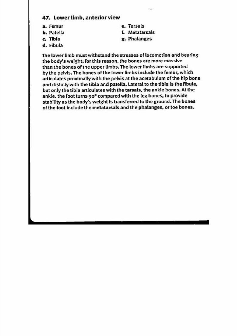
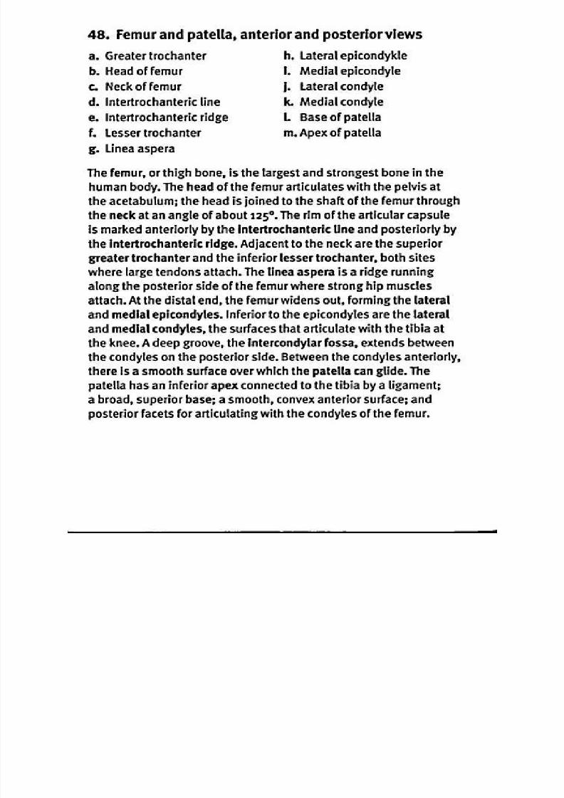
48. Femur and patella, anterior and posterior views
a. Greater trochanter
b. Head of femur
c. Neck of femurd. Intertrochanteric line
e. Intertrochanteric ridge
f. L esser trochanter
g. Linea aspera
h. Lateral epicondykle
I. Medial epicondyle
j. Lateral condylek. Medial condyle
L Base of patella
m . Apex of patella
The femur, or thigh bone, is the largest and strongest bone in thehuman body. The head of the femur articulates with the pelvis at
the acetabulum; the head is joined to the shaft of the femur through
the neck at an angle of about125°. The rim of the articular capsule
is marked anteriorly by the Intertrochanteric line and posteriorly by
the intertrochanteric ridge. Adjacent to the neck are the superior
greater trochanter and the inferior lesser trochanter, both sites
where large tendons attach. The Linea aspera is a ridge running
along the posterior side of the femur where strong hip muscles
attach. At the distal end, the femur widens out, forming the lateral
and medial epicondyles. Inferior to the epicondyles are the lateral
and medial condyles, the surfaces that articulate with the tibia at
the knee. A deep groove, the intercondylar fossa, extends between
the condyles on the posterior side. Between the condyles anteriorly,
there is a smooth surface over which the patella can glide. The
patella has an inferior apex connected to the tibia by a ligament;
a broad, superior base; a smooth, convex anterior surface; and
posterior facets for articulating with the condyles of the femur.
8/13/2019 113264016 Anatomia Omului
http://slidepdf.com/reader/full/113264016-anatomia-omului 97/298
r
Anterior view Posterior view
q
S Y S T E M
Tibia and fibula anterior and posterior views 9
KAPLAN) MEDICAL

8/13/2019 113264016 Anatomia Omului
http://slidepdf.com/reader/full/113264016-anatomia-omului 98/298
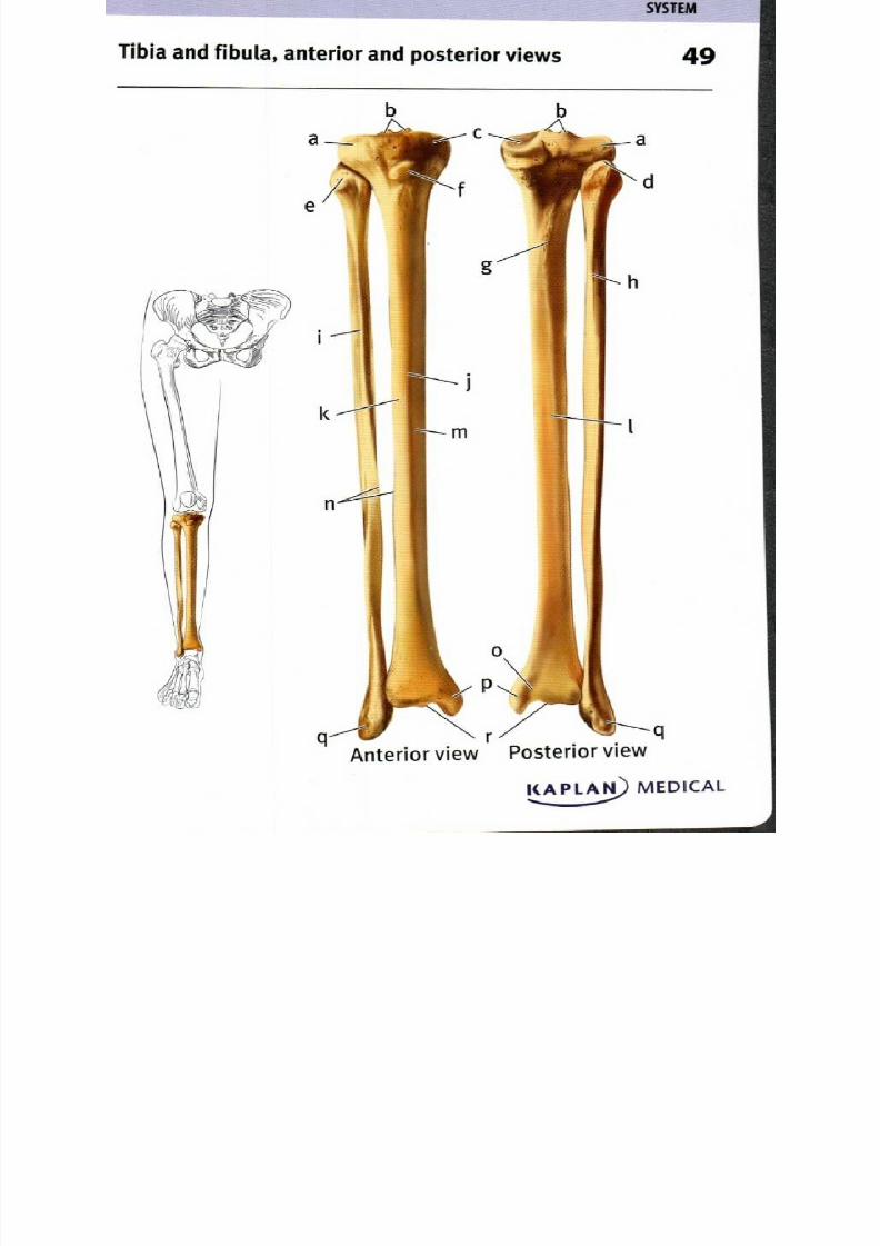
49. Tibia and fibula, anterior and posterior views
a. L ateral tibial condyle . La teral surface of tibial shaft
b. Intercondylar em inence Posterior surface of tibia
c. Medial tibial condyle . Medial surface of tibial shaftd. Ap ex of fibula Interosseous borders
e. Hea d of fibula . M alleolar groove
1. T ibial tubero sity . M edial m alleolus
g. Soleal line . L ateral m alleolus
h. M edial crest of fibula . Inferior articular su rface
I. An terior border of fibula f tibia
I. Anterior border (crest)
of tibia
T he tibia, or shinbone, articulates with the lateral and m edial condyles
of the femu r at the lateral and m edial tibial condyles. Betw een
the cond yles, the intercondylar em inence provides attachm entfor cru ciate ligam ents. Anteriorly, the tibial tuberosity is a site of
attachm ent for the patellar ligam ent. The d istal end o f the tibia has
an In ferior articular surface that articulates with a prox imal tarsal
bone. Ad jacent to this is the m edial m alleolus, a large process that
lends stability to the ank le joint; the m alleolar groove is a tendon
passagew ay. The fibula, or calf bone, is a long, slender bone. The
head of the fibula articulates w ith the lateral tibial condyle, w hile the
inferior end of the tibia also articulates with a flat region on the side
of the fibula. T he lateral m alleolus is a fibular process that continues
inferiorly beyond the articulation with the tibia, providing lateral
supp ort for the ankle joint. Alon g the shaft of both bones, prom inent
crests, borders, and lines ma rk the attachm ent sites for m uscles
or the interosseous m em brane that helps stabilize the positions of
the two bones and provides additional m uscle attachm ent sites.
8/13/2019 113264016 Anatomia Omului
http://slidepdf.com/reader/full/113264016-anatomia-omului 99/298

8/13/2019 113264016 Anatomia Omului
http://slidepdf.com/reader/full/113264016-anatomia-omului 100/298
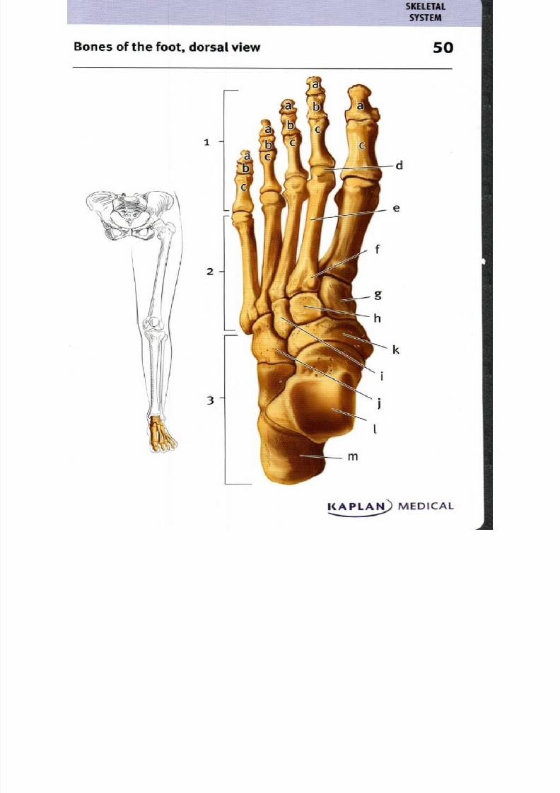
50. Bones of the foot, dorsal view
1. Phalanges . F irst (m edial) cuneiform
2. Metatarsals . Second (interm ediate)
3. Tarsals uneiform
a. Distal phalanges . Third (lateral) cuneiform
b. M iddle phalanges . Cuboid
c. Proxim al phalanges . Navicular
d. Head of m etatarsal Talus
e. Shaft of m etatarsal. C alcaneus
f. Base of m etatarsal
T he bones of the foot include seven tarsal or an kle bones, five
m etatarsal or foot bones, and 14 p halanges or toe bones. The toes
each have d istal, m iddle and p roxim al phalanges, with the exception
of the m ost m edial great toe, which only has two phalanges
(like the thum b)—the distal and proxim al. Each m etatarsal has a
head that articulates with the proxim al phalanges, a shaft, and a
base that articulates w ith the tarsals. T he talus is a large tarsal
that articulates w ith the tibia at a process that also articulates w ith
the lateral m alleolus of the fibula. T he calcaneu s or heel bone is
the largest tarsal. T he navicu lar bone is anterior to the talus and
articulates with the m edial, interm ediate and lateral cuneiformbones, that in turn articulate with m etatarsal bones I— III. Anterior
to the calcaneou s and lateral to the navicular and cu neiform s is
the cuboid bone, which articulates with m etatarsals IV and V.
8/13/2019 113264016 Anatomia Omului
http://slidepdf.com/reader/full/113264016-anatomia-omului 101/298
S Y S T E M
B on es of the foot, lateral view 1
KAPLAN) MEDICAL. . . .
8/13/2019 113264016 Anatomia Omului
http://slidepdf.com/reader/full/113264016-anatomia-omului 102/298
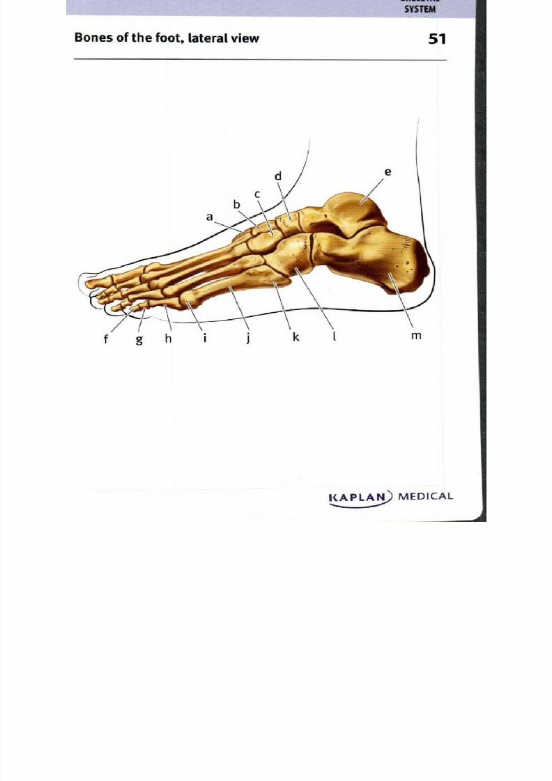
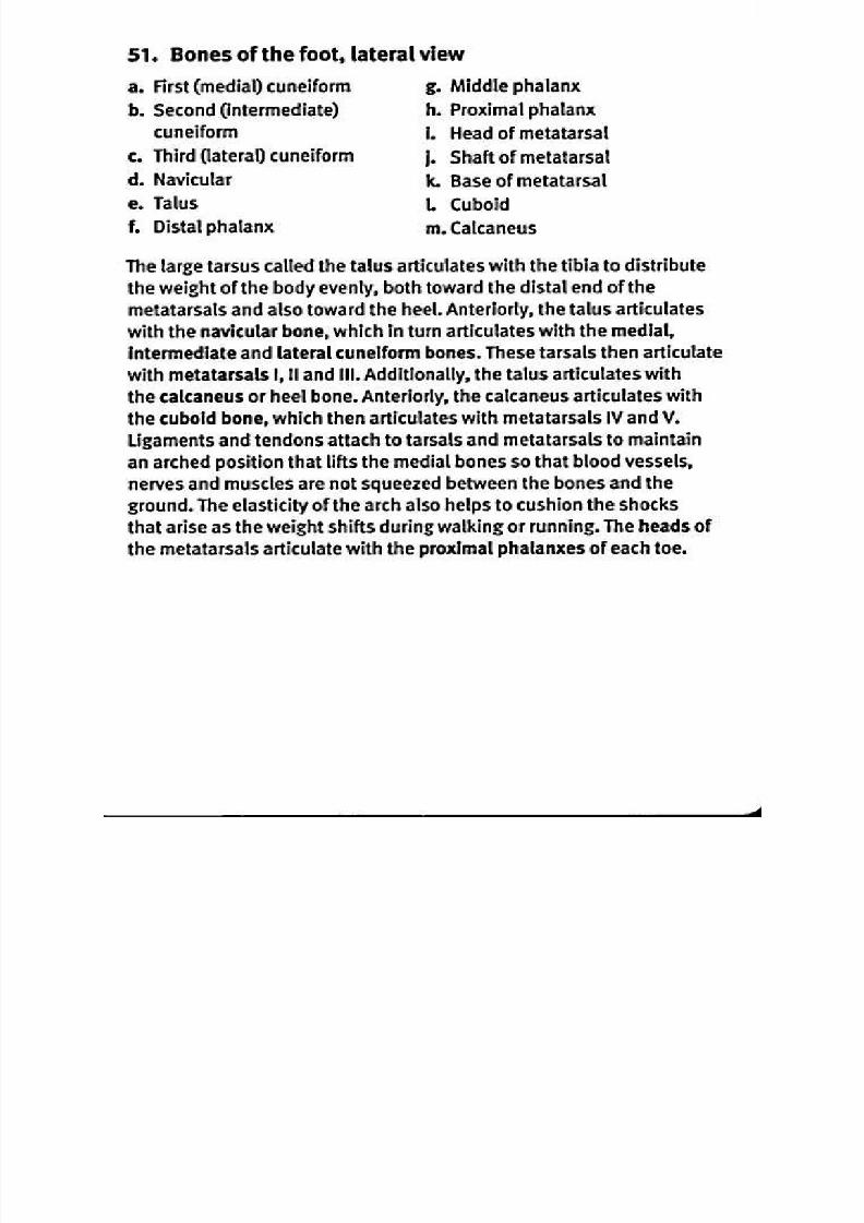
51. Bones of the foot, lateral view
a. F irst (m edial) cuneiform . M iddle phalanx
b. Second (intermediate) . Proxim al phalanx
cuneiform . H ead of m etatarsalc. Third (lateral) cuneiform . Shaft of metatarsal
d. Navicular . Base of m etatarsal
e. Talus Cuboid
1. Distal pha lanx . C alcaneus
The large tarsu s called the talus articulates w ith the tibia to distributethe weight of the body evenly, both toward the d istal end of the
m etatarsals and also toward the heel. An teriorly, the talus articulates
with the navicular bon e, which in turn articulates w ith the m edial,
interm ediate and lateral cuneiform bones. These tarsals then articulate
with m etatarsals I, II and III . Ad ditionally, the talus articulates with
the calcaneus or heel bone. Anteriorly, the calcaneu s articulates w ith
the cuboid bone, which then articulates with m etatarsals IV an d V .
L igam ents and tendons attach to tarsals and m etatarsals to m aintain
an a rched p osition that lifts the m edial bones so that blood vessels,
nerves and m uscles are not squeezed between the bones and the
grou nd . T he elasticity of the arch also helps to cushion the shocks
that arise as the weight shifts during w alking or run ning. The head s of
the m etatarsals articulate with the proxim al phalanxes of each toe.
8/13/2019 113264016 Anatomia Omului
http://slidepdf.com/reader/full/113264016-anatomia-omului 103/298

8/13/2019 113264016 Anatomia Omului
http://slidepdf.com/reader/full/113264016-anatomia-omului 104/298
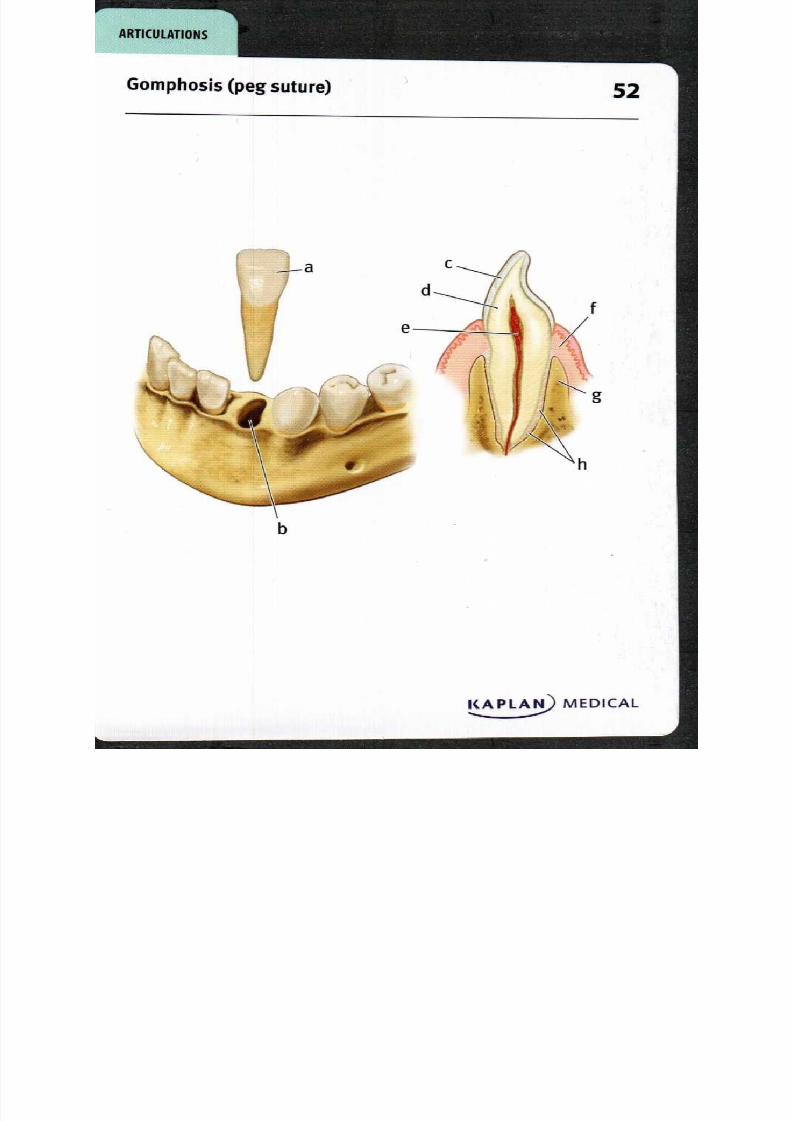
52. Go m phosis peg suture)
a Tooth
b Alveolar socket
c Enamel
d Dentin
e Pulp
f Gingiva
g Alveolar ridge
h Periodontal ligaments
A gomphosis is a fibrous synarthrotic (immovable) joint holding a tooth
in its alveolar socket in the maxilla or mandible. The bulk of the tooth iscomposed of dentin, a mineralized matrix secreted by cells found in the
pulp cavity. The exposed portion of the tooth is covered by a crystalline
calcium phosphate layer called enamel—the hardest substance in the
human body. The root of the tooth is bound in place by the periodontal
l igament; it is composed of collagen fibers extending from the dentin
of the tooth to the bone surrounding the root of the tooth. A bony
alveolar ridge forms the deep socket or alveolus where the peg-like
root of the tooth is inserted. Superficial to the bone is the gingiva,
mucosal tissue tightly bound to the bone surrounding the teeth; it
provides a smooth surface to reduce friction with food.
8/13/2019 113264016 Anatomia Omului
http://slidepdf.com/reader/full/113264016-anatomia-omului 105/298
R T I C U L T I O N S
Suture3
KAPLAN MEDICAL
8/13/2019 113264016 Anatomia Omului
http://slidepdf.com/reader/full/113264016-anatomia-omului 106/298
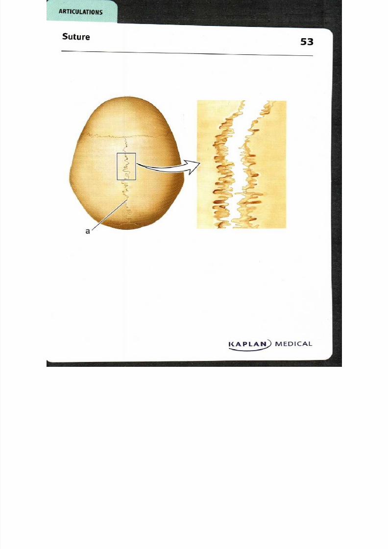
53. Suture
a. Sagittal suture
A suture is a fibrous synarthrotic (immovable) joint located between
the bones of the skull, in order to form a protective case for the brain
and sensory organs of the head. Cranial sutures include the sagittat
suture shown here, which connects the two parietal bones and extends
between the anterior coronal suture and the posterior lambdoid
suture. Further attachment between bones at the suture is provided
by collagen fibers that bind the bones in a firm but slightly flexible
manner. The bone edges at the sutures are interlocking in adults,
although they are slightly separated and are only connected by fibrous
connective tissue during development to allow both more flexibility
of the skull during birth and room for growth as the brain increases in
size during the early postnatal period.
8/13/2019 113264016 Anatomia Omului
http://slidepdf.com/reader/full/113264016-anatomia-omului 107/298
8/13/2019 113264016 Anatomia Omului
http://slidepdf.com/reader/full/113264016-anatomia-omului 108/298
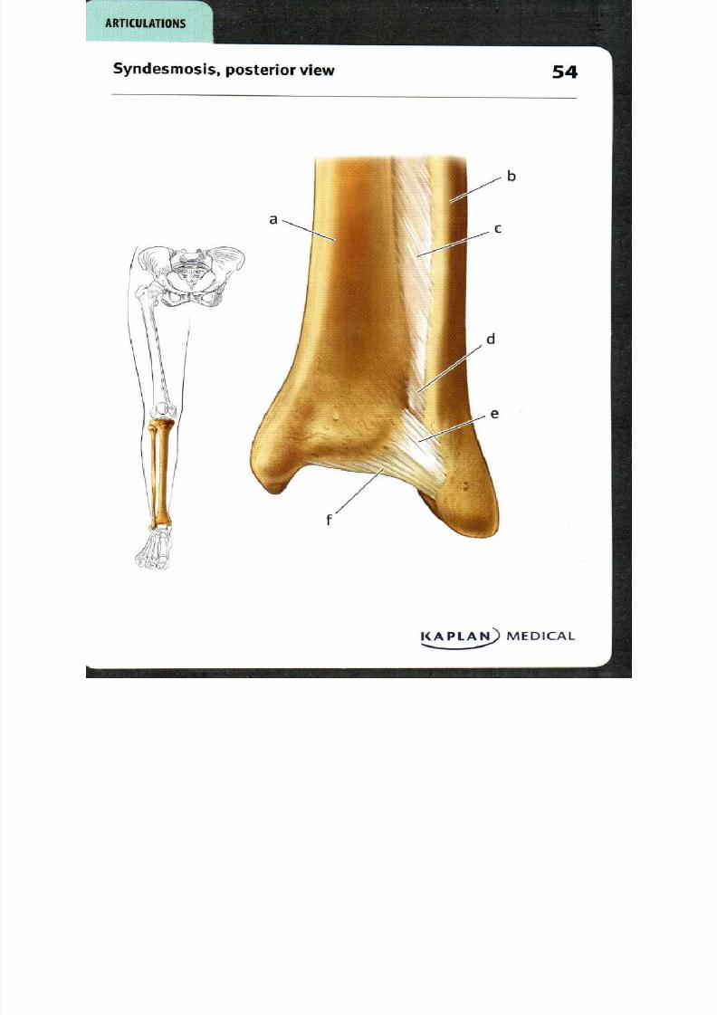
54. Syndesm osis, posterior view
a Tibia . Posterior tibiofibular
b Fibula igament
c Interosseous membrane Transverse tibiofibular
d Interosseous ligament igament
A syndesmosis is a fibrous amphiarthrotic (slightly moveable) joint
where the articulation between the bones is strengthened considerably
by a ligament or network of collagen fibers that connects them. The
syndesmosis between the tibia and fibula permits a small amount of
movement between them. The interosseous membrane is composed
of collagen fibers that connect the interosseous borders along most of
the length of the tibia and fibula; it is continuous with the interosseousligament, composed of fibers which connect the rough surfaces where
the tibia and fibula meet. The anterior, posterior and the deeper
transverse tibiofibular ligaments are strong bands of collagen that
extend from the distal end of the tibia to the lateral malleolus of the
fibula. The strength of the tibiofibular articulation is critical for the
strength of the ankle joint.
8/13/2019 113264016 Anatomia Omului
http://slidepdf.com/reader/full/113264016-anatomia-omului 109/298
Synchondrosis 55
KAPLAN) MEDICAL,...
A R T I C U L A T I O N S
8/13/2019 113264016 Anatomia Omului
http://slidepdf.com/reader/full/113264016-anatomia-omului 110/298
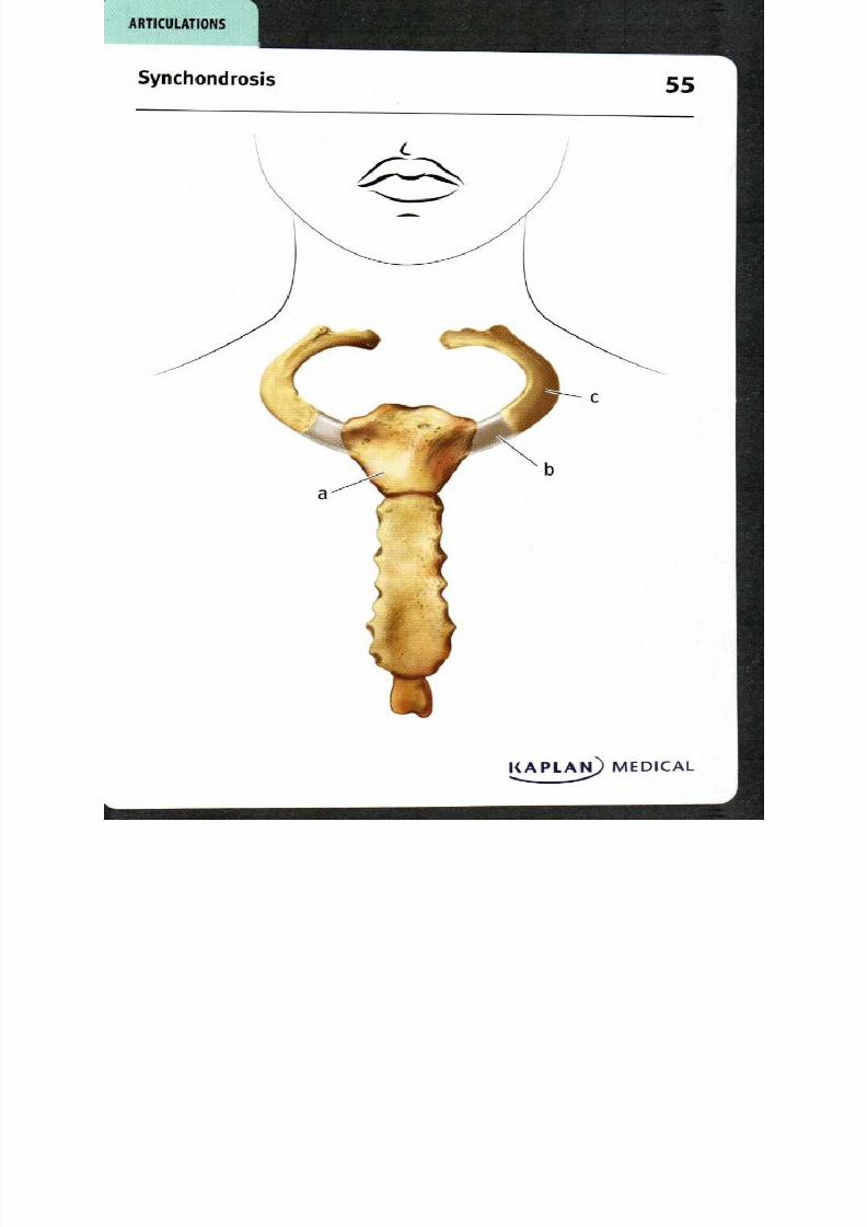
55. Synchondrosis
a Sternum: manubrium
b First costal cartilage
c First rib
A synchondrosisis a fibrous synarthrotic (immovable) joint where
the two articulating bones are joined by cartilage. While there are
many examples of synchondroses in the developing skeleton, such as
growth plates in the long bones that become completely ossified in the
adult, the sternocostal joint between the first rib and the manubrium
of the sternum remains a synchondrosis throughout adult life. The
costal cartilage of rib i is hyaline cartilage that is continuous with the
rib laterally and with the sternum medially. For other ribs, the costalcartilage is continuous with the rib laterally, but either fits into a
depression on the sternum (ribs 2-7), connects with the costal cartilage
on other ribs (ribs 8-so), or ends in the body wall (ribs 11-12).

8/13/2019 113264016 Anatomia Omului
http://slidepdf.com/reader/full/113264016-anatomia-omului 111/298
Symphysis 56
1
A R T I C U L A T I O N S
a
c
KAPLAN MEDICAL
8/13/2019 113264016 Anatomia Omului
http://slidepdf.com/reader/full/113264016-anatomia-omului 112/298
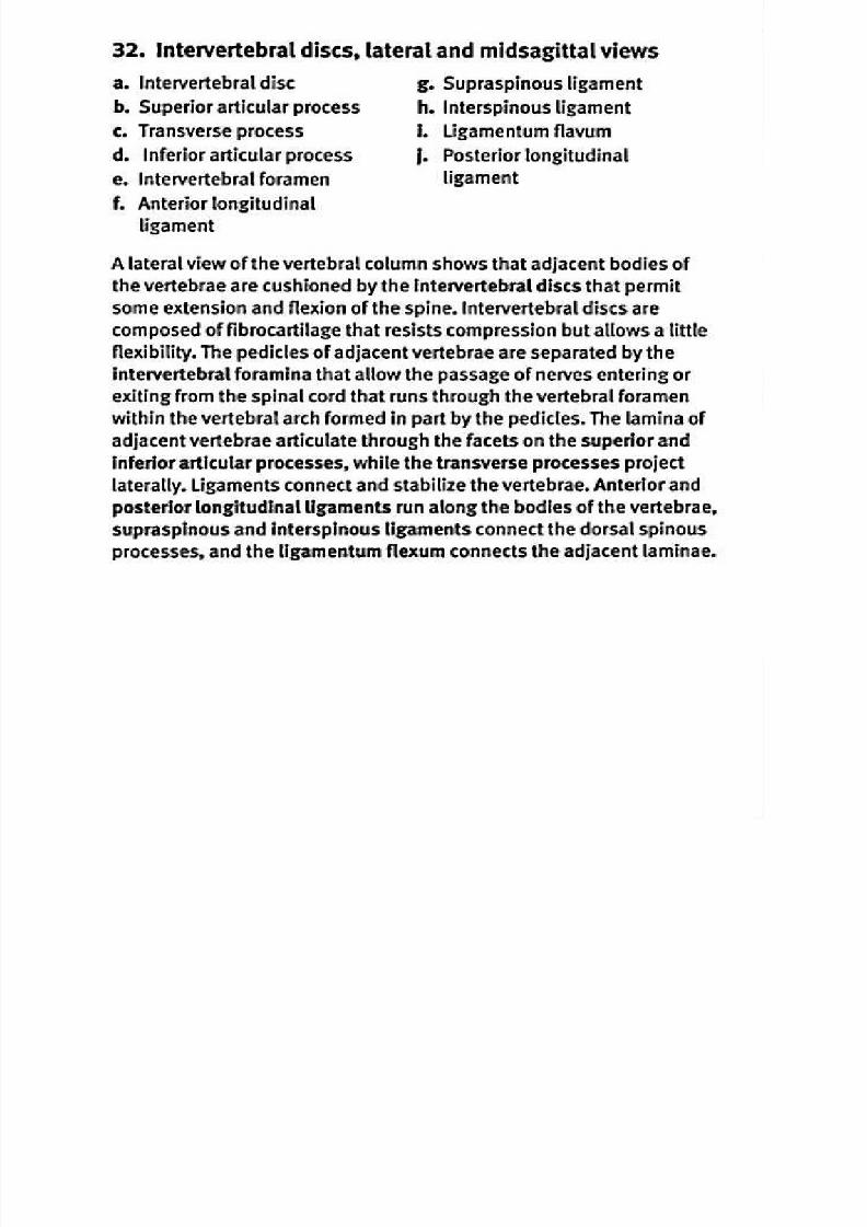
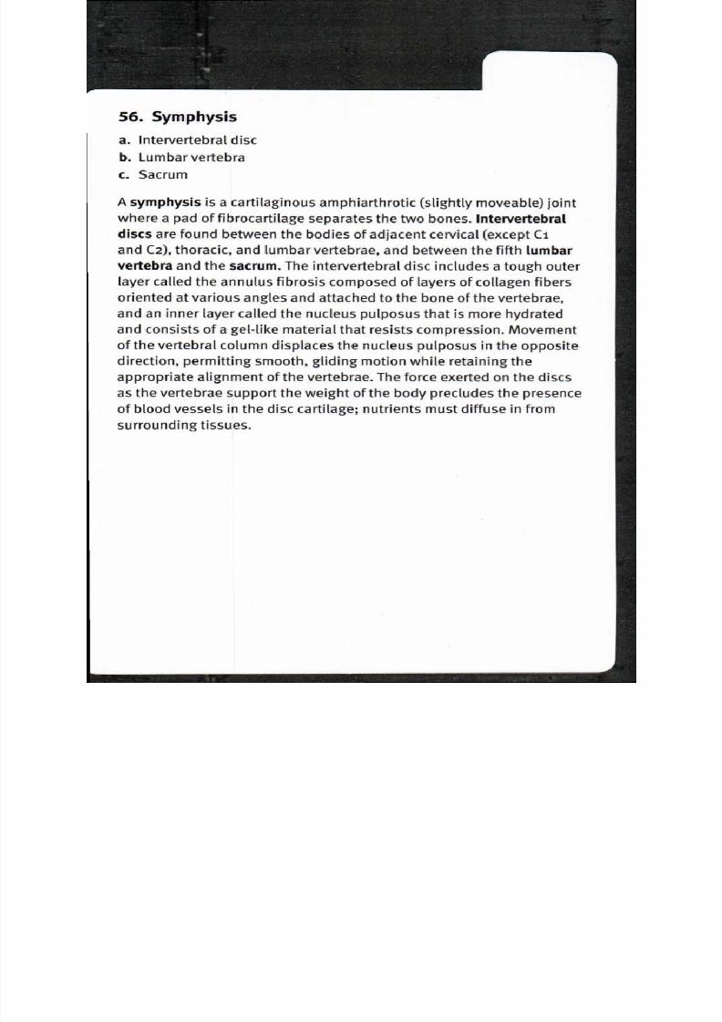
56. Symphysis
a Intervertebral disc
b Lumbar vertebra
c Sacrum
A symphysis is a cartilaginous amphiarthrotic (slightly moveable) joint
where a pad of fibrocartilage separates the two bones. Intervertebral
discs are found between the bodies of adjacent cervical (except Ci
and C2), thoracic, and lumbar vertebrae, and between the fifth lumbar
vertebra and the sacrum. The intervertebral disc includes a tough outer
layer called the annulus fibrosis composed of layers of collagen fibers
oriented at various angles and attached to the bone of the vertebrae,
and an inner layer called the nucleus pulposus that is more hydratedand consists of a gel-like material that resists compression. Movement
of the vertebral column displaces the nucleus pulposus in the opposite
direction, permitting smooth, gliding motion while retaining the
appropriate alignment of the vertebrae. The force exerted on the discs
as the vertebrae support the weight of the body precludes the presence
of blood vessels in the disc cartilage; nutrients must diffuse in from
surrounding tissues.

8/13/2019 113264016 Anatomia Omului
http://slidepdf.com/reader/full/113264016-anatomia-omului 113/298
A R T I C U L A T I O N S
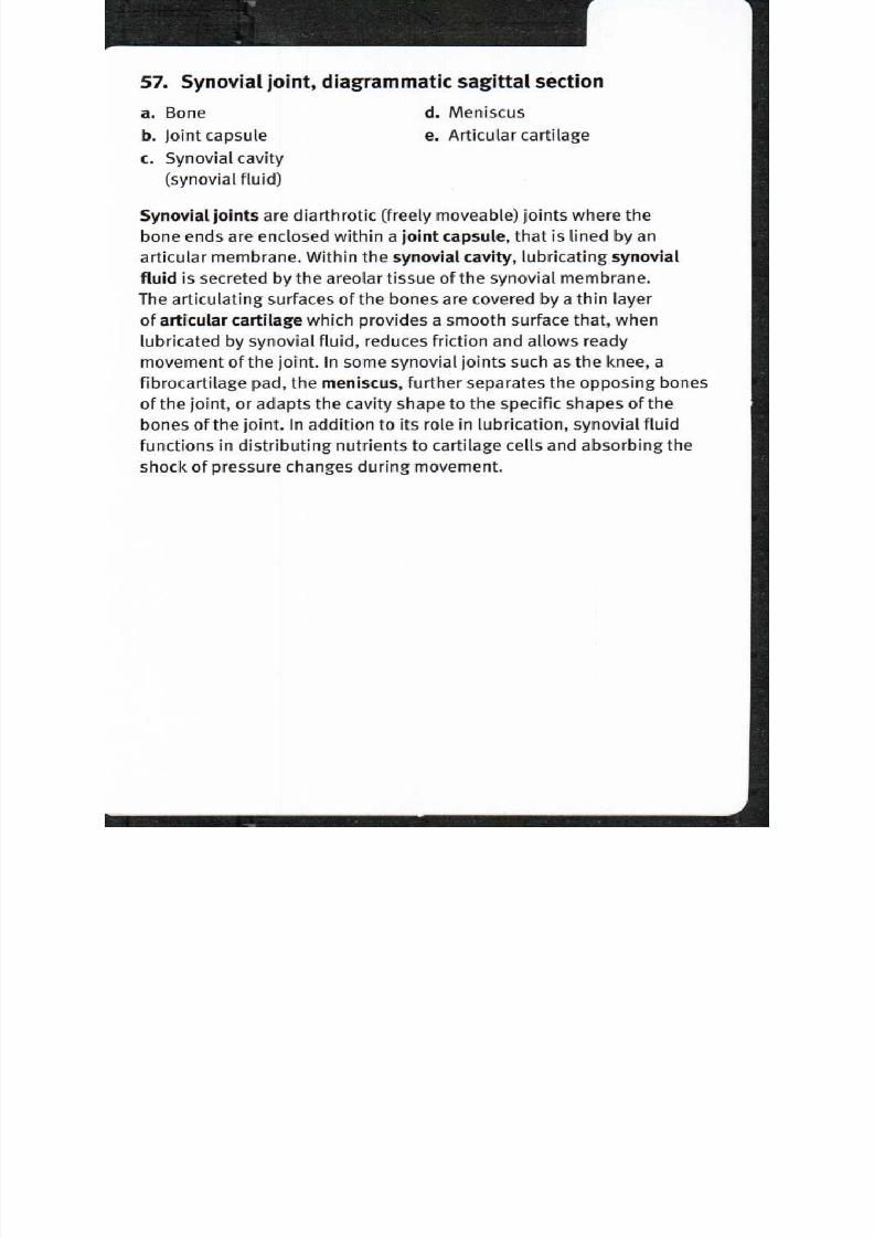
Synovial joint, diagrammatic sagittal section 7
KAPLA MEDICAL. . . . .
8/13/2019 113264016 Anatomia Omului
http://slidepdf.com/reader/full/113264016-anatomia-omului 114/298
57. Synovial joint, diagrammatic sagittal section
a Bone Meniscus
b Joint capsule . Articular cartilage
c Synovial cavity
(synovial fluid)
Synovial joints are diarthrotic (freely moveable) joints where the
bone ends are enclosed within a joint capsule, that is lined by an
articular membrane. Within the synovial cavity, lubricating synovial
fluid is secreted by the areolar tissue of the synovial membrane.
The articulating surfaces of the bones are covered by a thin layer
of articular cartilage which provides a smooth surface that, when
lubricated by synovial fluid, reduces friction and allows ready
movement of the joint. In some synovial joints such as the knee, a
fibrocartilage pad, the meniscus, further separates the opposing bones
of the joint, or adapts the cavity shape to the specific shapes of the
bones of the joint. In addition to its role in lubrication, synovial fluid
functions in distributing nutrients to cartilage cells and absorbing the
shock of pressure changes during movement.
8/13/2019 113264016 Anatomia Omului
http://slidepdf.com/reader/full/113264016-anatomia-omului 115/298
Tendon sheath
58
R T I C U L T I O N S
b
c
—dCC)
e
f

8/13/2019 113264016 Anatomia Omului
http://slidepdf.com/reader/full/113264016-anatomia-omului 116/298
f.
g .
58. Tendon sheath
a Distal phalanx
b Tendon insertion, flexor
digitorum profundus muscle
c Tendon, flexor digitorum
profundus muscle
d Middle phalanx
e Fibrous digital sheath,
cruciate part
Proximal phalanx
Fibrous digital sheath,
annular part
h synovial sheath
i Tendon, flexor digitorumsuperficialis muscle
A tendon sheath is a tubular pocket lined with syno vial sheath and
filled with synovial fluid, that may surround tendons where they run
along a bony surface. The synovial fluid reduces friction and acts asa shock absorber. Tendon sheaths in the fingers surround the long
tendons of the f lexor d igitorum profundus and the flexor digitorum
superficialis that insert upon and flex the distal and middle phalanges,
respectively; the origins of these muscles are near the elbow. As its
name implies, the flexor digitorum superficialis is closer to the surface
as it crosses the palm but, since it inserts on the middle phalanx, the
fibers split, allowing the flexor digitorum profundus to pass from
deeper to more superficial, on its way to the distal phalanx. The tendon
sheath is thicker along the shaft of the phalanges, and thinner and
more flexible at the interphalangeal joints. Thickenings of the sheath
capsule are termed annular where the fibers are parallel and go
around the sheath, and cruciate where the fibers cross over each other.
The arrangement of these fibers ensures that the sheath will not be
pinched during flexion of the fingers.

8/13/2019 113264016 Anatomia Omului
http://slidepdf.com/reader/full/113264016-anatomia-omului 117/298
Bursa 59
A R T I C U L A T I O N S
Sagittal section view
Lateral view
KAPLA MEDICAL

8/13/2019 113264016 Anatomia Omului
http://slidepdf.com/reader/full/113264016-anatomia-omului 118/298
8/13/2019 113264016 Anatomia Omului
http://slidepdf.com/reader/full/113264016-anatomia-omului 119/298
ARTICULATIONS
Gliding joint 0

8/13/2019 113264016 Anatomia Omului
http://slidepdf.com/reader/full/113264016-anatomia-omului 120/298
60. Gliding joint
a Superior articular process
b Vertebrae
c Inferior articular process
A gliding joint is a synovial joint where the two opposing surfaces
glide past one another. The movement is usually slight, and rotation
is prevented by the shape of the capsule and/or the arrangement of
ligaments. Articulations between the superior and inferior articular
processes of adjacent vertebrae are gliding joints. The articulating
surfaces of these processes are covered with cartilage. When the back
is flexed or rotated, small movements occur at these joints but not
between the bodies of the vertebrae. As one moves the torso to the
right or left, the superior articular process glides smoothly along the
lateral surface of the inferior articular process of the adjacent vertebra
to the superior side. The process on the left moves in an oppositedirection to that on the right. Infraspinous and supraspinous ligaments
prevent rotation, while the anterior and posterior longitudinal
ligaments hold the vertebral bodies stable relative to one another. This
also prevents the occurrence of larger movements that could injure the
spinal cord.

8/13/2019 113264016 Anatomia Omului
http://slidepdf.com/reader/full/113264016-anatomia-omului 121/298
CAPLAN) MEDICAL
R T I C U L T I O N S
Hinge joint 61

8/13/2019 113264016 Anatomia Omului
http://slidepdf.com/reader/full/113264016-anatomia-omului 122/298

8/13/2019 113264016 Anatomia Omului
http://slidepdf.com/reader/full/113264016-anatomia-omului 123/298
KAPLAN MEDICAL
--
R T I C U L T I O N S
Rotating joint 2

8/13/2019 113264016 Anatomia Omului
http://slidepdf.com/reader/full/113264016-anatomia-omului 124/298
62. Rotating joint
a Ulna
b Radius
A rotating joint is a synovial joint that rotates one bone in relation to
another. At the articulation between the proximal ends of the ulna and
radius, movement of the bones is limited to the rotation of the radialshaft; this allows the distal end of the radius to roll across the anterior
surface of the ulna. Pronation refers to this movement of the hand
from the anatomical position with the palm facing front to the opposite
orientation with palm facing back; supination is the opposite motion.
Similarly, movement of the hand from the palm up position to the
opposite orientation with the palm facing down is also pronation; the
opposite motion is also supination.

8/13/2019 113264016 Anatomia Omului
http://slidepdf.com/reader/full/113264016-anatomia-omului 125/298
R T IC U L T I O N S
all and socket joint

8/13/2019 113264016 Anatomia Omului
http://slidepdf.com/reader/full/113264016-anatomia-omului 126/298
63. Ball and socket joint
a lium
b Femur
A ball and socket joint is a synovial joint where the rounded head of
one bone moves within a cup-shaped depression in the other. This
permits a wide range of motion, including both angular and rotational
movements, at this type of joint. In the hip joint, the "ball" is therounded head of the femur, which is offset about 125° from the shaft of
the femur. The head of the femur rests within the acetabulum, which is
the depression located at the junction between the ilium, ischium and
pubis bones. The joint capsule extends from the lateral and inferior
surfaces of the pelvis and joins the femur at the intertrochanteric line
and intertrochanteric crest; thus the entire head and neck of the femur
are enclosed within the capsule. The hip joint is extremely strong and
stable because of the extent of the socket, the strong articular capsule,
strong supporting ligaments, and the bulk of the surrounding muscles.

8/13/2019 113264016 Anatomia Omului
http://slidepdf.com/reader/full/113264016-anatomia-omului 127/298
RT ICUL T IONS
ondyloid join t
KAPLAJ MEDIC L
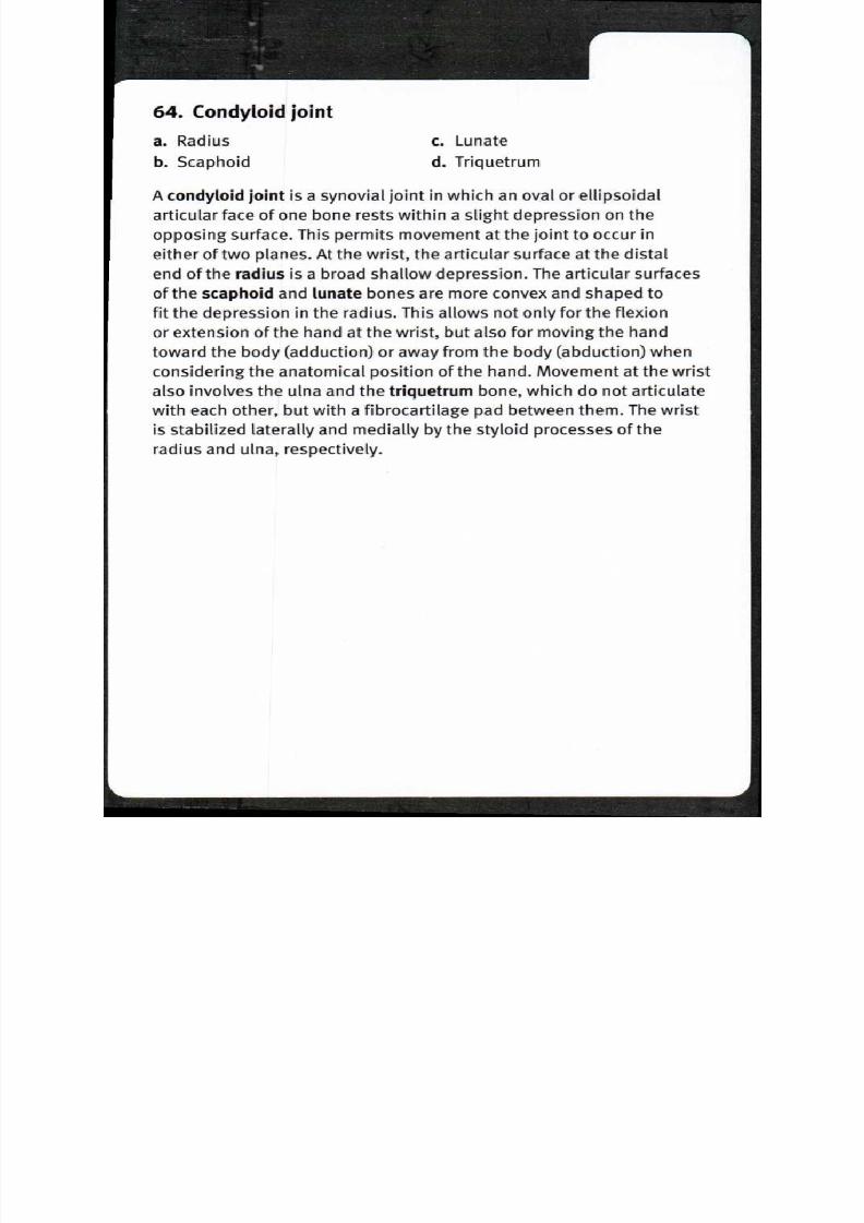
8/13/2019 113264016 Anatomia Omului
http://slidepdf.com/reader/full/113264016-anatomia-omului 128/298
64. Condyloid joint
a Radius . Lunate
b Scaphoid Triquetrum
A condyloid joint is a synovial joint in which an oval or ellipsoidal
articular face of one bone rests within a slight depression on the
opposing surface. This permits movement at the joint to occur in
either of two planes. At the wrist, the articular surface at the distalend of the radius is a broad shallow depression. The articular surfaces
of the scaphoid and lunate bones are more convex and shaped to
fit the depression in the radius. This allows not only for the flexion
or extension of the hand at the wrist, but also for moving the hand
toward the body (adduction) or away from the body (abduction) when
considering the anatomical position of the hand. Movement at the wrist
also involves the ulna and the triquetrum bone, which do not articulate
with each other, but with a fibrocartilage pad between them. The wrist
is stabilized laterally and medially by the styloid processes of the
radius and ulna, respectively.
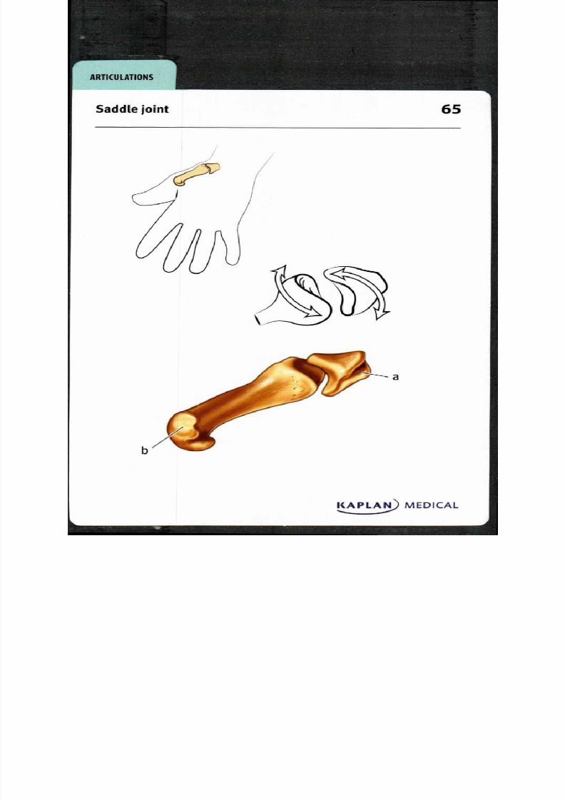
8/13/2019 113264016 Anatomia Omului
http://slidepdf.com/reader/full/113264016-anatomia-omului 129/298

8/13/2019 113264016 Anatomia Omului
http://slidepdf.com/reader/full/113264016-anatomia-omului 130/298
65. Saddle joint
a Trapezium
b First metacarpal
A saddle joint is a synovial joint where opposing articular faces are
convex along one axis and concave along the other. This allows angular
motion in a variety of directions, but prevents rotation. A saddle joint
exists in the carpometacarpal joint at the base of the thumb, at the
articulation between the trapezium and metacarpal. The range of
movements allowed at this joint include flexion and extension in the
plane of the palm, abduction and adduction in a plane at a right angle
to the palm, circumduction and opposition. It is the latter motion that
is important in the concept of the "opposable thumb," the evolution of
which is thought to have allowed humans to develop fine motor skillsand tool making capability.

8/13/2019 113264016 Anatomia Omului
http://slidepdf.com/reader/full/113264016-anatomia-omului 131/298
A R T I C U L A T I O N S
Temporomandibular joint, sagittai section 6

8/13/2019 113264016 Anatomia Omului
http://slidepdf.com/reader/full/113264016-anatomia-omului 132/298
66. Temporomandibular joint, sagittal section
a Zygomatic process of Articular disc
temporal bone Articular capsule
b Articular surface, mandibular . Head of mandible
fossa of temporal bone Mandible
The temporomandibular joint is a synovial joint with the unusualcharacteristic of having an articular disk dividing the articular capsule
into two parts. The lower joint compartment is formed between
the head of the mandible and the articular disk; movements are
rotational—opening and closing the jaw. As the jaw closes, the coronoid
process of the mandible slides into a cavity medial to the zygomatic
process of the temporal bone. The upper joint compartment is formed
between the mandibular fossa of the temporal bone and the articular
disk; movements here are translational—moving the jaw forwards and
backwards or side-to-side. The great mobility of the mandible enables
flexibility while chewing or talking, but also results in a joint that can
be easily dislocated by forceful forward or lateral displacement of the
mandible.
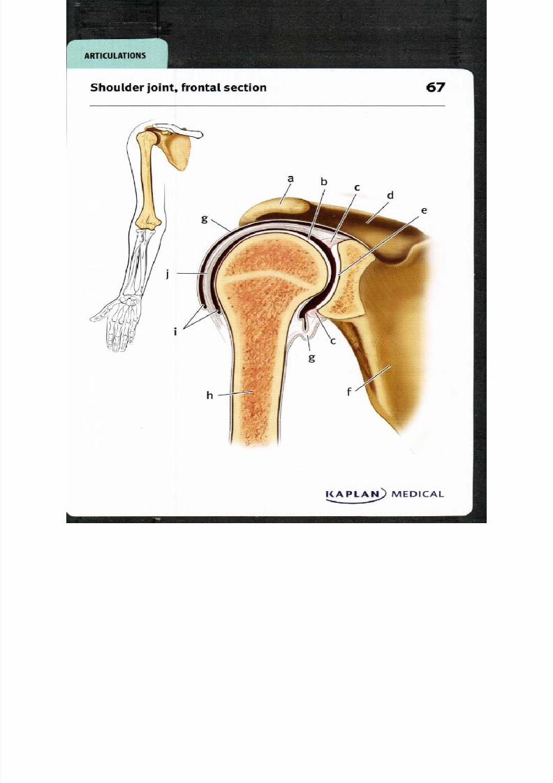
8/13/2019 113264016 Anatomia Omului
http://slidepdf.com/reader/full/113264016-anatomia-omului 133/298
A R T I C U L A T I O N S
Shoulder joint, frontal section 7
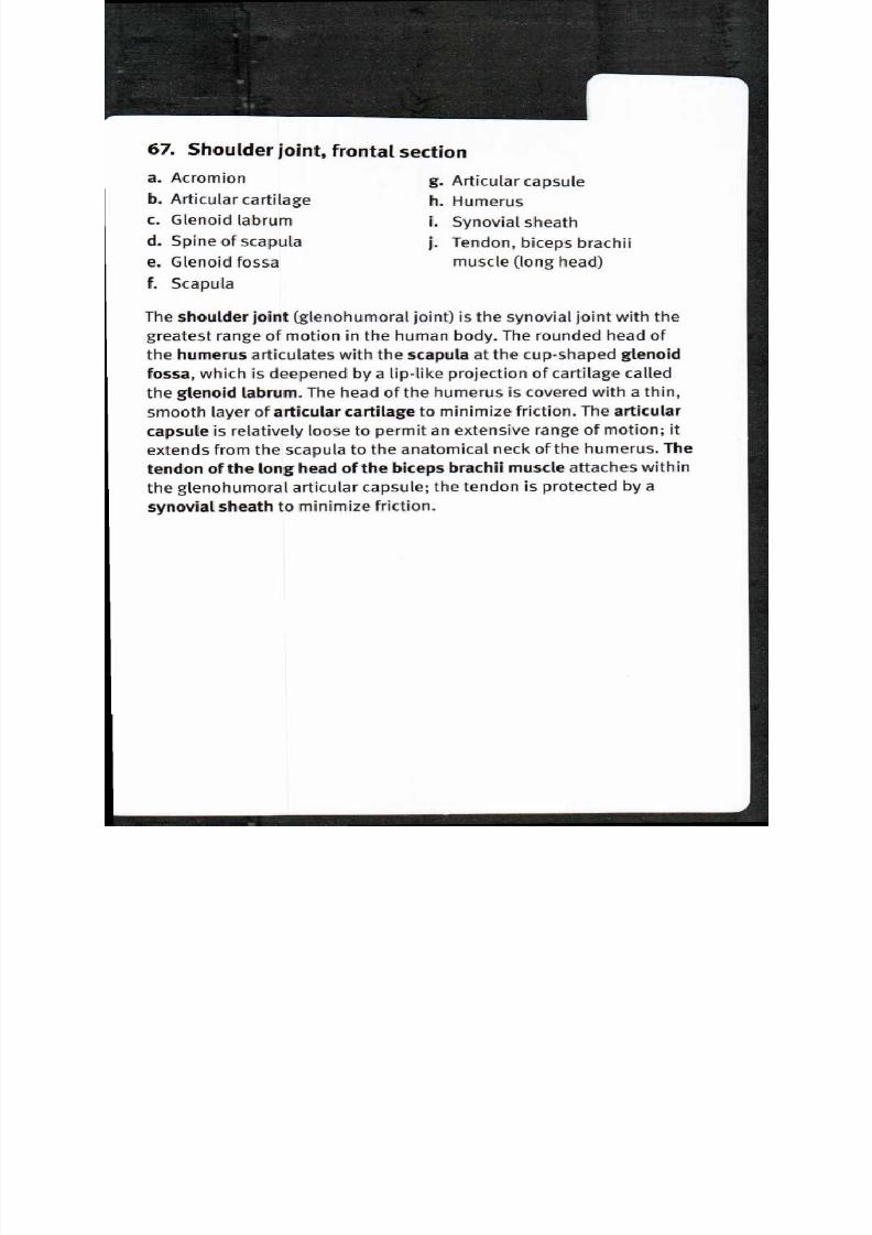
8/13/2019 113264016 Anatomia Omului
http://slidepdf.com/reader/full/113264016-anatomia-omului 134/298

8/13/2019 113264016 Anatomia Omului
http://slidepdf.com/reader/full/113264016-anatomia-omului 135/298
68
Isl MEDC L
R T I C U L T I O N S
Shoulder ligaments, anterior view

8/13/2019 113264016 Anatomia Omului
http://slidepdf.com/reader/full/113264016-anatomia-omului 136/298
68. Shoulder ligaments, anterior view
a Acromion
b Acromioclavicular ligament
c Coracoacromial ligament
d Coracoclavicular ligment,
trapezoid ligamente Coracoclavicular ligament,
conoid ligament
f Clavicle
g Coracohumeral ligament
h Transverse humeral ligament
i Humerus
j Articular capsulek Coracoid process of scapula
L Scapula
The shoulder joint is partly stabilized by the bones of the pectoral
girdle—particularly where the acromion and coracoid process of the
scapula extend laterally, superior to the head of the humerus. Another
measure of stability for the shoulder comes from ligaments. The
acromioclavicular, coracoacromial and coracoclavicular ligaments
connect the two processes of the scapula with the clavicle. The
coracohumoral ligament arises on the coracoid process and passes
across the joint to the greater tubercle of the humerus. The transverse
humoral ligament passes between the greater and lesser tubercles
of the humerus, forming a canal along the intertubercular groove
for the passage of the tendon of the biceps brachii. But by far, the
majority of the stability of the shoulder joint comes from the muscles
that move the humerus—especially the muscles collectively known as
the "rotator cuff", including the supraspinatus, the infraspinatus, the
subscapularis, and the teres minor.

8/13/2019 113264016 Anatomia Omului
http://slidepdf.com/reader/full/113264016-anatomia-omului 137/298
R T I C U L T I O N S
Elbow joint, sagittal section 9
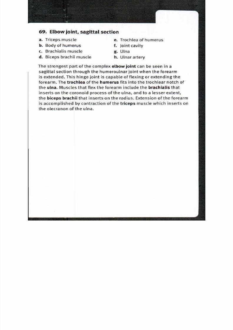
8/13/2019 113264016 Anatomia Omului
http://slidepdf.com/reader/full/113264016-anatomia-omului 138/298
69. Elbow joint, sagittal section
a Triceps muscle
b Body of humerus
c Brachialis muscle
d Biceps brachii muscle
e Trochlea of humerus
f Joint cavity
g Ulna
h Ulnar artery
The strongest part of the complex elbow joint can be seen in a
sagittal section through the humeroulnar joint when the forearm
is extended. This hinge joint is capable of flexing or extending the
forearm. The trochlea of the humerus fits into the trochlear notch of
the ulna. Muscles that flex the forearm include the brachialis that
inserts on the coronoid process of the ulna, and to a lesser extent,
the biceps brachii that inserts on the radius. Extension of the forearm
is accomplished by contraction of the triceps muscle which inserts on
the olecranon of the ulna.
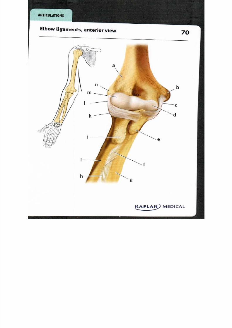
8/13/2019 113264016 Anatomia Omului
http://slidepdf.com/reader/full/113264016-anatomia-omului 139/298
KAPLA
R T IC U L T IO N S
Elbow ligaments anterior view 7

8/13/2019 113264016 Anatomia Omului
http://slidepdf.com/reader/full/113264016-anatomia-omului 140/298
70. Elbow ligaments, anterior view
a Humerus . Interosseous membraeb Medial epicondyle . Radius
c Ulnar collateral ligament Radial tuberosity
d Trochlea of humerus Radial annular ligament
e Ulnar tuberosity Radial collateral ligament
f Oblique cord Capitulum of humerus
g Ulna Lateral epicondyle
The elbow joint is very stable for several reasons: the humerus and
ulna interlock, the articular capsule is very thick, and several strong
ligaments reinforce the joint. The ulnar collateral ligament extends
from the medial epicondyle of the humerus anteriorly to the coronoid
processes of the ulna and posteriorly to the olecranon. The annular
ligament binds the head of the radius with the radial notch of the ulna.The radial collateral ligament extends from the lateral epicondyle
of the humerus to the annular ligament. Although the radioulnar
articulation allows rotational movement of the radius around the ulna,
the interosseous membrane allows only limited movement between
the two bones. The fibers of the oblique cord pass obliquely between
the bones just inferior to the ulnar and radial tuberosities.
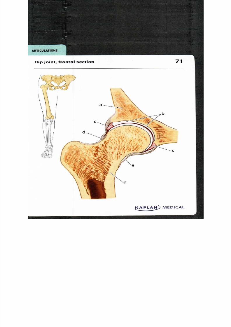
8/13/2019 113264016 Anatomia Omului
http://slidepdf.com/reader/full/113264016-anatomia-omului 141/298
71ip joint, frontal section
A R T I C U L A T I O N S

8/13/2019 113264016 Anatomia Omului
http://slidepdf.com/reader/full/113264016-anatomia-omului 142/298
71. Hip joint, frontal section
a Coxal bone Zona orbicularis of capsule
b Articular cartilage . Articular capsule
c Acetabular labrum Femur
The hip joint is a sturdy synovial joint (ball and socket joint) between
the globular head of the femur and the cup-like acetabulum of the
coxal bone. A thin layer of articular cartilage covers both bone surfaces
to reduce friction. A fibrocartilage lip called the acetabular labrum
extends the edge of the acetabulum to increase its depth; it is located
slightly beyond the widest diameter of the femoral head to hold it
firmly in place. The articular capsule is strong and dense; it completely
encloses the head and neck of the femur, and extends beyond the
edge of the acetabulum. The capsule includes longitudinal bands of
fibers that stretch between the hip bone and the femur to strengthen
the joint; the zona orbicularis of the capsule includes deep circular
fibers that form a collar to hold the head of the femur tightly in the
socket. The joint permits a range of movements: flexion and extension,
adduction and abduction, circumduction and rotation.

8/13/2019 113264016 Anatomia Omului
http://slidepdf.com/reader/full/113264016-anatomia-omului 143/298
R T I C U L T I O N S
Pelvic ligaments, posterior view 2
KAPLA MEDICAL

8/13/2019 113264016 Anatomia Omului
http://slidepdf.com/reader/full/113264016-anatomia-omului 144/298
72. Pelvic ligaments, posterior view
a Iliac crest . Femur
b Posterior superior iliac spine . Sacrospinous ligament
c Iliolumbar ligament Superficial dorsal
d Fifth lumbar vertebra acrococcygeal ligament
e Supraspinous ligament Sacrotuberous ligament
f Short dorsal sacroiliac Articular capsule of the
ligaments ip joint
g Long dorsal sacroiliac
ligament
Strong ligaments are necessary to stabilize the pelvis. The
supraspinous ligament that runs along the edges of the vertebral
dorsal spinous processes continues along the median sacral crest. In
addition, the iliolumbar ligament connects the fifth lumbar vertebra
both to the sacrum and the iliac crest. The sacroiliac joint is stabilized
by the horizontal short dorsal sacroiliac ligament between the sacrum
and the tuberosity of the ilium and by the oblique long dorsal sacroiliac
ligament connecting the sacrum to the posterior superior iliac spine.
The sacrotuberous ligament is connected at one end to the posterior
inferior iliac spine, the lower part of the sacrum, and the coccyx; the
other end attaches to the tuberosity and ramus of the ischium. Nearby,
the sacrospinous ligament extends from the sacrum and coccyx to the
spine of the ischium. The sacrococcygeal joint is stabilized by several
ligaments, including the superficial dorsal sacrococcygeal ligament.
At the hip joint, strong ligaments reinforce the articular capsule that
encloses the head and much of the neck of the femur.

8/13/2019 113264016 Anatomia Omului
http://slidepdf.com/reader/full/113264016-anatomia-omului 145/298
A R T I C U L A T I O N S
Knee joint, anterior view 3

8/13/2019 113264016 Anatomia Omului
http://slidepdf.com/reader/full/113264016-anatomia-omului 146/298
73. Kne e joint, anterior view
a Femur
b Patella
c Fibular collateral ligament
(lateral collateral ligament)
d Lateral meniscus
e Lateral condyle of tibia
f Fibula
g Tibia
h Tibial collateral ligament
(medial collateral ligament)
i Medial condyle of tibia
j Medial meniscus
k Patellar ligament
The articulation between the femur and tibia at the knee joint performs
a simple hinge function, primarily facilitating flexion and extension of
the lower leg. However, a small amount of medial and lateral rotation
(10° and 3o° respectively) is also possible. The medial and lateralcondyles of the femur articulate with the medial and lateral condyles
of the tibia; the medial and lateral menisci are fibrocartilage pads
that cushion and separate the bones within the joint. The patella is
held in place anterior to the distal portion of the femur by the patellar
ligament distally and the quadriceps muscle tendon proximally; it not
only protects the knee joint but also provides increased leverage for
the quadriceps muscle during knee extension. The patella resides at
its most superior location during full extension of the knee, and moves
as much as 7 cm inferiorly during flexion, until it is located between
the distal ends of the femoral condyles. To the side, the knee joint is
stabilized by the tibial medial) collateral ligament and the fibular
lateral) collateral ligament.
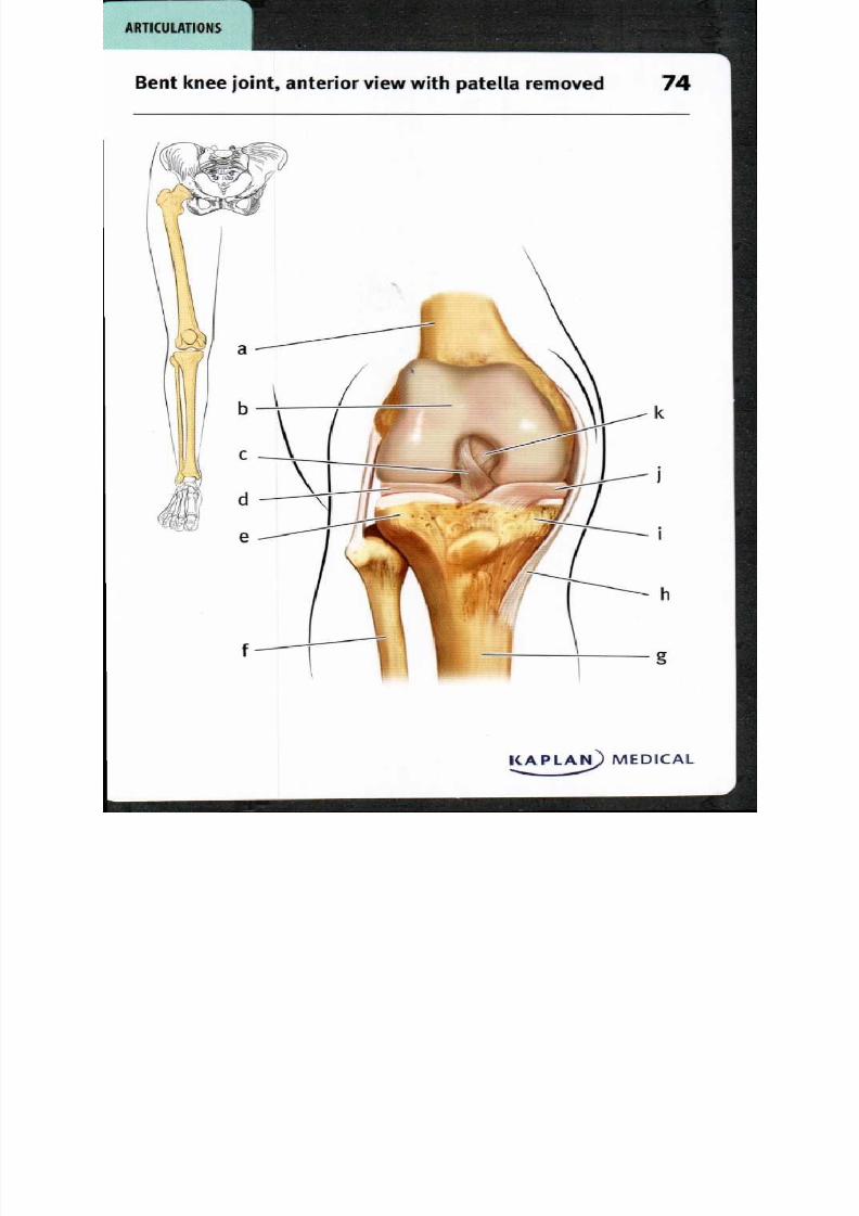
8/13/2019 113264016 Anatomia Omului
http://slidepdf.com/reader/full/113264016-anatomia-omului 147/298
KAPLAN MEDICAL
i
i
h
R T I C U L T I O N S
Bent knee joint, anterior view with patella removed 4 7

8/13/2019 113264016 Anatomia Omului
http://slidepdf.com/reader/full/113264016-anatomia-omului 148/298
74. Bent knee joint, anterior view with patella removed
a Femur
b Articular cartilage
c Anterior cruciate ligament
d Lateral meniscus
e Lateral condyle of tibia
1 Fibula
g Tibia
h Tibial collateral ligament
i Medial condyle of tibia
j Medial meniscus
k Posterior cruciate ligament
With the knee bent and the patella removed, the interior of the synovial
joint is revealed. The articular cartilage protecting the articular
surface of the femur extends superiorly behind the position of the
patella, as part of the femoropatellar joint. The two femoral condyles
are separated from the two tibial condyles by the medial and lateral
menisci. These pads of fibrocartilage fill in the space between the
convex surface of the femoral condyle and the flatter surface of the
tibial condyle; they act as durable shock absorbers and contributeto both stability and lubrication in the joint. Ligaments stabilize the
joint; the anterior cruciate ligament (ACL) extends between the lateral
condyle of the femur posteriorly and the intercondylar region of the
tibia anteriorly; the posterior cruciate ligament (PCL) connects the
posterior intercondylar region of the tibia with the medial condyle of
the femur anteriorly. The ACL resists forces pushing the tibia forward,
while the PCL resists forces pushing the tibia posteriorly relative to the
femur. Excessive abduction or adduction motion at the knee joint is
limited by the fibular (lateral) and tibial medial) collateral ligaments.

8/13/2019 113264016 Anatomia Omului
http://slidepdf.com/reader/full/113264016-anatomia-omului 149/298
R T I C U L T I O N S
Knee joint, sagittal section 5
h

8/13/2019 113264016 Anatomia Omului
http://slidepdf.com/reader/full/113264016-anatomia-omului 150/298
75. Knee joint, sagittal section
a Femur . Lateral meniscus, anterior
b Lateral menniscus, orn
posterior horn . Infrapatellar fat pad
c Fibula Patella
d Tibia . Articular cartilage
e Patellar ligament
A sagittal section through the lateral condyles of the femur and tibia
reveals the anterior-posterior relationships of the fully-extended
knee joint. Articular cartilage covers the convex articular surface
of the femur and the flatter articular surface of the tibia. Between
the two articular cartilages lies the C-shaped lateral meniscus, with
posterior and anterior horns positioned to fill the area between the
curving articular surfaces. Popliteal ligaments strengthen the back
of the synovial articular capsule. The fibula articulates with the tibial
epicondyle laterally and posteriorly; a collateral ligament extends
from the fibula to the lateral epicondyle of the femur to strengthen the
joint. Anteriorly, the patella moves along the femoral articular surface
when the quadriceps muscle flexes the knee; the articular cartilage
of the patella is one of the thickest due to the intense stresses of this
movement. The quadriceps muscle tendon attaches on the superior
surface and is continuous with the inferior patellar ligament. The
infrapatellar fat pad absorbs shocks and fills in the space below and
behind the patella.
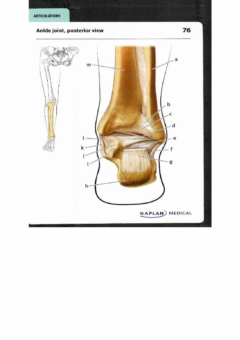
8/13/2019 113264016 Anatomia Omului
http://slidepdf.com/reader/full/113264016-anatomia-omului 151/298
I
A R T I C U L A T I O N S
k
i
Ankle joint, posterior view 6
KAPLAN MEDICAL. ...

8/13/2019 113264016 Anatomia Omului
http://slidepdf.com/reader/full/113264016-anatomia-omului 152/298
76. Ankle joint, posterior view
a Fibula
b Posterior tibiofibular
ligament
c Transverse tibiofibular
ligament
d Posterior talofibular
ligament
e Posterior talocalcaneal
ligament
f Calcaneofibular ligament
g Calcaneal (Achilles) tendon
h Calcaneus
i Medial talocalcaneal
ligament
I Talus
k Deltoid ligament,
tibiocalcaneal part
I Deltoid ligament, tibiotalar
part
m Tibia
The posterior view of the ankle joint shows several ligaments that
stabilize the articulations between the leg and ankle bones. The
largest tendon in the human body is the calcaneal tendon (also known
as the Achilles tendon) that connects three lower leg muscles—the
gastrocnemius, the soleus, and the plantaris—with their insertion on
the calcaneus, the largest of the tarsal bones. Above the ankle, the
distal articulation of the tibia and fibula are stabilized by the posteriorand transverse tibiofibular ligaments. Laterally, the ankle joint is
stabilized by connections between the lateral malleolus of the fibula
and the tarsal bones, including the talofibular and calcaneofibular
ligaments. On the medial side, the ankle is stabilized by ligaments
connecting the medial malleolus of the tibia with tarsal bones such as
the calcaneus and the talus, including the tibiocalcaneal and tibiotalar
parts of the deltoid ligament. Articulations between the tarsal bones
are also stabilized by ligaments, such as the medial and posterior
talocalcaneal ligaments connecting the talus and calcaneus.
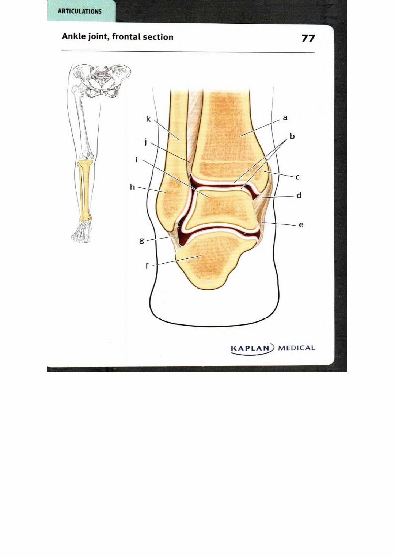
8/13/2019 113264016 Anatomia Omului
http://slidepdf.com/reader/full/113264016-anatomia-omului 153/298
c
d
e
A R T I C U L A T I O N S
Ankle joint, frontal section 7
KAPLAN MEDICAL
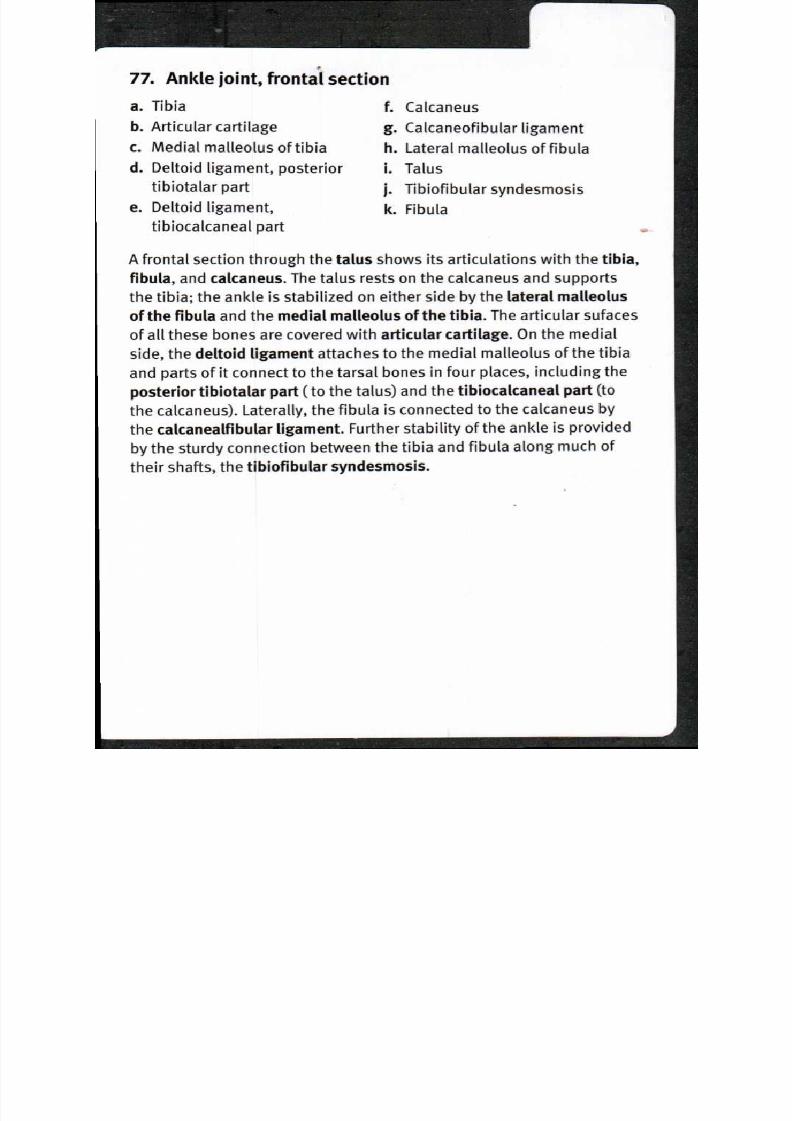
8/13/2019 113264016 Anatomia Omului
http://slidepdf.com/reader/full/113264016-anatomia-omului 154/298
77. Ankle joint, frontal section
a Tibia
b Articular cartilage
c Medial malleolus of tibia
d Deltoid ligament, posterior
tibiotalar part
e Deltoid ligament,
tibiocalcaneal part
f Calcaneus
g Calcaneofibular ligament
h Lateral malleolus of fibula
i Talus
j Tibiofibular syndesmosis
k Fibula
A frontal section through the talus shows its articulations with the tibia,
fibula, and calcaneus. The talus rests on the calcaneus and supports
the tibia; the ankle is stabilized on either side by the lateral malleolus
of the fibula and the medial malleolus of the tibia. The articular sufaces
of all these bones are covered with articular cartilage. On the medial
side, the deltoid ligament attaches to the medial malleolus of the tibia
and parts of it connect to the tarsal bones in four places, including theposterior tibiotalar part (to the talus) and the tibiocalcaneal part (to
the calcaneus). Laterally, the fibula is connected to the calcaneus by
the calcanealfibular ligament. Further stability of the ankle is provided
by the sturdy connection between the tibia and fibula along much of
their shafts, the tibiofibular syndesmosis.

8/13/2019 113264016 Anatomia Omului
http://slidepdf.com/reader/full/113264016-anatomia-omului 155/298
Superficial muscles of the body, anterior view

8/13/2019 113264016 Anatomia Omului
http://slidepdf.com/reader/full/113264016-anatomia-omului 156/298
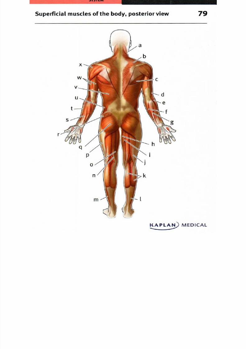
8/13/2019 113264016 Anatomia Omului
http://slidepdf.com/reader/full/113264016-anatomia-omului 157/298
Superficial muscles of the body posterior view 9
KAPLA MEDICAL

8/13/2019 113264016 Anatomia Omului
http://slidepdf.com/reader/full/113264016-anatomia-omului 158/298
79. Superficial muscles of the body, posterior view
a. Sternocleidomastoid . Soleus
b. Trapezius . Semitendinosus
c. Teres Major . Semimembranosus
d. Brachioradialis . Iliotibial tract
e. Extensor carpi radialis longus q . Gluteus maximus
f. Flexor carpi ulnaris . Gluteus medius
g. Extensor carpi ulnaris . Extensor digitorum
h. Gracilis . Extensor carpi radialis brevis
I Adductor magnus . External abdominal oblique
j. Biceps femoris . Latissimus dorsi
k. Gastrocnemius . Triceps
L Calcaneal tendon . Deltoid
More than 600 muscles carry out the actions of moving body parts;
in many cases, their names are descriptive regarding the location,
origin, insertion, action, shape, etc. Thus, the two trapezius muscles
together form a trapezoid shape between the neck, shoulders, and
thoracic vertebrae; they act to shrug the shoulders and to pull the
shoulder blades toward the vertebrae. The flexor carpi ulnaris and
extensor carpi u lnaris are two muscles that act to flex or extend
the wrist (carpus); they originate on the ulna. The triceps muscle isnamed because it has three heads that originate on either the scapula
or the humerus; the muscle has a single tendon that inserts on the
olecranon of the ulna so that it acts to extend the forearm. The gluteal
muscles in the butt are named for their size—the gluteus maximus
is larger than the gluteus medius or the deeper gluteus minimus.
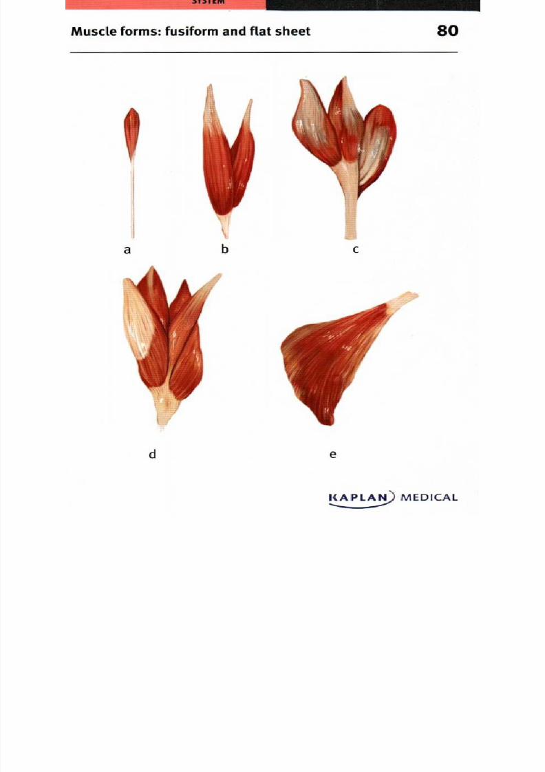
8/13/2019 113264016 Anatomia Omului
http://slidepdf.com/reader/full/113264016-anatomia-omului 159/298
M uscle forms : fusiform an d flat sheet 0
d
KAPLAN) MEDICAL

8/13/2019 113264016 Anatomia Omului
http://slidepdf.com/reader/full/113264016-anatomia-omului 160/298
80. M uscle form s: fusiform an d f lat sheet
Fusiform: road, flat sheet:
a. Basic fusiform, Palm aris . Latissimus dorsi
longus
b. Bicipital, Biceps fem oris
c. Tricipital, Triceps surae
(gastrocnemius and soleus)
d. Quadriceps femoris
Fusiform muscles are wide in the middle and taper at both ends. The
patmaris longus is a slender, fusiform muscle that originates on
the humerus and ends in a long tendon that inserts on the palmar
fascia. A bicipital muscle has two heads or origins, as the biceps
femoris with the long head originating on the ischium and the shorthead on the femur; the muscle inserts at the knee and both heads
act to flex the knee. The triceps surae is a tricipital muscle with
three heads; this composite muscle consists of the gastrocnemius
with two heads originating on the femur and the soleus which
originates on the tibia; the triceps surae inserts on the calcaneus
and acts in plantar flexion of the foot. The quadriceps femoris is a
quadricipital muscle with four heads originating on the ilium and
femur; the muscle inserts on the patella and extends the knee. Other
muscles occur in broad, flat sheets, such as the latissimus dorsi
which is a triangular, flat muscle that originates along the thoracic
and lumbar vertebrae as well as the sacrum and ilium; it inserts on
the humerus and acts to extend, adduct and rotate the shoulder.
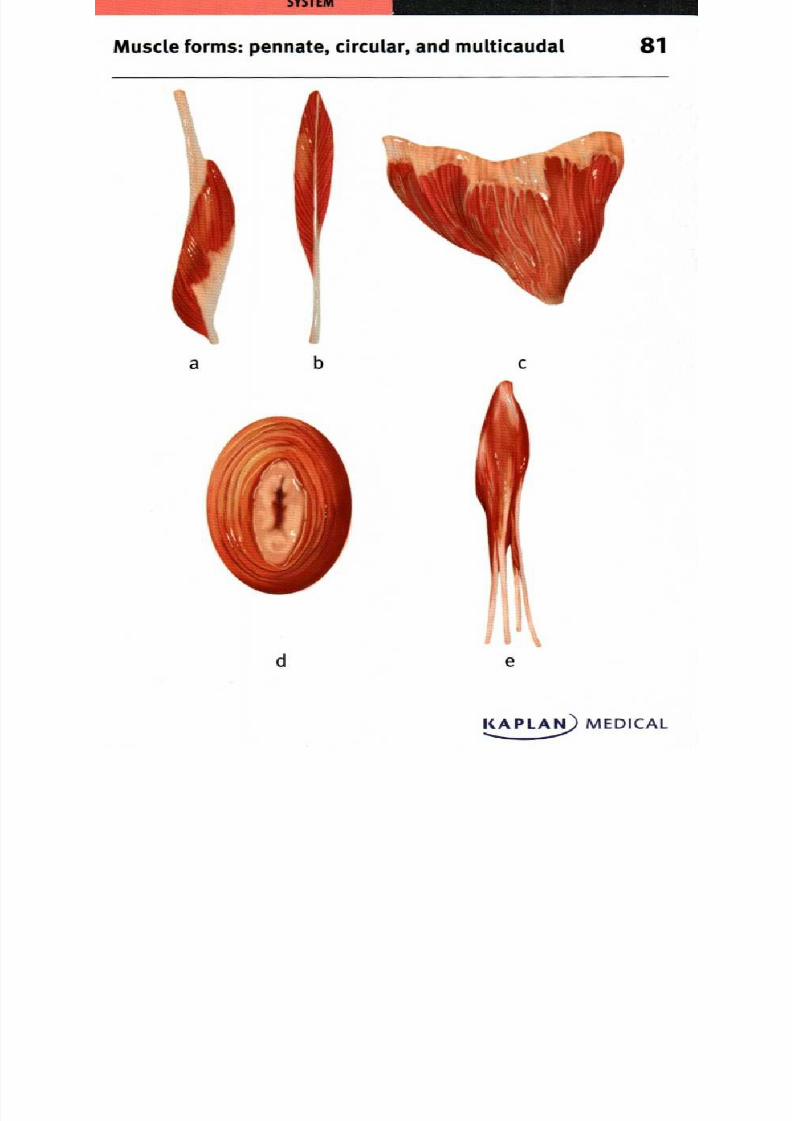
8/13/2019 113264016 Anatomia Omului
http://slidepdf.com/reader/full/113264016-anatomia-omului 161/298
Muscle forms: pennate, circular, and multicaudal 1
a b c
d e
KAPLAN MEDICAL
8/13/2019 113264016 Anatomia Omului
http://slidepdf.com/reader/full/113264016-anatomia-omului 162/298
81. Muscle forms: pennate, circular, and multicaudal
Pennate:
a. Unipennate,
semimembranosus
b. Bipennate, tibialis anterior
c. Multipennate, deltoid
Circular:
d. External sphincter
ani, deep portion
Multicaudal:
e. Flexor digitorum profundus
In pennate muscles, the contracting fibers attach to the tendon at an
oblique angle; they provide more stability and force, but the tendon
is not moved as far as when the fibers are parallel to the tendon. All
the muscle fibers are on the same side of the tendon in unipennate
muscles, such as the semimembranosus muscle which extends
between the ischium and the tibia. Bipennate muscles are more
common and have muscle fibers on both sides of the tendon, including
the tibialis anterior which is a shin muscle that flexes the foot. In
multipennate muscles, the fibers attach on both sides of the tendon,
and the tendon branches within the muscle; an example is the deltoid
muscle that forms the rounded shape of the shoulder. Circular, or
sphincter, muscles are arranged concentrically around an opening; a
good example is the deep portion of the external sphincter ani muscle
which closes off the anal canal at its orifice. Multicaudal musclesattach at multiple sites; the flexor digitorum profundus is a single
muscle that inserts on the phalanges of the four fingers to flex them.

8/13/2019 113264016 Anatomia Omului
http://slidepdf.com/reader/full/113264016-anatomia-omului 163/298
S Y S T E M
Muscle forms: cylindrical, triangular, 2quadrilateral, biventral, multiventral
a
d
KAPLAN) MEDICAL
8/13/2019 113264016 Anatomia Omului
http://slidepdf.com/reader/full/113264016-anatomia-omului 164/298
82. Muscle forms: cylindrical, triangular, quadrilateral,
biventral, m ultiven tral
a. Cylindrical teres major . Biventral, digastric
b. Triangular deltoid . Multiventral, rectus
c. Quadrilateral, pronator bdominis
quadratus
Named from the Latin word for round or cylindrical the teres major
muscle extends between the scapula and humerus; it adducts
and rotates the arm. The triangular deltoid muscle covers the
shoulder joint, extending from the clavicle and scapula to the
humerus; different parts of the muscle act to flex, extend or rotate
the humerus. The pronator quadratus muscle is a square, or
quadrilateral, muscle that extends between the ulna and radius at
their distal end; it acts to pronate the forearm. The digastric muscleconsists of two bellies with different origins that unite in a single
tendon that inserts on the hyoid bone; the longer, posterior belly
originates on the mastoid process of the temporal bone, while the
anterior belly arises on the mandible. The rectus abdominis muscle
has multiple insertions; it arises on the pubis bone and three
portions of the muscle insert on the fifth, sixth and seventh ribs.
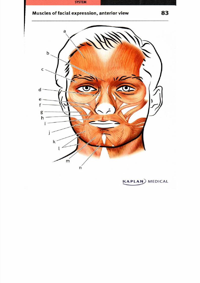
8/13/2019 113264016 Anatomia Omului
http://slidepdf.com/reader/full/113264016-anatomia-omului 165/298
M uscles of facial expression anterior view
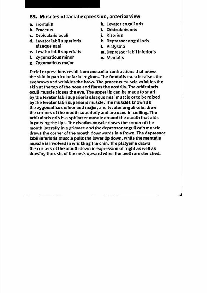
8/13/2019 113264016 Anatomia Omului
http://slidepdf.com/reader/full/113264016-anatomia-omului 166/298
83. Muscles of facial expression, anterior view
a. Frontalis . Levator anguli oris
b.Procerus . Orbicularis oris
c. Orbicularis oculi . Risorius
d. Levator labii superioris . Depressor anguli oris
alaeque nasi Platysma
e. Levator labii superioris . Depressor labii inferioris
f. Zygomaticus minor . Mentalis
g. Zygomaticus major
Facial expressions result from muscular contractions that move
the skin in particular facial regions. The frontalis muscle raises the
eyebrows and wrinkles the brow. The procerus muscle wrinkles the
skin at the top of the nose and flares the nostrils. The orbicularis
oculi muscle closes the eye. The upper lip can be made to snarl
by the levator [Ail superioris alaeque nasi muscle or to be raised
by the levator [Ail superioris muscle. The muscles known as
the zygomaticus minor and major, and levator anguli oris, draw
the corners of the mouth superiorly and are used in smiling. The
orbicularis oris is a sphincter muscle around the mouth that aids
in pursing the lips. The risorius muscle draws the corner of the
mouth laterally in a grimace and the depressor anguli oris muscledraws the corner of the mouth downwards in a frown. The depressor
labii inferioris muscle pulls the lower lip down, while the mentalis
muscle is involved in wrinkling the chin. The platysma draws
the corners of the mouth down in expression of fright as well as
drawing the skin of the neck upward when the teeth are clenched.

8/13/2019 113264016 Anatomia Omului
http://slidepdf.com/reader/full/113264016-anatomia-omului 167/298
Muscles of facial expression, lateral view 4
KAPLAN) MEDICAL
8/13/2019 113264016 Anatomia Omului
http://slidepdf.com/reader/full/113264016-anatomia-omului 168/298
84. Muscles of facial expression, lateral view
a. Frontalis part,
occipitofrontalis
b. Orbicularis oculi
c. Procerus
d. Nasalis
e Levator labii superioris
alaeque nasi
1. Levator labii superioris
g. Orbicularis oris
h. Depressor labii inferioris
i. Depressor anguli oris
j. Risorius
k. Platysma
1. Zygomaticus major
m . Zygomaticus minor
n. Zygomatic arch
Muscles controlling facial expression often originate on bones
and insert on the skin of the face. The frontalis portion of the
occipitofrontalis muscle covers the forehead from above the hairline
to the eyebrows. The orbicularis oculi muscle arises on the frontal
bone and circles the eye to the temple and the cheek. The procerusmuscle extends from the nasal bone to the skin of the medial
forehead, while the nasalis muscle extends from the maxilla to the
nasal bone and compresses the nasal cartilage. The levator labii
superioris alaeque nasi extends from the maxilla to the upper lip
and the levator labii superioris muscle arises on the margin of the

8/13/2019 113264016 Anatomia Omului
http://slidepdf.com/reader/full/113264016-anatomia-omului 169/298

8/13/2019 113264016 Anatomia Omului
http://slidepdf.com/reader/full/113264016-anatomia-omului 170/298
85. Superficial muscles of mastication, lateral view
a. Temporalis . Buccinator
b. Zygomatic arch Mandible
c. O rbicularis oris . Masseter
M astication, or chewing, involves the jaw open ing and closing,
accom plished by m uscles that m ove the m andible at the
tem perom andibular joint. The tem poralis m uscle originates on the
tem poral bone, passes m edial to the zygom atic arch and inserts
on the m andible on the anterior and m edial aspects of the coronoidprocess. T he m asseter m uscle arises on the zygom atic arch, passes
lateral to the broad surface of the m andible, and inserts along the
angle and lower part of the ramu s of the man dible. T he temp oralis
and m asseter both elevate and retract the m andible. T he buccinator
m uscle is a qu adrilateral facial m uscle located between the m axilla
and m andible. It inserts on the orbicularis oris m uscle and a cts to
flatten the cheek against the teeth, acting as an auxiliary m astication
m uscle holding the food betw een the teeth during m astication.
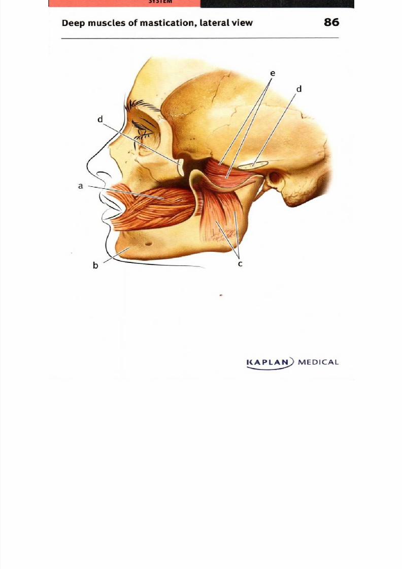
8/13/2019 113264016 Anatomia Omului
http://slidepdf.com/reader/full/113264016-anatomia-omului 171/298
Deep muscles of mastication, lateral view 6
-
KAPLAN) MEDICAL

8/13/2019 113264016 Anatomia Omului
http://slidepdf.com/reader/full/113264016-anatomia-omului 172/298
86. Deep muscles of mastication, lateral view
a. Buccinator muscle Zygomatic arch (cut)
b. Mandible . Lateral pterygoid musclec. Medial pterygoid muscle
The deep muscles of mastication are attached on the medial aspect
of the mandible. During mastication, the jaw is opened by action of
the lateral pterygoid muscle. The lateral pterygoid originates on the
sphenoid bone and inserts on the coronoid process of the mandible
and on the articular disc of the temperomandibular joint. The lateral
pterygoids act both to lower the mandible and to bring it forward.
Closing of the jaw during chewing involves elevation of the mandible
by actions of the masseter, temporalis, and medial pterygoid muscles.
The medial pterygoid has two heads, one originating on the sphenoid
bone and the other from the palatine bone. The muscle inserts on the
medial side of the ramus of the mandible to elevate it. The medial
pterygoid and masseter work together, respectively, on the medial and
lateral aspects of the mandible to raise it. Both the medial and lateral
pterygoid muscles can also act to move the mandible side-to-side.

8/13/2019 113264016 Anatomia Omului
http://slidepdf.com/reader/full/113264016-anatomia-omului 173/298
M uscles of the neck anterior view 7
KAPLAN MEDIC L

8/13/2019 113264016 Anatomia Omului
http://slidepdf.com/reader/full/113264016-anatomia-omului 174/298
87. Muscles of the neck, anterior view
a. Hyoid bone
b. Superior belly of omohyoid
muscle
c. Inferior belly of om ohyoid
muscle
d. Scapula
e. Manubrium of sternum
f. Clavicle
g. Sternocleidomastoid muscle
h. Sternohyoid muscle
i. Mastoid process of temporal
bone
Muscles in the neck control the position of the larynx, contribute
to mastication and respiration, and support activities of the tongueand pharynx. The inferior belly of the omohyoid muscle arises on
the scapula, runs parallel to the clavicle while being held in position
by connective tissue, and the tendon then turns superiorly where it
becomes the superior belly. The superior belly of the omohyoid inserts
on the hyoid bone and acts to depress the larynx. The stemohyoid
muscle also acts to depress the larynx; it originates on the manubriumof the sternum and inserts on the hyoid. The two heads of the
stemocleidomastoid muscle arise on the sternum and clavicle and
manubrium of the sternum; the muscle inserts on the mastoid process
of the temporal bone. Acting separately, the sternocleidomastoids
on each side rotate the head; acting together, they flex the neck
and assist in respiration along with the scalene muscles.

8/13/2019 113264016 Anatomia Omului
http://slidepdf.com/reader/full/113264016-anatomia-omului 175/298
88uprahyoid and infrahyoid m usclesof the nec k anterior view
KAPLA MEDICAL••
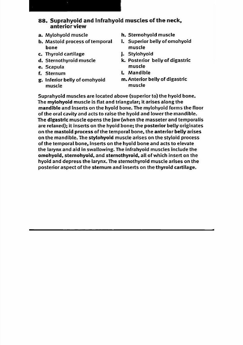
8/13/2019 113264016 Anatomia Omului
http://slidepdf.com/reader/full/113264016-anatomia-omului 176/298
88. Suprahyoid and infrahyoid muscles of the neck,
anterior v iew
a. Mylohyoid muscle . Sternohyoid muscleb. Mastoid process of temporal . Superior belly of omohyoid
bone uscle
c. Thyroid cartilage . Stylohyoid
d. Sternothyroid muscle . Posterior belly of digastric
e. Scapula uscle
1 Sternum Mandibleg. Inferior belly of omohyoid . Anterior belly of digastric
muscle uscle
Suprahyoid muscles are located above (superior to) the hyoid bone.
The mylohyoid muscle is flat and triangular; it arises along the
mandible and inserts on the hyoid bone. The mylohyoid forms the floor
of the oral cavity and acts to raise the hyoid and lower the mandible.
The digastric muscle opens the jaw (when the masseter and temporalis
are relaxed); it inserts on the hyoid bone; the posterior belly originates
on the mastoid process of the temporal bone, the anterior belly arises
on the mandible. The stylohyoid muscle arises on the styloid process
of the temporal bone, inserts on the hyoid bone and acts to elevate
the larynx and aid in swallowing. The infrahyoid muscles include the
omohyoid, stemohyoid, and stemothyroid, all of which insert on the
hyoid and depress the larynx. The sternothyroid muscle arises on the
posterior aspect of the sternum and inserts on the thyroid cartilage.
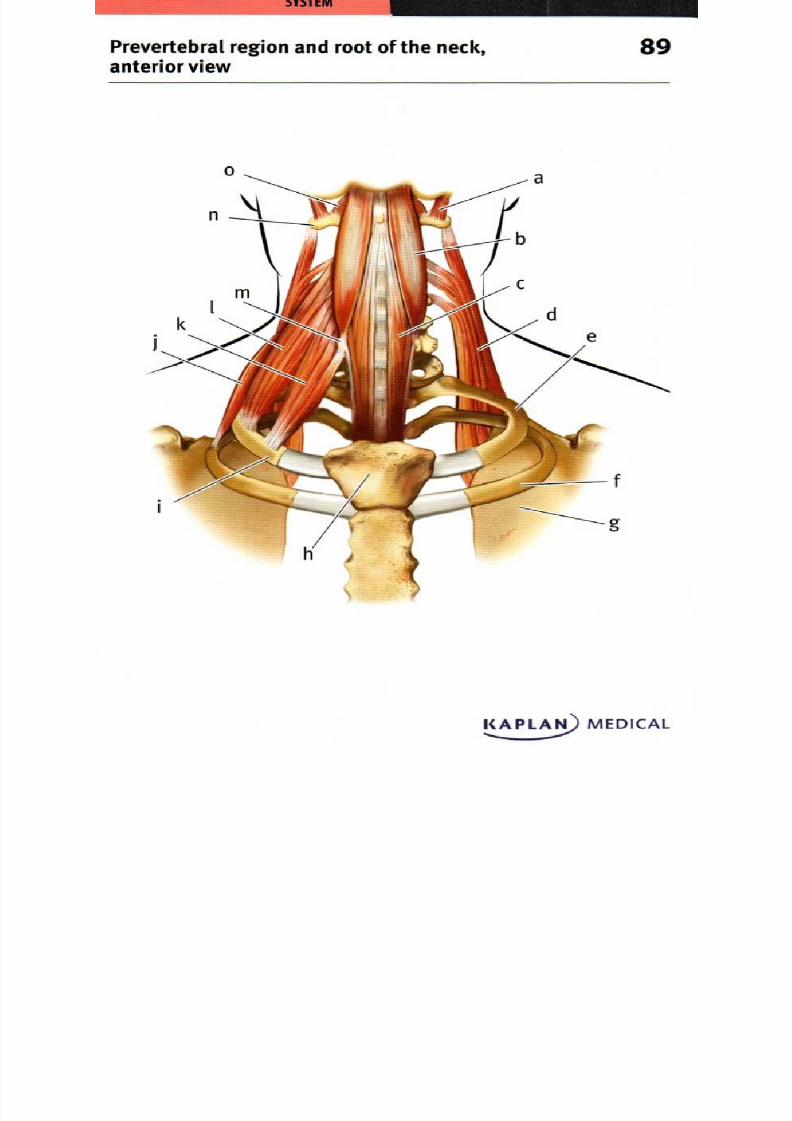
8/13/2019 113264016 Anatomia Omului
http://slidepdf.com/reader/full/113264016-anatomia-omului 177/298
Prevertebral region and root of the neck 9anterior view
KAPLAN) MEDICAL

8/13/2019 113264016 Anatomia Omului
http://slidepdf.com/reader/full/113264016-anatomia-omului 178/298
89. Pr evertebr al region an d root of the n eck an terior view
a. Rectus capitis lateralis
muscle
b. Longus capitis muscle
c. Longus colli muscle
d. Levator scapulae muscle
e. Left rib
1 Second rib
g. Scapula
h. Sternum
i. Right rib
j. Posterior scalene muscle
k. Middle scalene muscle
1 Anterior scalene muscle
m . Carotid tubercle of sixth
cervical vertebra
n. Transverse process of atlas
o. Rectus capitis anterior muscle
The scalene muscles of the neck arise on the transverse processes
of the cervical vertebrae and insert on the first two ribs; they act to
rotate the neck and to assist in respiration. The anterior scalene
muscle originates on C3-6 and inserts on the first rib; the middle
scalene originates on C2-7 and also inserts on the first rib. The
posterior scalene muscle arises on C4-6 and inserts on the second
rib. The neck is flexed at the joint between the atlas and the occipital
bone by several muscles that arise on the vertebrae and insert
on the occipital bone. The longus capitus muscle arises from the
transverse processes of C3-6, and inserts on the occipital bone toflex the neck. The small rectus capitis anterior and lateralis muscles
both originate on the atlas Ci) and insert on the occipital bone to
flex the atlanto-occipital joint. The longus colli muscle originates
both on the transverse processes of C3-7 and the vertebral body
ofTi-3, inserts on C1-4, and acts to flex and rotate the neck. The
levator scapulae muscle arises on C1-4 and inserts on the scapula
to either raise the scapula or incline the neck toward that side.

8/13/2019 113264016 Anatomia Omului
http://slidepdf.com/reader/full/113264016-anatomia-omului 179/298
Muscles of the neck, lateral view 90
KAPLAN)MEDICAL

8/13/2019 113264016 Anatomia Omului
http://slidepdf.com/reader/full/113264016-anatomia-omului 180/298
90. Muscles of the neck, lateral view
a. Hyoid bone
b. Superior belly of omohyoidmuscle
c. Sternothyroid muscle
d. Sternocleidomastoid muscle
e. Inferior belly of om ohyoid
muscle
1 Clavicle
g. First rib
h. Sternum
I Occipital bone
j. Trapezius muscle
k. Levator scapulae muscle
I Middle scalene muscle
m . Anterior scalene muscle
n. Scapula
The trapezius is a broad, superficial muscle that originates along
the dorsal midline, from the occipital bone, ligaments along the
cervical vertebrae, or thoracic vertebrae. The muscle inserts on the
acromion process and spine of the scapula as well as part of the
clavicle. Its actions may include extension of the neck, elevation of
the clavicle, or a variety of movements of the scapula. The levator
scapulae extends between the cervical vertebrae and the scapula;
it also elevates the scapula. The stemocleidomastoid muscle
extends from the sternum and clavicle to the temporal bone and
acts to flex or rotate the neck. The omohyoid and stemothyroid
muscles arise on the scapula or sternum, respectively, and act
to depress both the hyoid bone and the larynx. The anterior andmiddle scalene muscles originate on the cervical vertebrae and
insert on the first rib; they rotate the neck and aid in respiration.
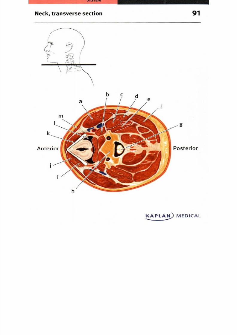
8/13/2019 113264016 Anatomia Omului
http://slidepdf.com/reader/full/113264016-anatomia-omului 181/298
91eck, transverse section
KAPLAN) MEDICAL......
8/13/2019 113264016 Anatomia Omului
http://slidepdf.com/reader/full/113264016-anatomia-omului 182/298
91. Neck, transverse section
a. Sternocleidomastoid muscle . Body of C5 vertebrab. Anterior scalene muscle . Sternothyroid muscle
c. Middle scalene muscle . Sternohyoid muscle
d. Posterior scalene muscle . Thyroid cartilage
e. Levator scapulae muscle Pharynx
1 Trapezius muscle . Platysma
g. Spinal cord
A transverse section of the neck shows the central spinal cord lying
posterior to the body of the C5 vertebra, and the anterior pharynx
lying behind the larynx and its thyroid cartilage. The superficial
platysma is the most anterior of the muscles; lying between it and the
thyroid cartilage are the stemothyroid and stemohyoid muscles that
depress the larynx. Anteriolaterally, the stemocleidomastoid muscle
extends from the sternum and clavicle to the mastoid process. Deep
to this, the anterior, m iddle, and p osterior scalene m uscles extend
from the cervical vertebrae to the first or second rib and act to flex the
neck. Alongside the scalene m uscles, the levator scapu lae m uscle
acts to lift the scapu la or bend the neck . O n the posterior aspect
of the neck , the broad, flat trapezius m uscle is m ost superficial.
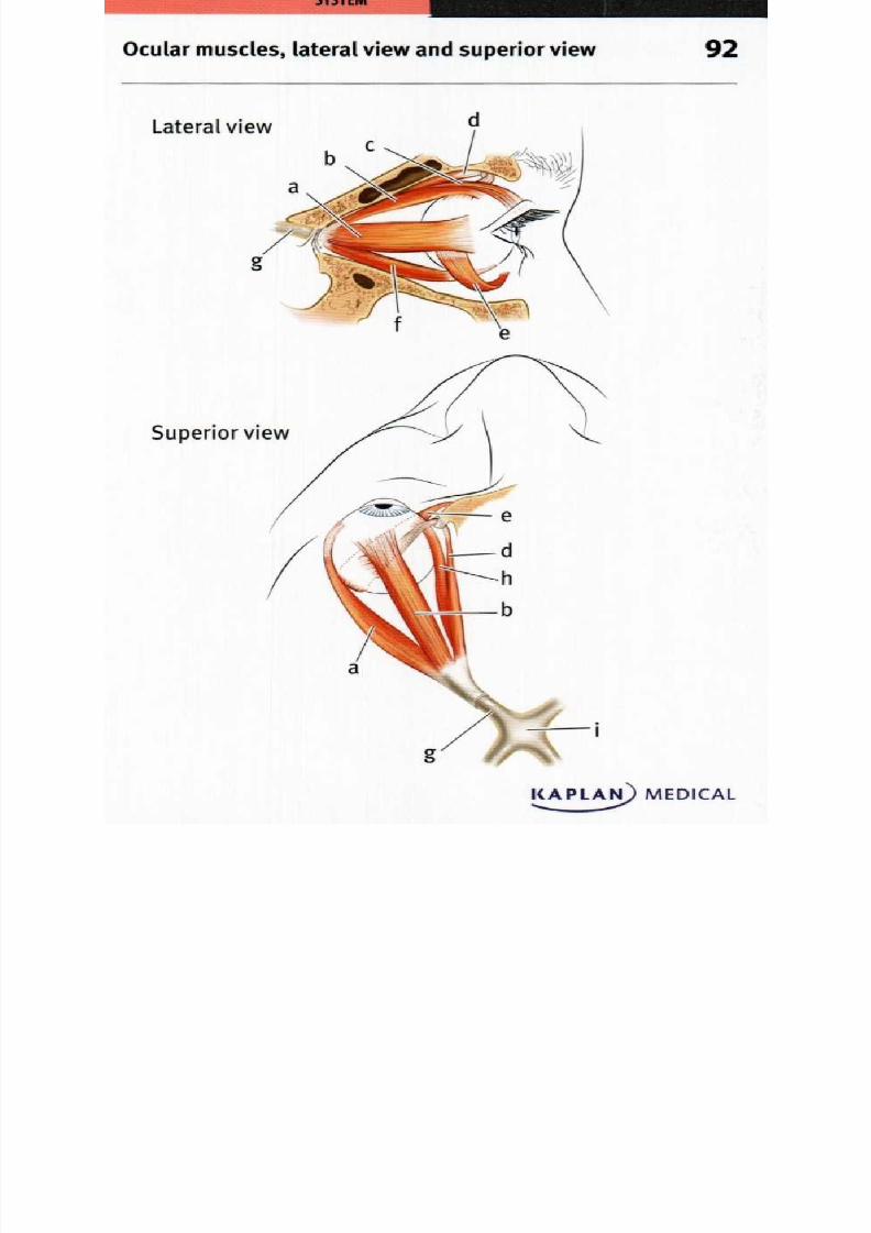
8/13/2019 113264016 Anatomia Omului
http://slidepdf.com/reader/full/113264016-anatomia-omului 183/298
Lateral view
Superior view
i
KAPLAN) MEDICAL......
Ocular muscles, lateral view and superior view 2

8/13/2019 113264016 Anatomia Omului
http://slidepdf.com/reader/full/113264016-anatomia-omului 184/298
92 Ocular muscles, lateral view and superior view
a. Lateral rectus muscle . Inferior oblique muscle
b. Superior rectus muscle . Inferior rectus muscle
c. Levator palpebrae superioris . Optic nerve
muscle Medial rectus muscle
d. Superior oblique muscle . Optic chiasma
The extrinsic eye muscles work together to produce movements of
the eyeball. Four rectus muscles originate on the sphenoid bone
near the optic nerve, and insert on the surface of the eyeball. They
are the superior rectus muscle to move the eyeball to look up, the
inferior rectus muscle to move the eyeball to look down, the medial
rectus muscle to rotate the eyeball medially, and the lateral rectus
muscle to rotate the eyeball laterally. The superior oblique muscle
primarily rotates the eye medially, while the inferior oblique laterally
rotates it; in addition, actions of the oblique muscles include
depression and abduction, or elevation and abduction, respectively.
The levator palpabrae superioris muscle extends from the sphenoid
bone to the eyelid and acts to elevate and retract the eyelid.

8/13/2019 113264016 Anatomia Omului
http://slidepdf.com/reader/full/113264016-anatomia-omului 185/298
Intrinsic muscles of the tongue, sagittal section 3
KAPLAMEDICAL--.
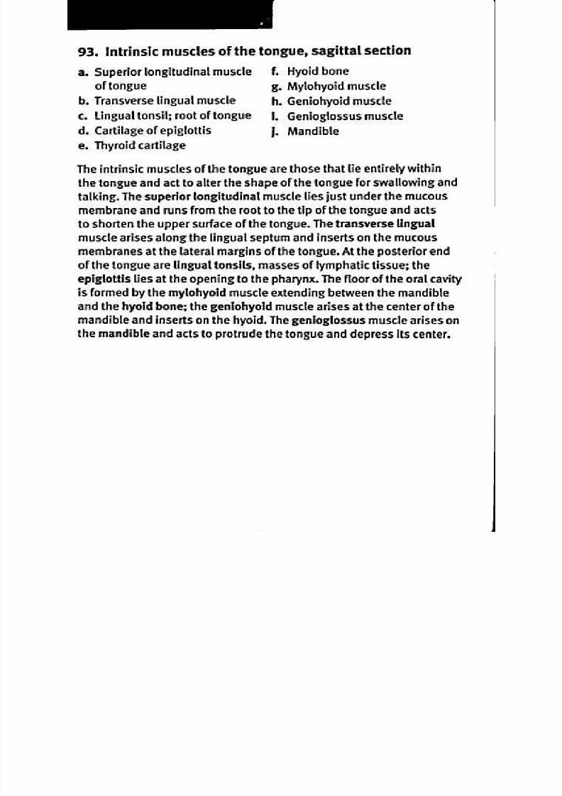
8/13/2019 113264016 Anatomia Omului
http://slidepdf.com/reader/full/113264016-anatomia-omului 186/298
93. Intrinsic muscles of the tongue, sagittal section
a. Superior longitudinal muscle Hyoid bone
of tongue . M ylohyoid m uscleb. Transverse lingual muscle . Geniohyoid muscle
c. Lingual tonsil; root of tongue . Genioglossus muscle
d. Cartilage of epiglottis Mandible
e. Thyroid cartilage
The intrinsic muscles of the tongue are those that lie entirely withinthe tongue and act to alter the shape of the tongue for swallowing and
talking. The superior longitudinal muscle lies just under the mucous
membrane and runs from the root to the tip of the tongue and acts
to shorten the upper surface of the tongue. The transverse lingual
muscle arises along the lingual septum and inserts on the mucous
membranes at the lateral margins of the tongue. At the posterior end
of the tongue are lingual tonsils, masses of lymphatic tissue; the
epiglottis lies at the opening to the pharynx. The floor of the oral cavity
is formed by the mylohyoid muscle extending between the mandible
and the hyoid bone; the geniohyoid muscle arises at the center of the
mandible and inserts on the hyoid. The genioglossus muscle arises on
the mandible and acts to protrude the tongue and depress its center.
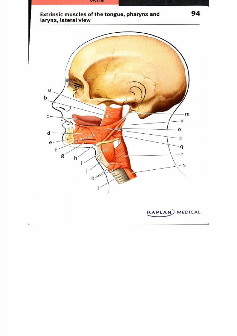
8/13/2019 113264016 Anatomia Omului
http://slidepdf.com/reader/full/113264016-anatomia-omului 187/298
Extrinsic muscles of the tongue, pharynx and
larynx, lateral view
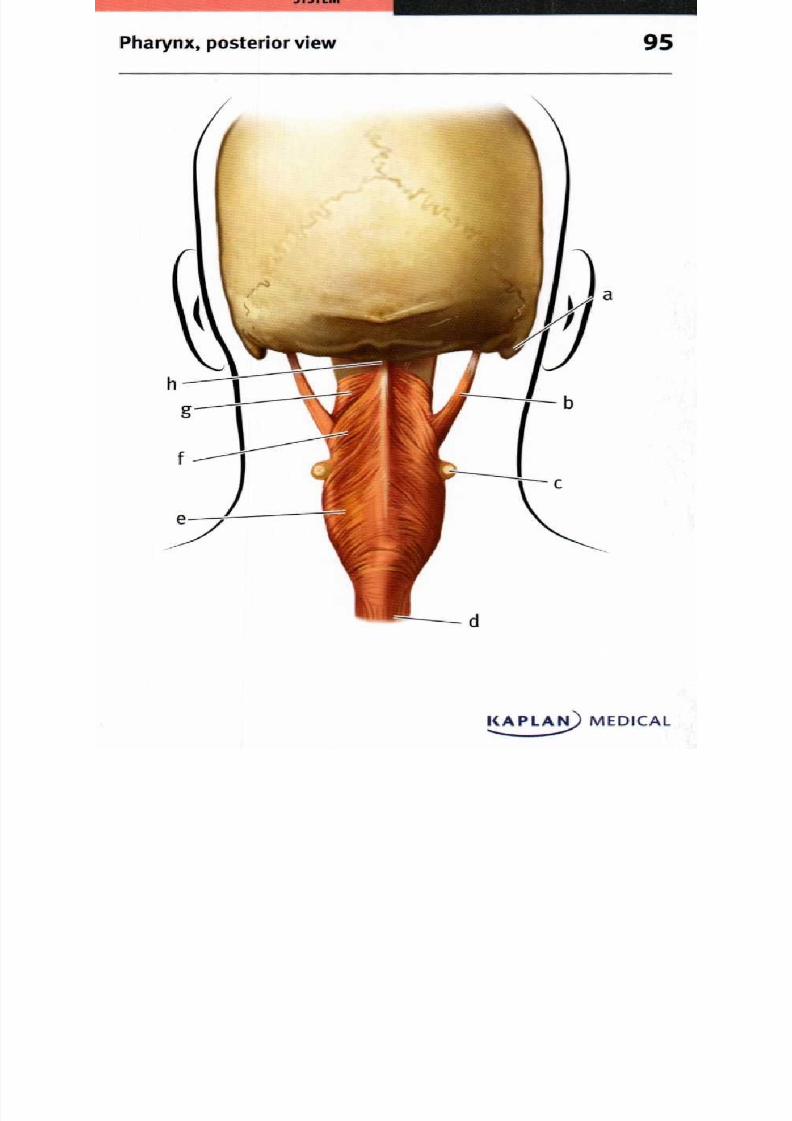
8/13/2019 113264016 Anatomia Omului
http://slidepdf.com/reader/full/113264016-anatomia-omului 188/298
Pha rynx pos terior view 5
KAPLA) MEDICAL

8/13/2019 113264016 Anatomia Omului
http://slidepdf.com/reader/full/113264016-anatomia-omului 189/298
Superficial shoulder muscles, anterior view

8/13/2019 113264016 Anatomia Omului
http://slidepdf.com/reader/full/113264016-anatomia-omului 190/298
S Y S T E M
Muscles of the shoulder, scapula and arm, 7anterior view
CAPLAN) MEDICAL

8/13/2019 113264016 Anatomia Omului
http://slidepdf.com/reader/full/113264016-anatomia-omului 191/298
Deep m uscles of the shou lder and arm anterior view 8
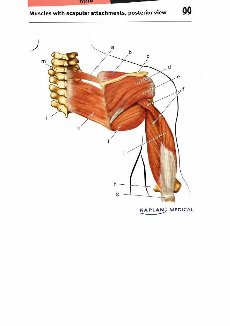
8/13/2019 113264016 Anatomia Omului
http://slidepdf.com/reader/full/113264016-anatomia-omului 192/298
M uscles w ith scapu lar attachm ents posterior view
I APLA) MEDICAL....
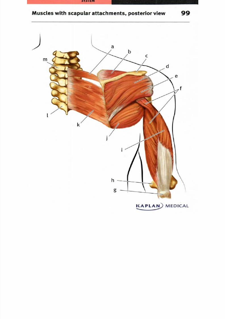
8/13/2019 113264016 Anatomia Omului
http://slidepdf.com/reader/full/113264016-anatomia-omului 193/298
Muscles with scapular attachments, posterior view 9
KAPLA) MEDICAL

8/13/2019 113264016 Anatomia Omului
http://slidepdf.com/reader/full/113264016-anatomia-omului 194/298

8/13/2019 113264016 Anatomia Omului
http://slidepdf.com/reader/full/113264016-anatomia-omului 195/298
Deep muscles of the back posterior view 1
KAPLAN MEDIC L
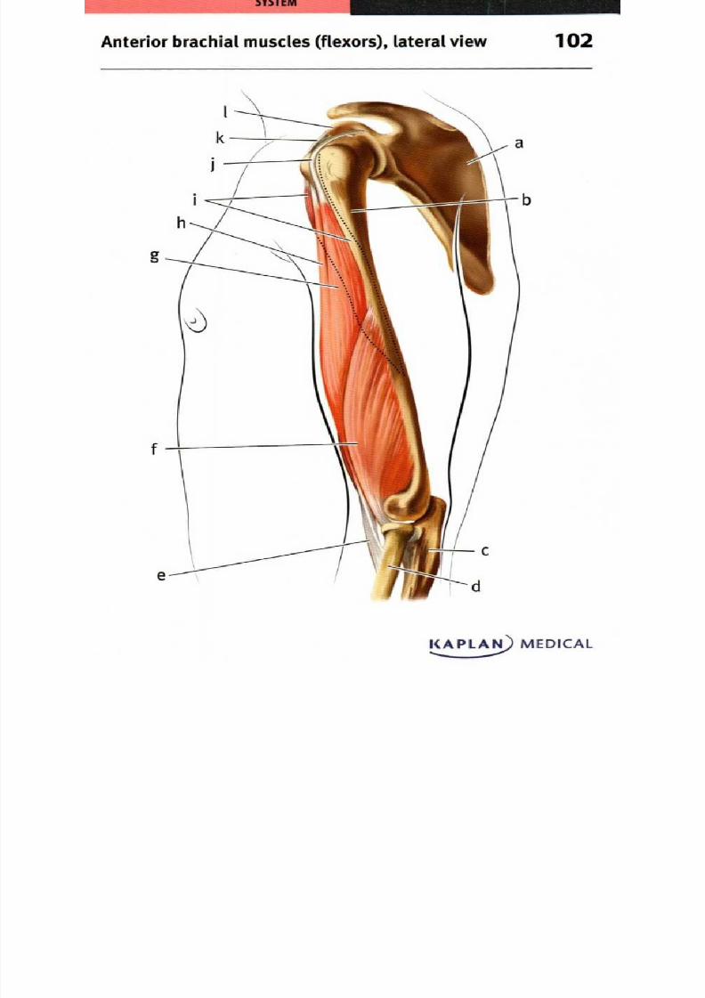
8/13/2019 113264016 Anatomia Omului
http://slidepdf.com/reader/full/113264016-anatomia-omului 196/298
Anterior brachial muscles (flexors), lateral view
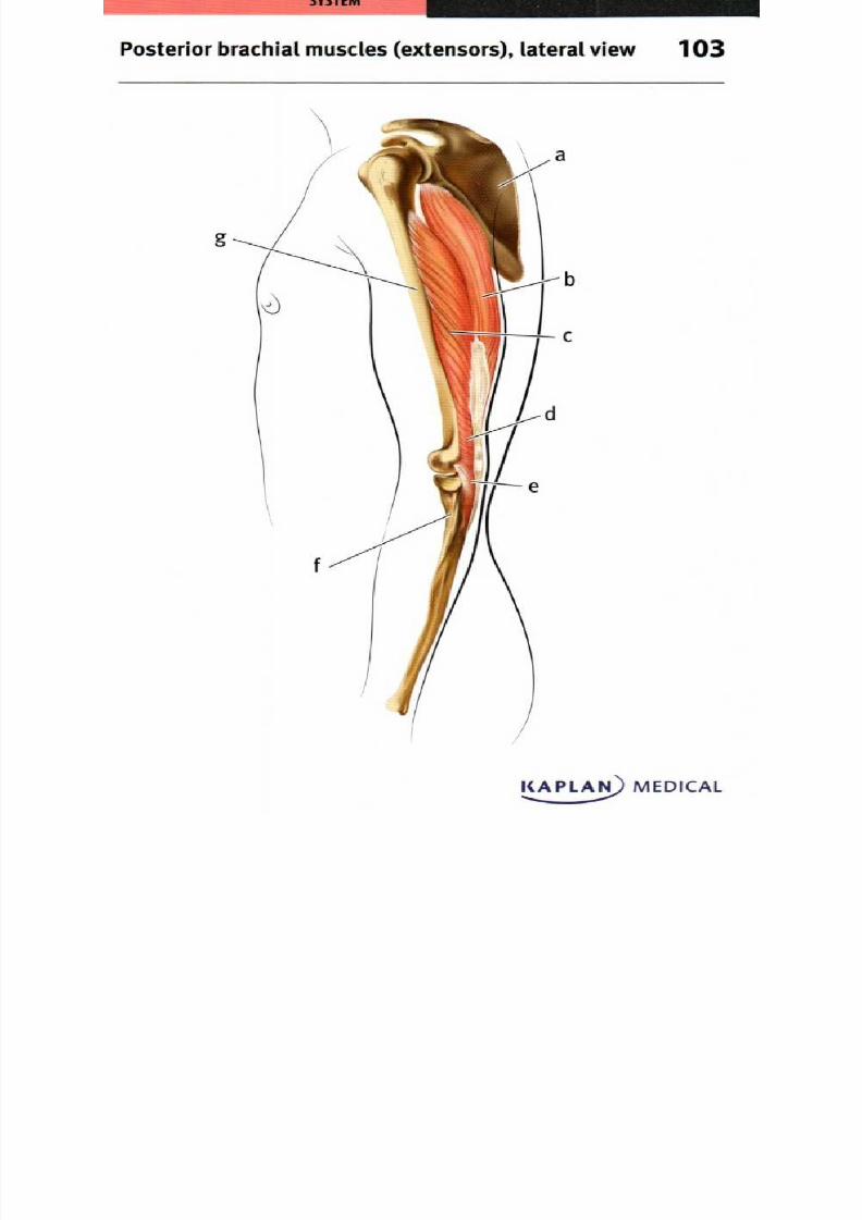
8/13/2019 113264016 Anatomia Omului
http://slidepdf.com/reader/full/113264016-anatomia-omului 197/298
Posterior brachial muscles (extensors), lateral view 03
KAPLAii) MEDICAL..
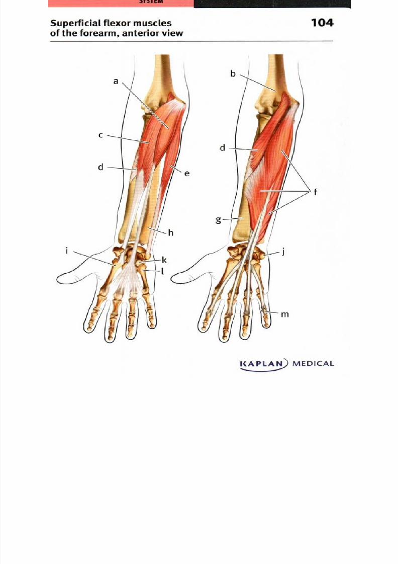
8/13/2019 113264016 Anatomia Omului
http://slidepdf.com/reader/full/113264016-anatomia-omului 198/298
104uperficial flexor muscles
f the forearm, anterior view
KAPLAN) MEDICAL
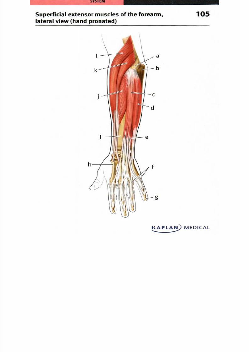
8/13/2019 113264016 Anatomia Omului
http://slidepdf.com/reader/full/113264016-anatomia-omului 199/298
Superficial extensor muscles of the forearm, 05lateral view hand pronated)
ICAl...._1 1 MEDICAL
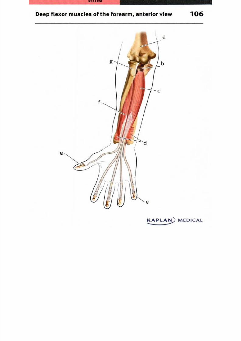
8/13/2019 113264016 Anatomia Omului
http://slidepdf.com/reader/full/113264016-anatomia-omului 200/298
Deep flexor muscles of the forearm, anterior view 06
KAPLAJ) MEDICAL
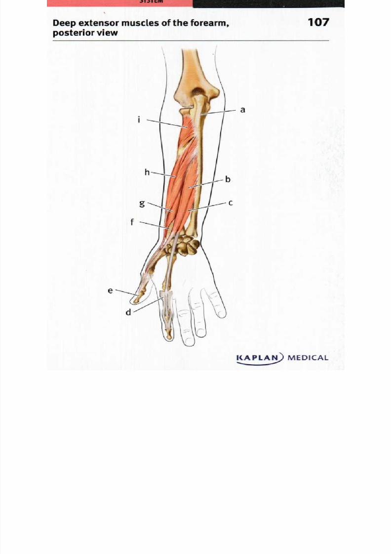
8/13/2019 113264016 Anatomia Omului
http://slidepdf.com/reader/full/113264016-anatomia-omului 201/298
Deep extensor muscles of the forearm, 07posterior view
I(APLAN MEDICAL

8/13/2019 113264016 Anatomia Omului
http://slidepdf.com/reader/full/113264016-anatomia-omului 202/298
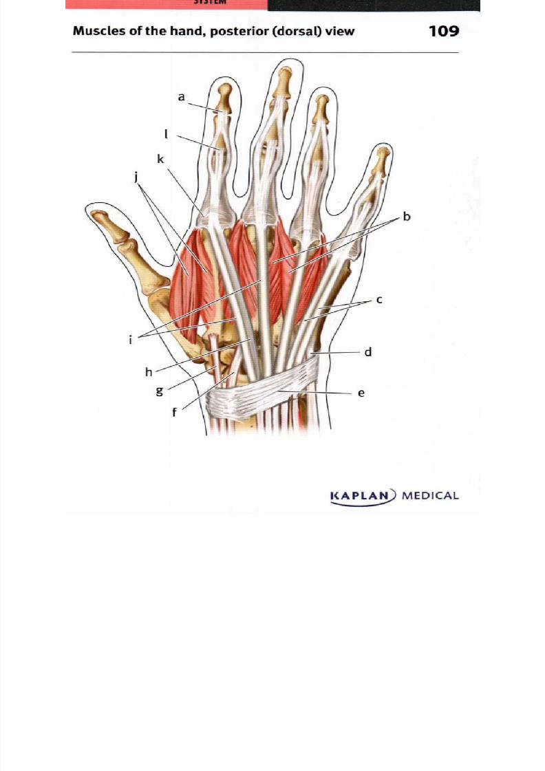
8/13/2019 113264016 Anatomia Omului
http://slidepdf.com/reader/full/113264016-anatomia-omului 203/298
Muscles of the hand, posterior dorsal) view 9
I_ NMEDCL
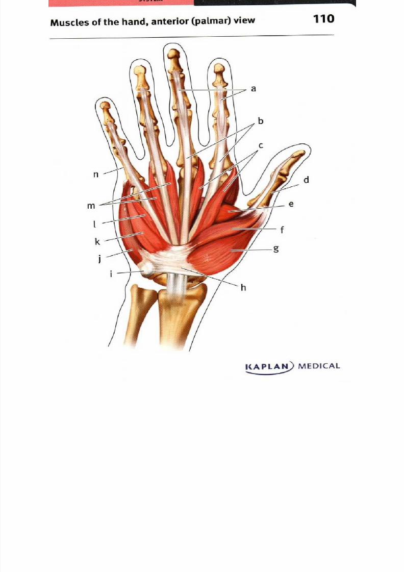
8/13/2019 113264016 Anatomia Omului
http://slidepdf.com/reader/full/113264016-anatomia-omului 204/298
uscles of the hand, anterior palmar) view 1
KAPLA MEDIC L

8/13/2019 113264016 Anatomia Omului
http://slidepdf.com/reader/full/113264016-anatomia-omului 205/298
Intercostal muscles, anterior view 11
KAPLAN) MEDICAL. ...

8/13/2019 113264016 Anatomia Omului
http://slidepdf.com/reader/full/113264016-anatomia-omului 206/298
Diaphragm anterior view 12
K PL MEDIC L
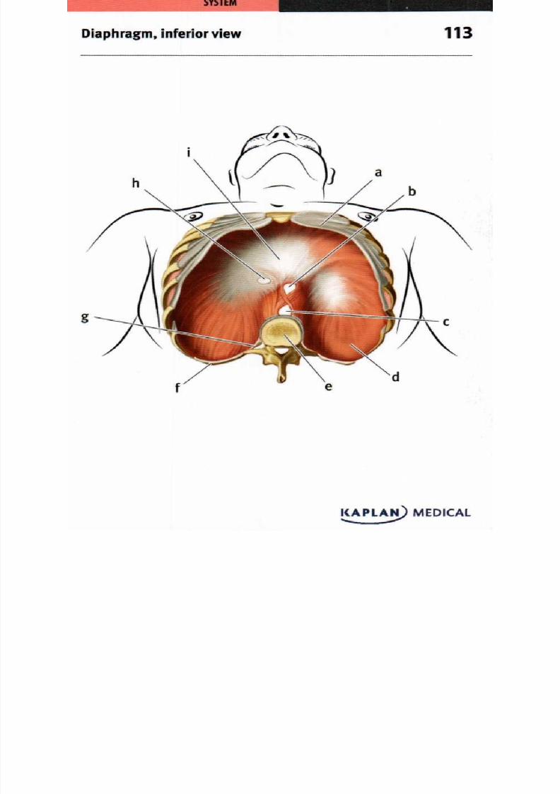
8/13/2019 113264016 Anatomia Omului
http://slidepdf.com/reader/full/113264016-anatomia-omului 207/298

8/13/2019 113264016 Anatomia Omului
http://slidepdf.com/reader/full/113264016-anatomia-omului 208/298
Superficial abdominal muscles, anterior view 14
KAPLAI I MEDICAL

8/13/2019 113264016 Anatomia Omului
http://slidepdf.com/reader/full/113264016-anatomia-omului 209/298
Dee p abdo m inal m uscles anterior view 5
KAPLAN) MEDICAL....
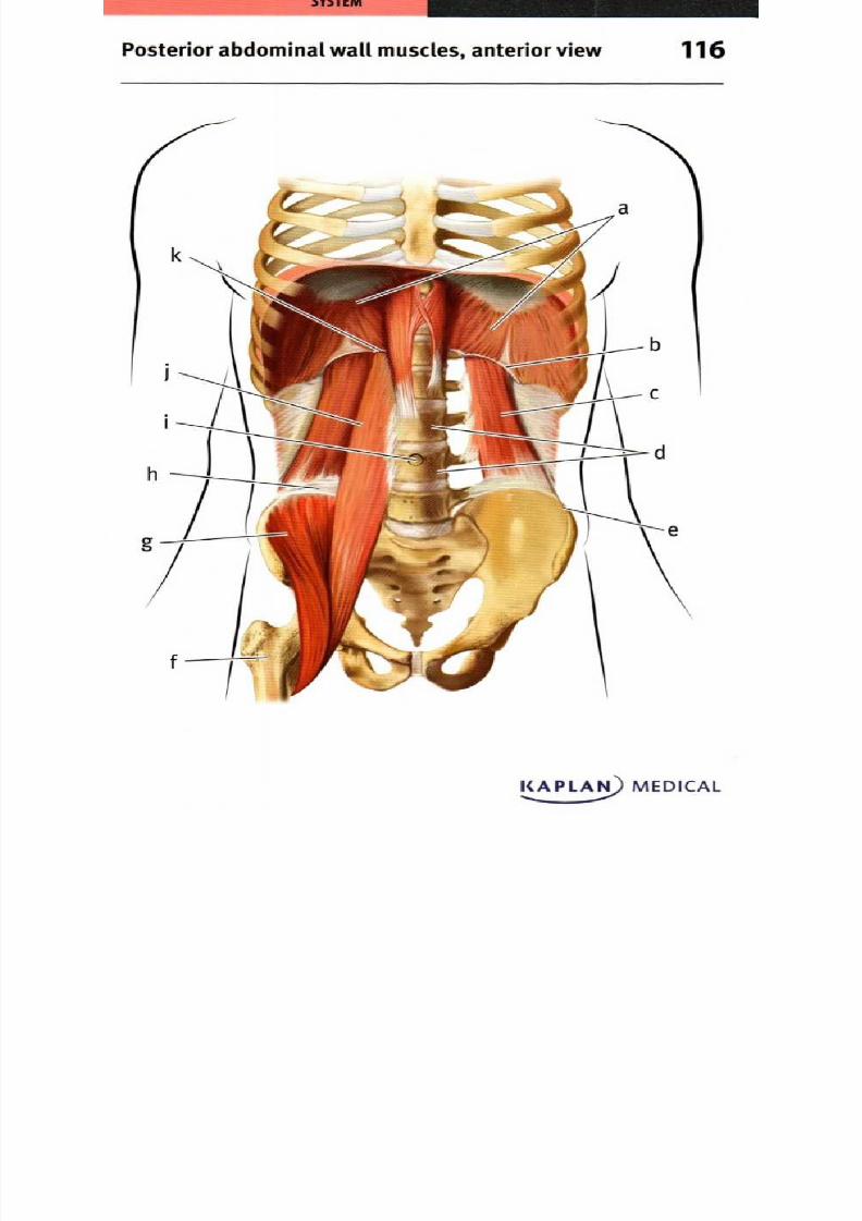
8/13/2019 113264016 Anatomia Omului
http://slidepdf.com/reader/full/113264016-anatomia-omului 210/298
Posterior abdominal wall muscles, anterior view 16
KAPLAN) MEDICAL

8/13/2019 113264016 Anatomia Omului
http://slidepdf.com/reader/full/113264016-anatomia-omului 211/298
b
Pelvic diaphragm, superior view 17
KAPLAts) MEDICAL

8/13/2019 113264016 Anatomia Omului
http://slidepdf.com/reader/full/113264016-anatomia-omului 212/298
Perineal muscles, inferior view 1 8
Male
h
KAPLAN) MEDICAL
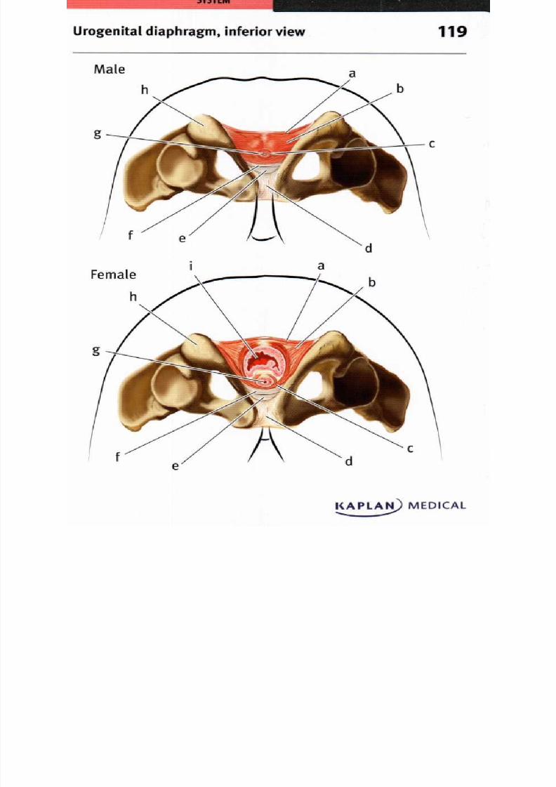
8/13/2019 113264016 Anatomia Omului
http://slidepdf.com/reader/full/113264016-anatomia-omului 213/298
Urogenital diaphragm, inferior view 19
CAPLAN) MEDICAL

8/13/2019 113264016 Anatomia Omului
http://slidepdf.com/reader/full/113264016-anatomia-omului 214/298
ower limb muscles, anterior view 20
KAPLAMEDICAL
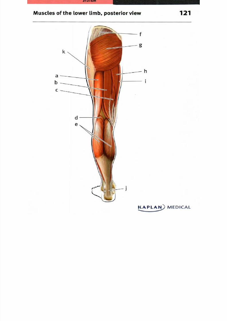
8/13/2019 113264016 Anatomia Omului
http://slidepdf.com/reader/full/113264016-anatomia-omului 215/298
Muscles of the lower limb, posterior view

8/13/2019 113264016 Anatomia Omului
http://slidepdf.com/reader/full/113264016-anatomia-omului 216/298
Superficial femoral muscles, anterior view

8/13/2019 113264016 Anatomia Omului
http://slidepdf.com/reader/full/113264016-anatomia-omului 217/298
Deep femoral muscles, anterior view
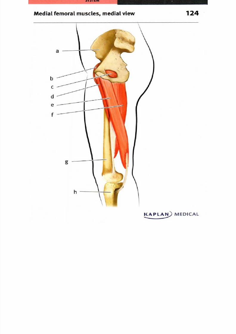
8/13/2019 113264016 Anatomia Omului
http://slidepdf.com/reader/full/113264016-anatomia-omului 218/298
Medial femoral muscles, medial view

8/13/2019 113264016 Anatomia Omului
http://slidepdf.com/reader/full/113264016-anatomia-omului 219/298
g
Lateral femoral muscles, lateral view 25
KAPLAN) MEDICAL
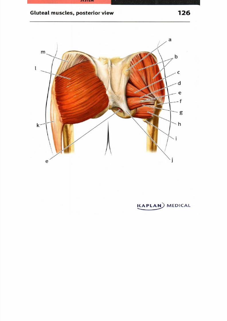
8/13/2019 113264016 Anatomia Omului
http://slidepdf.com/reader/full/113264016-anatomia-omului 220/298
k
Gluteal muscles, posterior view 26
KAPLAINI) MEDICAL
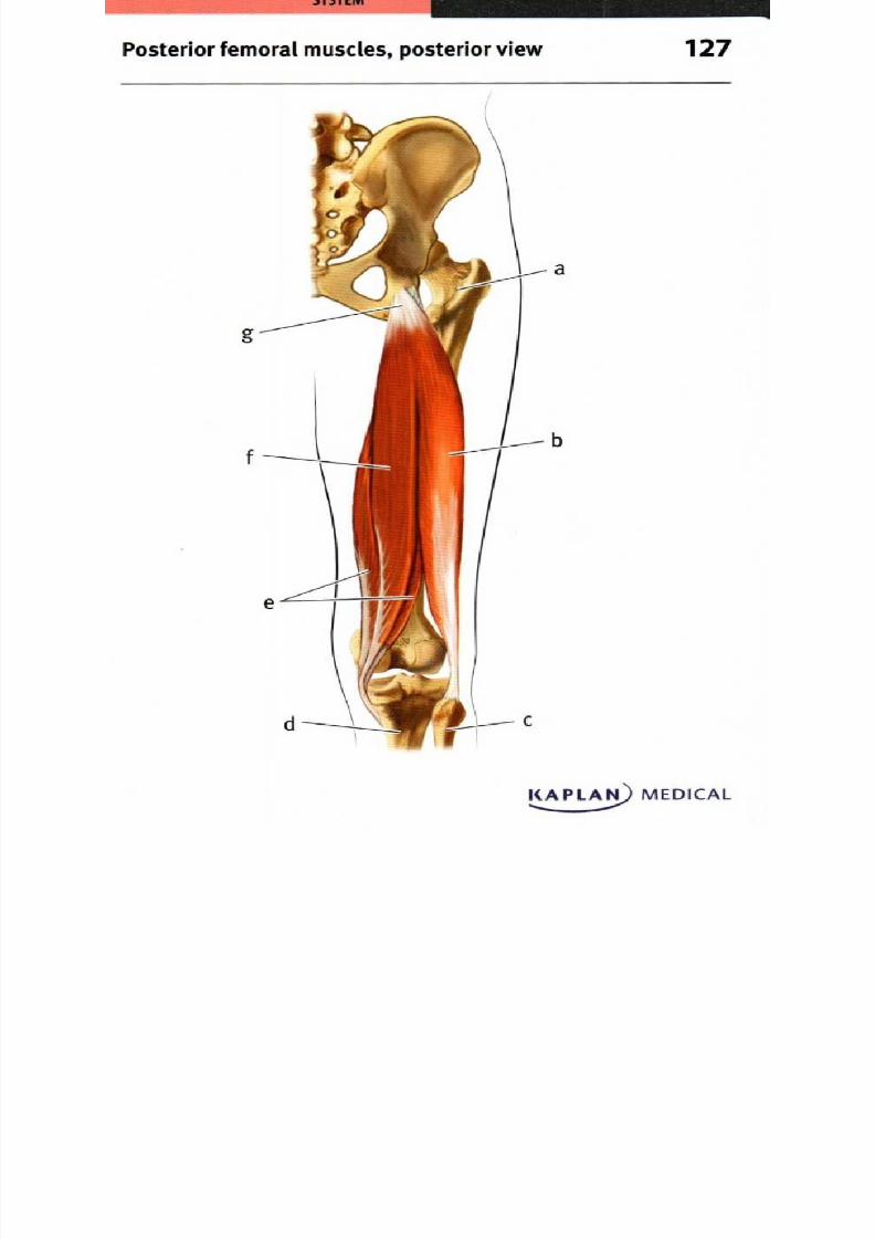
8/13/2019 113264016 Anatomia Omului
http://slidepdf.com/reader/full/113264016-anatomia-omului 221/298
g
Posterior femo ral m uscles posterior view 27
KAPLAN) MEDICAL

8/13/2019 113264016 Anatomia Omului
http://slidepdf.com/reader/full/113264016-anatomia-omului 222/298
Posterior thigh and gluteal muscles,
deep dissection, posterior view
128
KAPLAN) MEDICAL
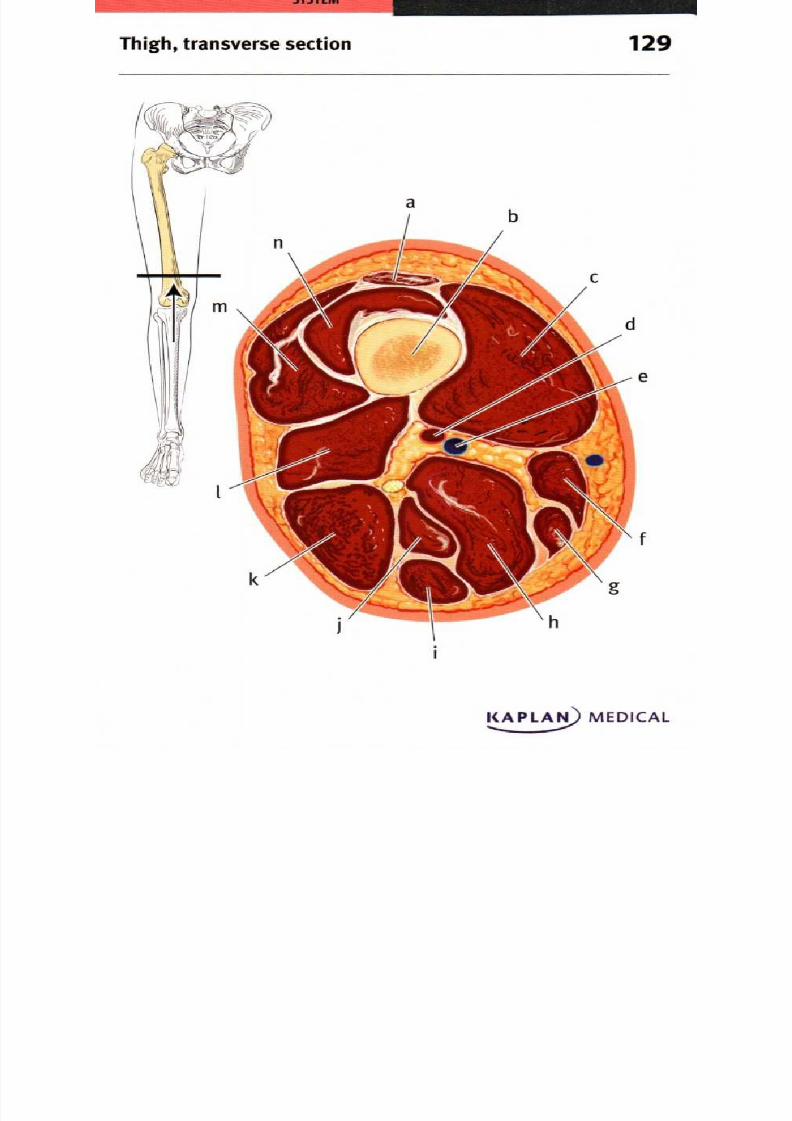
8/13/2019 113264016 Anatomia Omului
http://slidepdf.com/reader/full/113264016-anatomia-omului 223/298
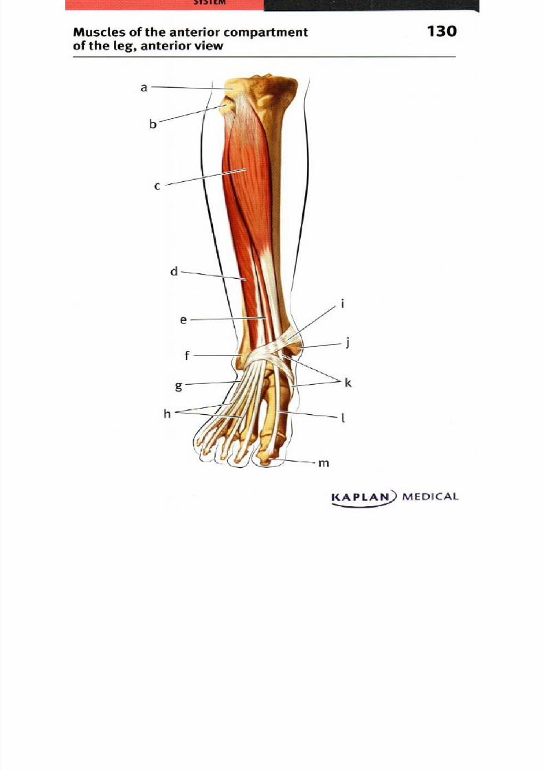
8/13/2019 113264016 Anatomia Omului
http://slidepdf.com/reader/full/113264016-anatomia-omului 224/298
M uscles of the anterior com partmen t 30of the leg anterior view
KAPLAN) MEDICAL.....
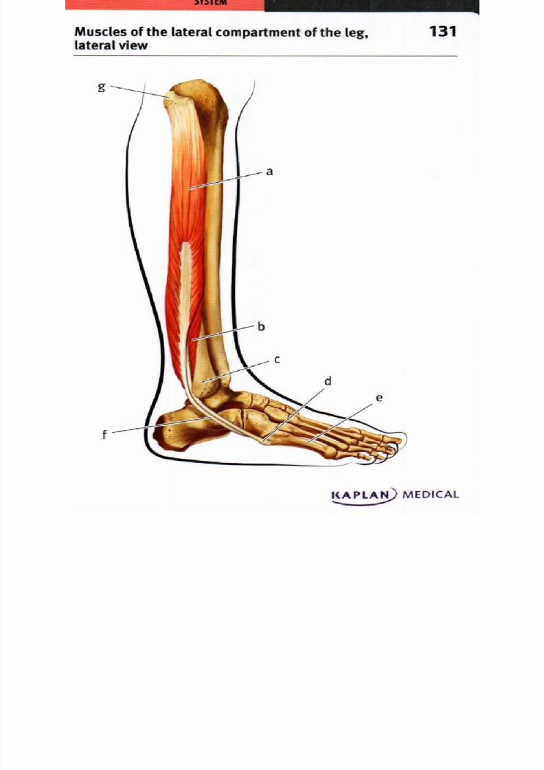
8/13/2019 113264016 Anatomia Omului
http://slidepdf.com/reader/full/113264016-anatomia-omului 225/298
g
f
Muscles of the lateral compartment of the leg, 31lateral view
KAPLAls MEDICAL
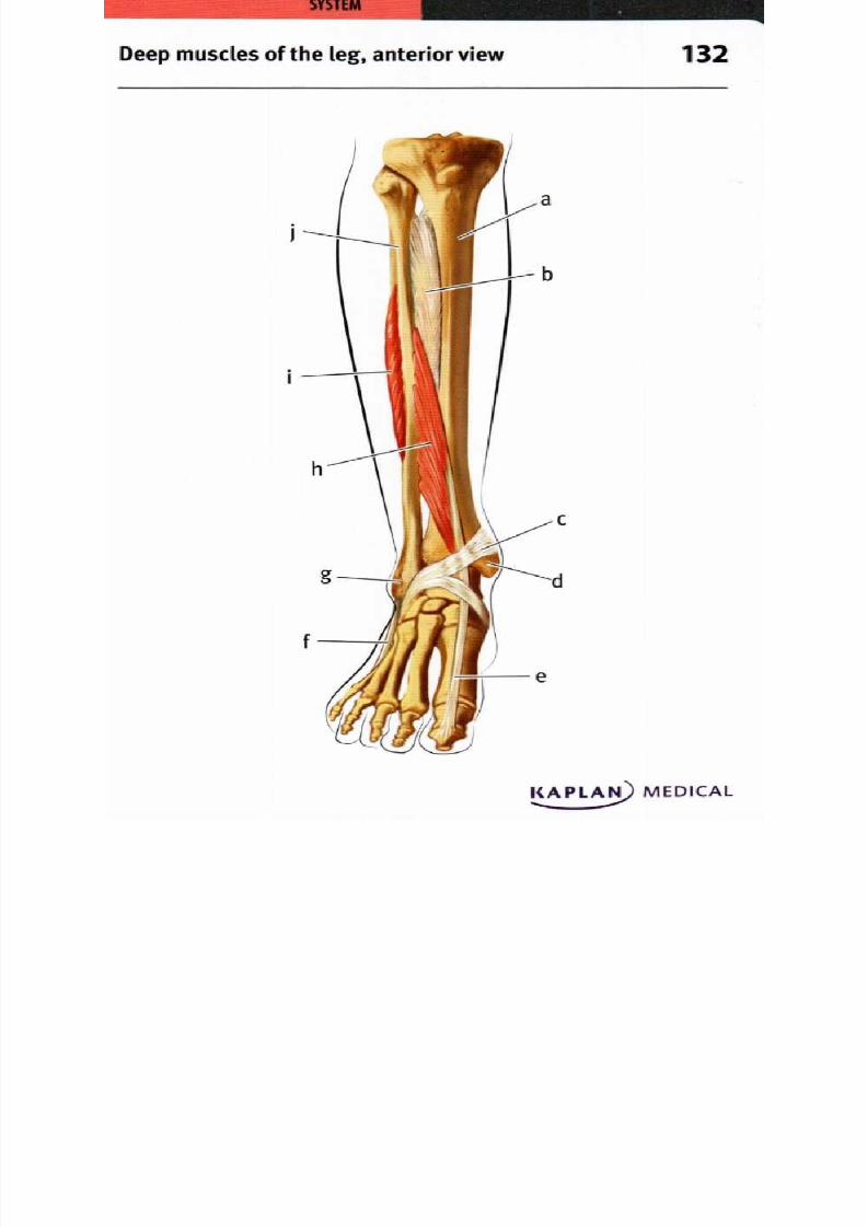
8/13/2019 113264016 Anatomia Omului
http://slidepdf.com/reader/full/113264016-anatomia-omului 226/298

8/13/2019 113264016 Anatomia Omului
http://slidepdf.com/reader/full/113264016-anatomia-omului 227/298
c
g
e
Muscles of the superficial posterior 33compartment of the leg, posterior view
KAPLA) MEDICAL
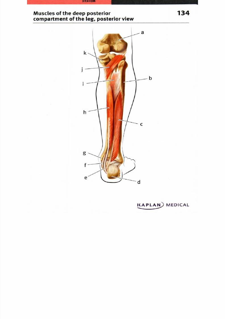
8/13/2019 113264016 Anatomia Omului
http://slidepdf.com/reader/full/113264016-anatomia-omului 228/298
Muscles of the deep posterior 34compartment of the leg, posterior view
KAPLA) MEDICAL

8/13/2019 113264016 Anatomia Omului
http://slidepdf.com/reader/full/113264016-anatomia-omului 229/298
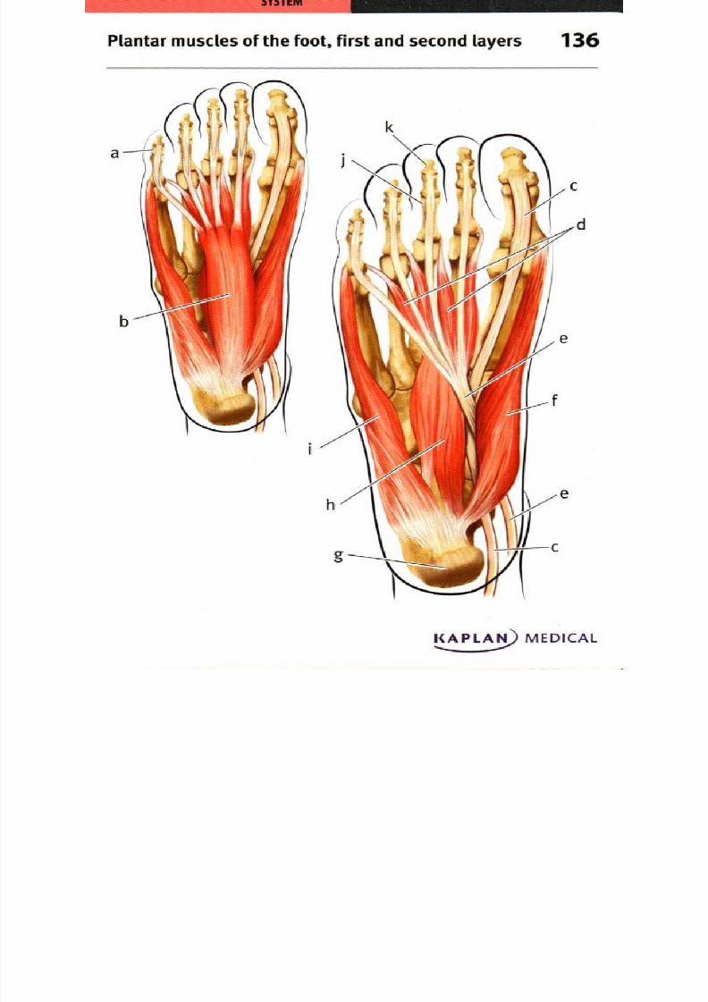
8/13/2019 113264016 Anatomia Omului
http://slidepdf.com/reader/full/113264016-anatomia-omului 230/298
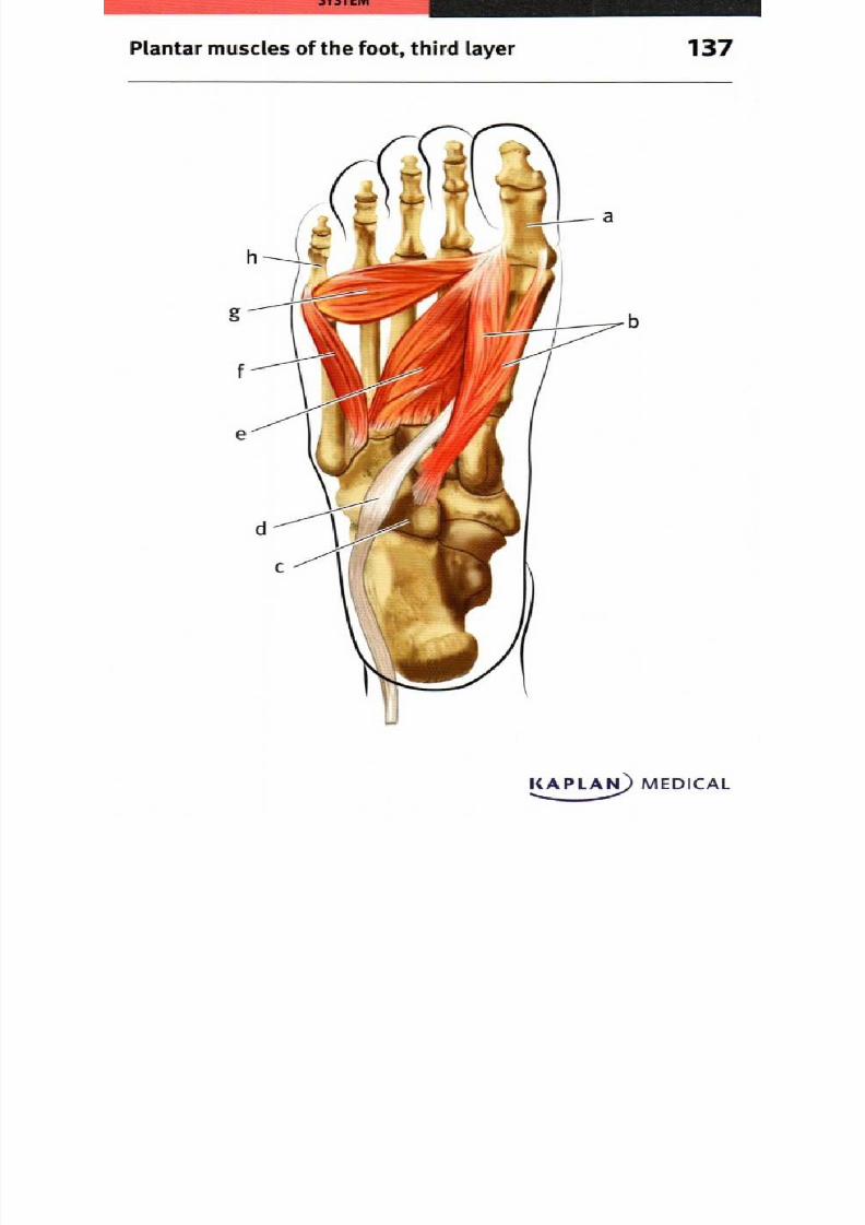
8/13/2019 113264016 Anatomia Omului
http://slidepdf.com/reader/full/113264016-anatomia-omului 231/298
Plantar muscles of the foot, third layer
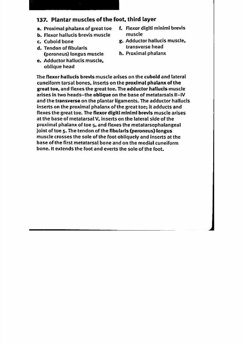
8/13/2019 113264016 Anatomia Omului
http://slidepdf.com/reader/full/113264016-anatomia-omului 232/298

8/13/2019 113264016 Anatomia Omului
http://slidepdf.com/reader/full/113264016-anatomia-omului 233/298
71. Hip joint, frontal section
a. C oxal bone . Z ona orbicularis of capsule
b. Articular cartilage . Articular capsule
c. Acetabular labrum Femur
T he hip joint is a sturdy syn ovial joint (ball and so cket joint) betw een
the globular head of the fem ur and the cu p•like acetabulum of the
coxal bone. A thin layer of articular cartilage covers both bone su rfaces
to reduc e friction. A fibrocartilage lip called the acetabular labrum
extends the edge of the acetabulum to increase its depth; it is locatedslightly beyond the widest diam eter of the femora l head to hold it
firm ly in place. The articular capsule is strong and dense; it com pletely
encloses the head and neck of the fem ur, and extends beyond the
edge of the acetabulum . The capsu le includes longitudinal bands of
fibers that stretch between the hip bone an d the fem ur to strengthen
the joint; the zona orbicularis of the capsule includes deep circular
fibers that form a collar to hold the head o f the femur tightly in the
socket. The joint perm its a range of m ovem ents: flexion an d extension,
addu ction and a bduction, circum duction and rotation.

8/13/2019 113264016 Anatomia Omului
http://slidepdf.com/reader/full/113264016-anatomia-omului 234/298
72. Pelvic ligaments, posterior view
a. Iliac crest
b. Posterior superior iliac spine
c. Iliolumbar ligament
d. Fifth lumbar vertebra
e Supraspinous ligament
f. Short dorsal sacroiliac
ligaments
g Long dorsal sacroiliac
ligament
h.
I
Ik.
I.
Femur
Sacrospinous ligament
Superficial dorsal
sacrococcygeal ligament
Sacrotuberous ligament
Articular capsule of the
hip joint
Strong ligaments are necessary to stabilize the pelvis. The
supraspinous ligament that runs along the edges of the vertebral
dorsal spinous processes continues along the median sacral crest. In
addition, the iliolumbar ligament connects the fifth lumbar vertebra
both to the sacrum and the iliac crest. The sacroiliac joint is stabilized
by the horizontal short dorsal sacroiliac ligament between the sacrum
and the tuberosity of the ilium and by the oblique long dorsal sacroiliac
ligament connecting the sacrum to the posterior superior iliac spine.
The sacrotuberous ligament is connected at one end to the posterior
inferior iliac spine, the lower part of the sacrum, and the coccyx; the
other end attaches to the tuberosity and ramus of the ischium. Nearby,
the sacrospinous ligament extends from the sacrum and coccyx to the
spine of the ischium. The sacrococcygeal joint is stabilized by several
ligaments, including the superficial dorsal sacrococcygeal ligament.
At the hip joint, strong ligaments reinforce the articular capsule that
encloses the head and much of the neck of the femur.

8/13/2019 113264016 Anatomia Omului
http://slidepdf.com/reader/full/113264016-anatomia-omului 235/298
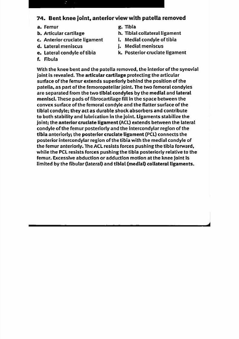
8/13/2019 113264016 Anatomia Omului
http://slidepdf.com/reader/full/113264016-anatomia-omului 236/298
74. Bent knee joint, anterior view with patella removed
a. Femur
b. Articular cartilage
c. Anterior cruciate ligament
d. Lateral meniscus
e. Lateral condyle of tibia
1 Fibula
g. Tibia
h. Tibial collateral ligament
I. Medial condyle of tibia
j. Medial meniscus
k. Posterior cruciate ligament
With the knee bent and the patella removed, the interior of the synovial
joint is revealed. The articular cartilage protecting the articular
surface of the femur extends superiorly behind the position of the
patella, as part of the femoropatellar joint. The two femoral condyles
are separated from the two tibial condyles by the medial and lateral
menisci. These pads of fibrocartilage fill in the space between the
convex surface of the femoral condyle and the flatter surface of the
tibial condyle; they act as durable shock absorbers and contributeto both stability and lubrication in the joint. Ligaments stabilize the
joint; the anterior cruciate ligament (ACL) extends between the lateral
condyle of the femur posteriorly and the intercondylar region of the
tibia anteriorly; the posterior cruciate ligament (PCL) connects the
posterior intercondylar region of the tibia with the medial condyle of
the femur anteriorly. The ACL resists forces pushing the tibia forward
while the PCL resists forces pushing the tibia posteriorly relative to the
femur. Excessive abduction or adduction motion at the knee joint is
limited by the fibular (lateral) and tibial (medial) collateral ligaments.

8/13/2019 113264016 Anatomia Omului
http://slidepdf.com/reader/full/113264016-anatomia-omului 237/298
75. Knee joint, sagittal section
a. Femur . L ateral m eniscus, anterior
b. L ateral m enniscus, orn
posterior horn . Infrapatellar fat pad
c. Fibula . Patella
d. Tibia . Articular cartilage
e. Patellar ligament
A sagittal section through the lateral condyles of the fem ur an d tibia
reveals the anterior-posterior relationships of the fully-extendedknee joint. Articular cartilage covers the con vex articular surfac e
of the fem ur and the flatter articular sur face of the tibia. Between
the two articular cartilages lies the C -shaped lateral m eniscus, with
posterior and anterior horns positioned to fill the area between the
curving articular surfaces. Popliteal ligam ents strengthen the back
of the syn ovial articular capsu le. T he fibula articulates with the tibialepicondyle laterally a nd posteriorly; a collateral ligam ent extends
from the fibula to the lateral epicond yle of the femu r to strengthen the
joint. An teriorly, the patella m oves along the fem oral articular surface
when the qua driceps m uscle flexes the knee; the articular cartilage
of the pa tella is one o f the thickest du e to the intense stresses of this
m ovem ent. The q uadriceps mu scle tendon attaches on the superior
surface and is continuous w ith the inferior patellar ligam ent. The
infrapatellar fat pad absorbs shock s and fills in the space below and
behind the pa tella.

8/13/2019 113264016 Anatomia Omului
http://slidepdf.com/reader/full/113264016-anatomia-omului 238/298
76. Ankle joint, posterior view
a. Fibula . Calcaneal (Achilles) tendon
b. Posterior tibiofibular Calcaneus
ligament . Medial talocalcaneal
c. Transverse tibiofibular igament
ligament . Talus
d. Posterior talofibular . Deltoid ligament,
ligament ibiocalcaneal part
e. Posterior talocalcaneal . Deltoid ligament, tibiotalar
ligament art
f. Calcaneofibular ligament . Tibia
The posterior view of the ankle joint shows several ligaments that
stabilize the articulations between the leg and ankle bones. The
largest tendon in the human body is the calcaneal tendon also known
as the Achilles tendon) that connects three lower leg muscles—thegastrocnemius, the soleus, and the plantaris—with their insertion on
the calcaneus, the largest of the tarsal bones. Above the ankle, the
distal articulation of the tibia and fibula are stabilized by the posterior
and transverse tibiofibular ligaments. Laterally, the ankle joint is
stabilized by connections between the lateral malleolus of the fibula
and the tarsal bones, including the talofibular and calcaneofibularligaments. On the medial side, the ankle is stabilized by ligaments
connecting the medial malleolus of the tibia with tarsal bones such as
the calcaneus and the talus, including the tibiocalcaneal and tibiotalar
parts of the deltoid ligament. Articulations between the tarsal bones
are also stabilized by ligaments, such as the medial and posterior
talocalcaneal ligaments connecting the talus and calcaneus.

8/13/2019 113264016 Anatomia Omului
http://slidepdf.com/reader/full/113264016-anatomia-omului 239/298
104. Superficial flexor muscles of the forearm, anterior view
a
b.
c.
d.
e
1
Palmaris longus muscle
Humerus
Flexor carpi radialis muscle
Pronator teres muscle
Flexor carpi ulnaris muscle
Flexor digitorum superficialis
muscle
g.
h.
I.
J.
k.
l.
m .
Radius
Ulna
Second metacarpal bone
Pisiform bone
Hamate bone
Fifth metacarpal bone
Middle phalanx
The palmaris longus muscle arises on the medial epicondyle of the
humerus and flexes the wrist by inserting on the palm and a band
of connective tissue in the wrist called the flexor retinaculum. The
flexor carpi radialis muscle arises on the medial epicondyle of the
humerus, inserts at the base of the second and third metacarpal
bones, and both flexes and abducts the wrist. The flexor carpi
ulnaris muscle originates on the medial epicondyle of the humerusas well as adjacent parts of the ulna; it inserts on the pisiform and
hamate carpal bones, and on the fifth metacarpal bone. The flexor
carpi ulnaris both flexes and adducts the wrist. The flexor digitorum
superficialis muscle arises on the medial epicondyle of the humerus
as well as adjacent surfaces of the ulna and radius; it inserts on the
middle phalanges of fingers 2-5 by long tendons and acts to flex thefingers at the joints between the metacarpals and proximal phalanges
as well as the joint between the proximal and middle phalanges.
The pronator teres muscle arises on the medial epicondyle of the
humerus as well as the coronoid process of the ulna, inserts on the
lateral, distal surface of the radius, and pronates the forearm.
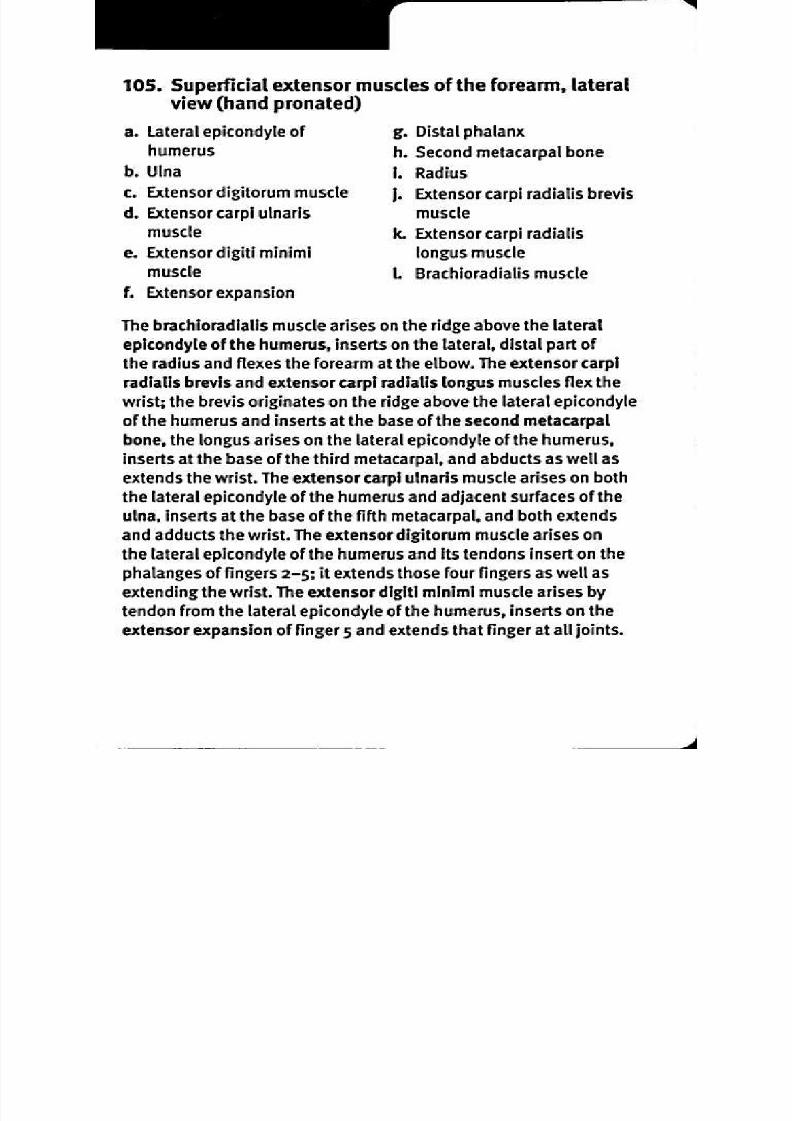
8/13/2019 113264016 Anatomia Omului
http://slidepdf.com/reader/full/113264016-anatomia-omului 240/298
105 Su pe rficial exten sor m us cles of the forearm , lateralv iew hand pronated)
a. Lateral epicondyle of . Distal phalanx
humerus . Second metacarpal bone
b. Ulna . Radius
c. Extensor digitorum muscle . Extensor carpi radialis brevis
d. Extensor carpi ulnaris uscle
muscle. Extensor carpi radialis
e. Extensor digiti minimi ongus muscle
muscle . Brachioradialis muscle
f. Extensor expansion
The brachioradialis muscle arises on the ridge above the lateral
epicondyle of the humerus, inserts on the lateral, distal part of
the radius and flexes the forearm at the elbow. The extensor carpi
radialis brevis and extensor carpi radialis longus muscles flex the
wrist; the brevis originates on the ridge above the lateral epicondyle
of the humerus and inserts at the base of the second metacarpal
bone, the longus arises on the lateral epicondyle of the humerus,
inserts at the base of the third metacarpal, and abducts as well as
extends the wrist. The extensor carpi ulnaris muscle arises on boththe lateral epicondyle of the humerus and adjacent surfaces of the
ulna, inserts at the base of the fifth metacarpal, and both extends
and adducts the wrist. The extensor digitorum muscle arises on
the lateral epicondyle of the humerus and its tendons insert on the
phalanges of fingers 2-5; it extends those four fingers as well as
extending the wrist. The extensor digiti minimi muscle arises by
tendon from the lateral epicondyle of the humerus, inserts on the
extensor expansion of finger 5 and extends that finger at all joints.
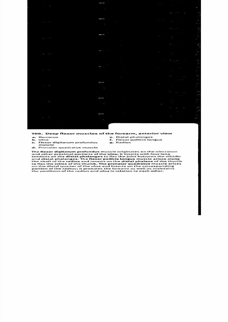
8/13/2019 113264016 Anatomia Omului
http://slidepdf.com/reader/full/113264016-anatomia-omului 241/298
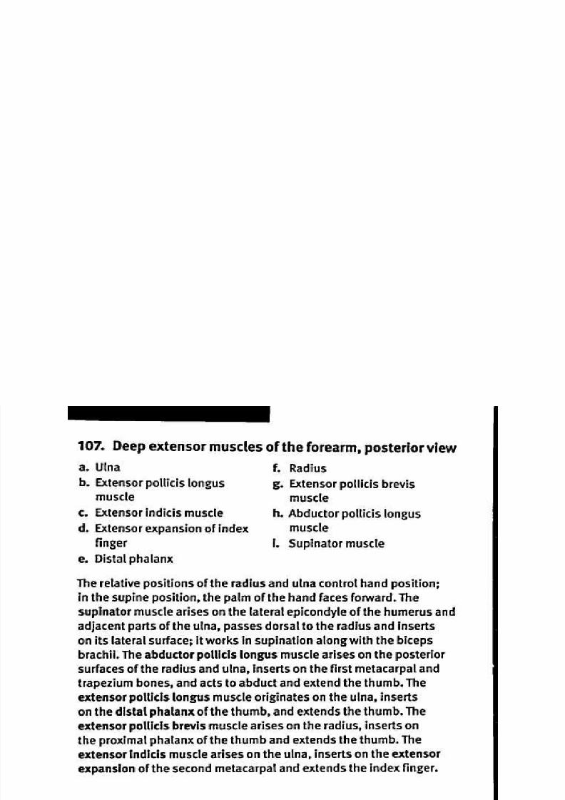
8/13/2019 113264016 Anatomia Omului
http://slidepdf.com/reader/full/113264016-anatomia-omului 242/298
107. Deep extensor muscles of th e forearm, posterior view
a. Ulna
b. Extensor pollicis longus
muscle
c. Extensor indicis muscle
d. Extensor expansion of index
finger
e. Distal phalanx
1 Radius
g. Extensor pollicis brevis
muscle
h. Abductor pollicis longus
muscle
I. Supinator muscle
The relative positions of the radius and ulna control hand position;in the supine position, the palm of the hand faces forward. The
supinator muscle arises on the lateral epicondyle of the humerus and
adjacent parts of the ulna, passes dorsal to the radius and inserts
on its lateral surface; it works in supination along with the biceps
brachii. The abductor pollicis longus muscle arises on the posterior
surfaces of the radius and ulna, inserts on the first metacarpal andtrapezium bones, and acts to abduct and extend the thumb. The
extensor pollicis longus muscle originates on the ulna, inserts
on the distal phalanx of the thumb, and extends the thumb. The
extensor pollicis brevis muscle arises on the radius, inserts on
the proximal phalanx of the thumb and extends the thumb. The
extensor indicis muscle arises on the ulna, inserts on the extensor
expansion of the second metacarpal and extends the index finger.

8/13/2019 113264016 Anatomia Omului
http://slidepdf.com/reader/full/113264016-anatomia-omului 243/298
108. Trans verse sect ions of the arm
a. Biceps brachii muscle
b. Brachialis muscle
c. Humerus
d. Triceps muscle, lateral head
e. Triceps muscle, long headf. Triceps muscle, medial head
g. Tendons of flexor digitorum
superficialis muscle
h. Tendon of flexor carpi radialis
muscle
i. Tendon of flexor pollicis
longus muscle
j. Tendon of abductor pollicis
longus muscle
k. Tendon of extensor pollicis
brevis muscle
m. Tendon of extensor carpi
radialis longus muscle
n. Tendon of extensor carpi
radialis brevis muscle
o. Tendon of extensor pollicis
longus muscle
p. Tendons of extensor
digitorum muscle
q. Tendon of extensor digiti
minimi muscle
r. Tendon of extensor carpi
ulnaris muscle
s. Ulna
t. Pronator quadratus muscle
u. Tendon of flexor carpi ulnaris
muscle
v. Tendons of flexor digitorum

8/13/2019 113264016 Anatomia Omului
http://slidepdf.com/reader/full/113264016-anatomia-omului 244/298
109. M usc les of the han d, po ster ior do rsal ) v iew
a. Insertion of lateral slips of
extensor digitorum muscleand interosseous and
lumbrical muscles
b. Third interosseous muscle
c. Tendons of extensor digiti
minimi muscle
d. Tendon of extensor carpiulnaris muscle
e. Extensor retinaculum
f. Tendon of extensor carpi
radialis brevis muscle
g. Tendon of extensor carpi
radialis longus muscleh. Tendon of extensor indicis
muscle
I. Tendons of extensor
digitorum muscle
j. First interosseous muscle
k. Extensor expansionL Insertion of central slip of
extensor digitorum muscle
The extensor retinaculum is a wide band of connective tissue along
the wrist; it holds tendons in place as they extend from muscles in
the forearm to their insertion points in the hand. Each of the four
tendons of the extensor digitorum muscle spreads out across the
joint at the base of the proximal phalanx to form a broad hood called
the extensor expansion, continues along the proximal phalanx, then
splits to form a central slip that inserts on the middle phalanx and
two lateral slips that pass along the side of the first interphalangealjoint. The two lateral slips rejoin and insert on the distal phalanx
along with tendons from the interosseous and lumbrical muscles.
The tendons of the extensor indicis and extensor digiti minimi
muscles parallel those of the extensor digitorum to insert on the
second and fifth finger, respectively. The tendons of the extensor
carpi radialis longus, extensor carpi radialis brevis, and extensor
carpi ulnaris insert on the base of the second, third, and fifth
metacarpals, respectively. Interosseous muscles are bipennate
muscles that originate along the sides of the metacarpals and
insert on the extensor expansions, acting to abduct the fingers.
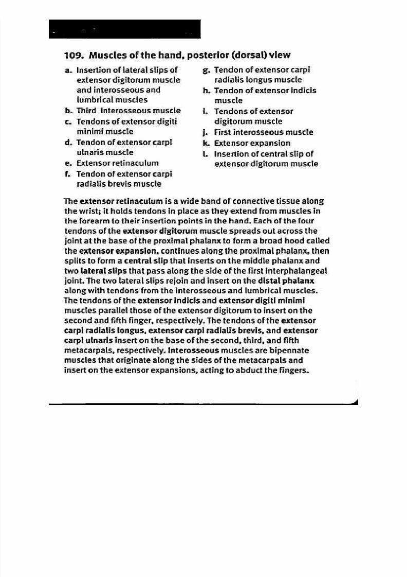
8/13/2019 113264016 Anatomia Omului
http://slidepdf.com/reader/full/113264016-anatomia-omului 245/298
110. Muscles of the hand, anterior (palmar) view
a. Tendons of flexor digitorum
profundus muscleb. Tendons of flexor digitorum
superficialis muscle
c. First and second lumbrical
muscles
d. Proximal phalanx
e. Adductor pollicis muscle1 Flexor pollicis brevis muscle
g. Abductor pollicis brevis
muscle
h . Flexor retinaculum
I. Pisiform boneI. Abductor digiti minimi muscle
k. Flexor digiti minimi brevis
muscle
1 Opponens digiti minimi
muscle
m. Fourth and fifth lumbricalmuscles
n. Proximal phalanx
The tendons of the flexor digitorum superficialis muscle are stabilized
at the wrist as they pass under the flexor retinaculum, and insert
on the middle phalanges. The tendons of the flexor digitorum
profundus muscle are deeper at the wrist and palm, but pass
through the superficial tendons to insert on the distal phalanx. On
the lateral (thumb) side of the hand, the adductor pollicis, flexor
pollicis brevis and abductor pollicis brevis muscles insert on the
proximal phalanx of the thumb to control its movements. On the
medial side of the hand, the abductor digiti minimi and flexor digiti
minimi muscles arise on the pisiform and hamate bones respectively,
insert on the proximal phalanx, and act to abduct or flex the little
finger. The opponens digit minimi muscle acts to bring the little
finger in opposition with the thumb. Four wormlike lumbrical muscles
extend between the tendons of the flexor digitorum profundus
and the extensor expansions (dorsal) to simultaneously flex themetacarpophalangeal joints and extend the interphalangeal joints.

8/13/2019 113264016 Anatomia Omului
http://slidepdf.com/reader/full/113264016-anatomia-omului 246/298
111. Intercostal muscles, anterior view
a. External intercostal muscle . Costal cartilage
b. First rib . Internal intercostal muscle
c. First thoracic vertebra
Respiration involves changes in pressure within the thoracic cavity
due to movements of the ribs and diaphragm that change the
volume of the cavity. The intercostal muscles extend between the
ribs and function in the respiratory movements of the ribs. Eleven
external intercostal muscles on each side arise from the inferiorborder of ribs 1-11 and act to lift the rib during inspiration. The
external intercostals pass obliquely forward and down to insert
on the next lower rib, ranging from the tubercles posteriorly to the
end of the ribs anteriorly, except that the lower two extend to the
costal cartilage and th e upper two don t quite reach th e end of the
rib. Eleven internal intercostal muscles on each side originate at
th e costal groove on th e interior, inferior surface of ribs 1-11, pass
obliquely down and laterally to insert on the superior margin of th e
next lower rib. Th ey bring th e ribs closer together during exh alation.

8/13/2019 113264016 Anatomia Omului
http://slidepdf.com/reader/full/113264016-anatomia-omului 247/298
112. D iaph ragm , an ter io r v iew
a. Xiphoid process . Medial arcuate ligament
b. Esophageal hiatus . Left crus of diaphragm
c. Diaphragm . Right crus of diaphragm
d. Aortic hiatus . Third lumbar vertebra
The diaphragm is a broad, thin dome-shaped muscle that separates
the thoracic cavity from the abdominal cavity. The muscle originates
laterally and anteriorly around the inferior margin of the rib cage
and costal cartilages, as well as the posterior aspect of the xiphoid
process of the sternum. Posteriorly, the diaphragm arises from the
medial arcuate ligament, allowing the psoas major muscle to pass
vertically along the body wall. Muscular fibers of the right crus of the
diaphragm arise on vertebrae I.1-3, while the left crus arises on 11-2;
the median arcuate ligament unites the two crura and passes over the
aortic hiatus. Openings in the diaphragm allow important structuresto be continuous between the two cavities; these openings include
the esophageal hiatus and the aortic hiatus. The diaphragm plays
an important role in respiration; contraction of the muscle causes
the "dome" to move inferiorly, expanding the thoracic cavity and
triggering inhalation by reducing intrathoracic pressure. Relaxation
of the diaphragm allows it to return to a convex shape that makes
the thoracic cavity smaller; elasticity of the lungs then expels air.
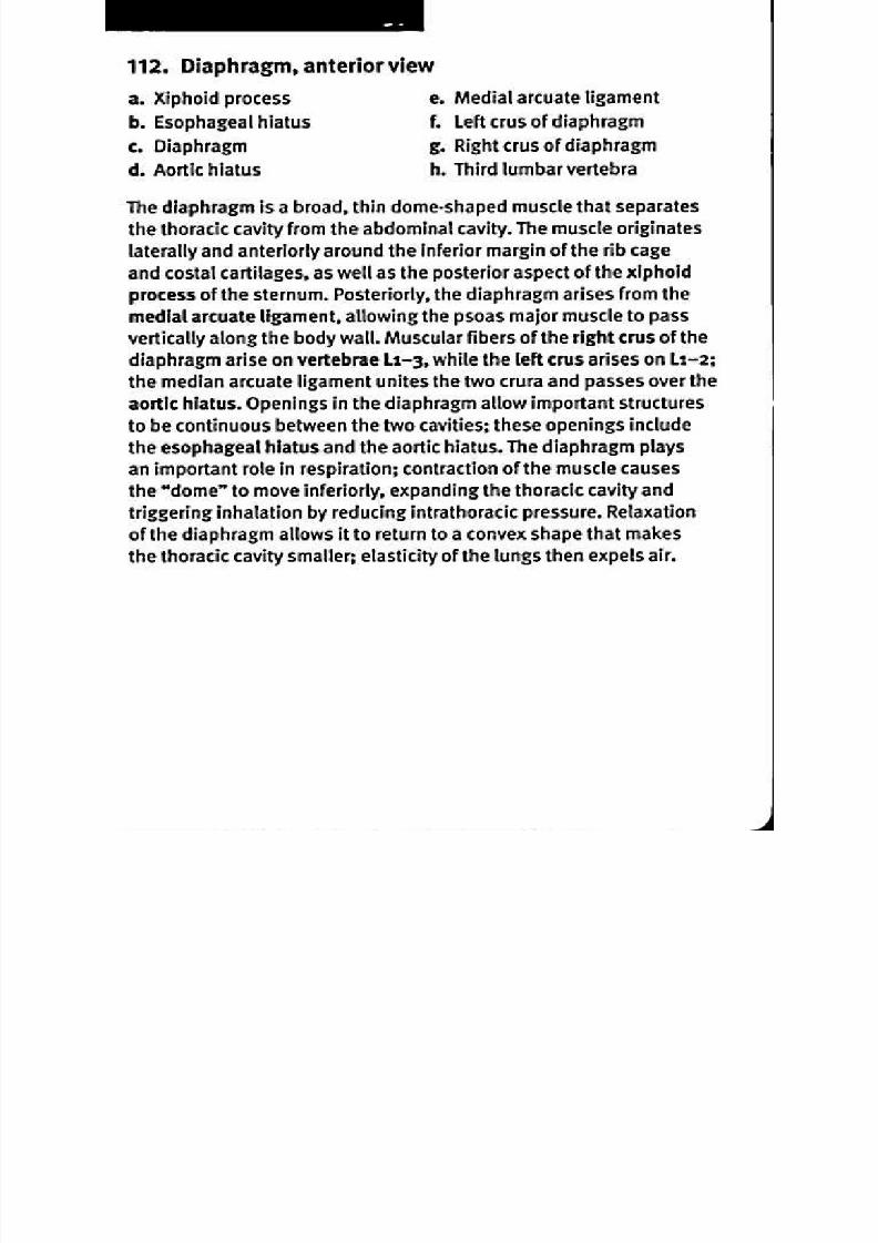
8/13/2019 113264016 Anatomia Omului
http://slidepdf.com/reader/full/113264016-anatomia-omului 248/298
113. Diaphragm, inferior view
a Costal cartilage . Lateral arcuate ligament and
b. Esophageal hiatus welfth rib
c. Aortic h iatus . Medial arcuate ligament
d. Diaphragm . Vena caval foramen
e First lumbar vertebra . Central tendon of diaphragm
Muscle fibers of the diaphragm arise from the medial arcuate ligament
attached to the transverse process of vertebra Li, the lateral arcuate
ligament along the twelfth rib, the interior surface of the anterior and
lateral parts of the inferior ribs and costal cartilage, and the posterior
surface of the sternum at the xiphoid process. Anterior to the lumbar
vertebrae, muscle fibers of the right and left crura arise and pass
to either side of the aortic hiatus, an opening behind the fibers of
the diaphragm that allows passage of the abdominal aorta from thethoracic cavity to the abdomen. Other openings in the diaphragm
include the esophageal hiatus for passage of the esophagus toward
the stomach, and the vena caval foramen that allows the posterior
vena cava to return toward the heart. The muscle fibers of the
diaphragm converge on the central tendon, which has no skeletal
attachment, but is fused on its superior surface with the pericardium.

8/13/2019 113264016 Anatomia Omului
http://slidepdf.com/reader/full/113264016-anatomia-omului 249/298
114. Superficial abdom inal muscles, anterior view
a Sternum
b. Cut edge of aponeurosis,external abdominal oblique
muscle
c. Aponeurosis, internal
abdominal oblique muscle
d. Internal abdominal oblique
musclee Inguinal ligament
f. Pubic tubercle
g Symphysis pubis
h . Spermatic cord
I. Umbilicus
I. External abdominal oblique
muscle
k. Aponeurosis, external
abdominal oblique muscle
L Fifth rib
The external abdominal oblique muscle is the most superficial of
the muscles in the torso wall. It is a broad, flat muscle that arises
on the anterior angles of the inferior eight ribs and inserts on the
pubic tubercle, the inguinal ligament, and the iliac crest. Thefibers of the broad, flat internal abdominal oblique muscle run
perpendicular and deep to the fibers of the external abdominal
oblique. The internal abdominal oblique arises on the fascia of the
lower back, the iliac crest and the inguinal ligament and inserts
on the inferior border of the lower three ribs and on the linea alba.
Both abdominal oblique muscles end in broad aponeuroses that
together form the anterior wall of the abdomen. Near the inguinal
ligament, a triangular hole in the aponeurosis provides for passage
of the spermatic cord (in males) or the round ligament of the uterus
(in females). Acting together, the two abdominal oblique muscles
on each side compress the abdomen, aiding in breathing and
defecation, or flex the trunk. Acting separately, they rotate the trunk.
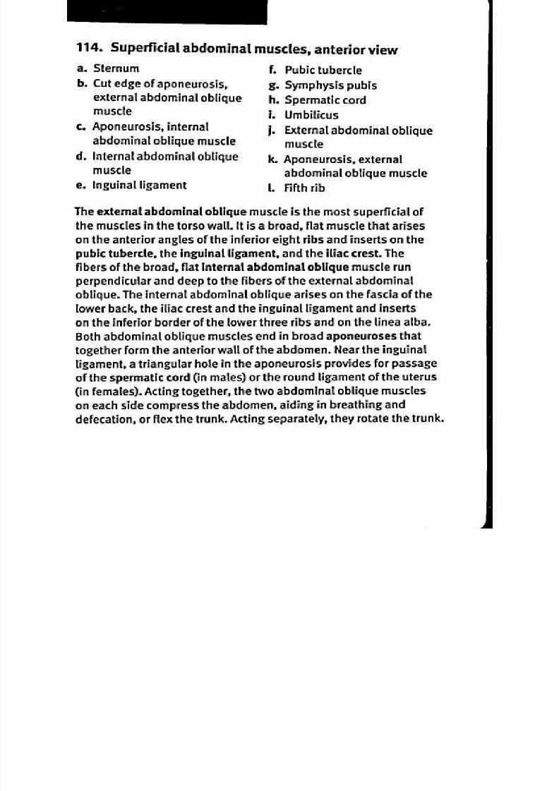
8/13/2019 113264016 Anatomia Omului
http://slidepdf.com/reader/full/113264016-anatomia-omului 250/298
115. Deep abdominal muscles, anterior view
a. Sternum
b. Fifth rib
c. Tendinous intersection
d. Rectus abdominis muscle
e. Arcuate line
1 Symphysis pubis
g. Aponeurosis of transversus
abdominis muscle
h. Iliac crest
i. Transversus abdominis
muscle
j. Umbilicus
k. Cut edges of rectus
abdominis muscle
The transversus abdominis muscle lies deep to the internal and
external abdominal oblique muscles and works with them to compress
the abdomen. The transversus abdominis originates on the iliac
crest, the inguinal ligament and the costal cartilage of ribs 7-12. The
aponeurosis of the transversus abdominis inserts on the linea alba.
The rectus abdominis muscle extends vertically along either side ofthe linea alba, and acts to depress the ribs, flex the spine and stabilize
the pelvis during walking. It originates along the symphysis pubis and
inserts on the costal cartilages of ribs 5 and the xiphoid process of
the sternum. Inferior to the arcuate line, the aponeurosis of the other
abdominal muscles is superior to the rectus abdominus; superficial to
the arcuate line, parts of the aponeurosis lie both superficial and deep
to the rectus abdominis, forming a sheath that encloses the muscle. At
several tendinous intersections, the rectus abdominis muscle is firmly
attached to the part of the sheath that stretches anterior to the muscle.

8/13/2019 113264016 Anatomia Omului
http://slidepdf.com/reader/full/113264016-anatomia-omului 251/298
116. Posterior abdominal wall muscles, anterior view
a. Diaphragm . Iliacus muscle
b. Lateral arcuate ligament . Iliolumbar ligament
c. Quadratus lumborum muscle i. Umbilicus
d. Lumbar vertebrae . Psoas major muscle
e. Iliac crest . Medial arcuate ligament
1. Femur
The quadratus lumborum muscle arises from the iliac crest and the
iliolumbar ligament, which extends between the transverse processes
of the 5th lumbar vertebra and the iliac crest. The quadratus lumborum
passes under the edge of the diaphragm at the lateral arcuate
ligament and inserts on the lowest rib and the transverse processes
of lumbar vertebrae 1-4; together, the two quadratus lumborum
muscles act to depress the rib cage and individually each flexes thespine laterally. The psoas major muscle originates on the transverse
processes and bodies of the lumbar vertebrae, passes under the
edge of the diaphragm at the medial arcuate ligament, and inserts on
the lesser trochanter of the femur. The iliacus muscle arises on the
concave superior surface of the ala of the ilium, and its fibers join the
tendon of the psoas major to insert on the lesser trochanter of thefemur. The psoas major and the iliacus are part of a group of muscles
known as hip flexors—they flex the hip and laterally rotate the thigh.

8/13/2019 113264016 Anatomia Omului
http://slidepdf.com/reader/full/113264016-anatomia-omului 252/298
117. Pelvic diaphragm, superior view
a. Coccyx . Symphysis pubis
b. C occygeus muscle . H iatus of urethra
c. Anal canal . Obturator internus m uscle,
d. Levator ani muscle, nd overlying obturator fascia
iliococcygeus . Tendinous arch for origin of
e. Levator ani muscle, evator ani muscle
pubococcygeus
f. Levator ani muscle,
puborectalis
L ooking down on th e pelvis from th e abdomen, one can see th e pelvic
diaph ragm th at separates th e pelvic cavity from th e perineal region
and supports the pelvic viscera including bladder and intestines.
T h e coccygeus muscle arises on th e spine of th e isch ium and th esacrospinous ligament, and widens to insert along th e coccyx. Th e
levator ani m uscle consists of th ree parts: th e iliococcygeus extends
from th e isch ial spine and adjacent tendinous arch of th e pelvic
fascia to the coccyx; th e pubococcygeus stretches from th e pubic
bone to the coccyx and surrounds th e urethra; and th e puborectalis
arises from th e symp h ysis pubis, surrounds th e anal canal andmeets with corresponding fibers from th e opposite side to support
th e rectum. Th e obturator intemus muscle is covered by an overlying
fascia; th e muscle originates on th e fascia as well as th e adjacent
margins of the pubis and isch ium. T h e obturator internus inserts on
th e greater troch anter of th e femur and rotates th e th igh laterally.

8/13/2019 113264016 Anatomia Omului
http://slidepdf.com/reader/full/113264016-anatomia-omului 253/298
118. Perinea( muscles, inferior view
a. Coccyx
b. Anococcygeal ligamentc. Levator ani muscle
d. Ischia' tuberosity
e. Ischiocavernosus muscle
f. Bulbocavernosus
(bulbospongiosus) muscle
g. Central tendinous point ofperineum
h. Inferior fascia urogenital
diaphragm
i. Superficial transverse
perinea( muscle
I External anal sphincter
muscle
k. Urogenital hiatus
The levator an muscle extends from the surface of the lower pelvis to
the coccyx and the midline; those fibers anterior to the coccyx join with
the fibers from the opposite side to form the anococcygeal ligament.
The external anal sph incter m uscle is an elliptically-shaped group of
muscle fibers that surrounds the anus; external fibers arise on the
anococcygeal ligament and insert on the central tendinous point of the
perineum and deeper fibers form a complete sphincter. The superficial
transverse perinea( muscle also inserts on the central tendinous
point of the perineum. The ischiocavemosus muscle arises on the
ischial tuberosity and ramus and inserts on the pubic symphysis.
The ishiocavernosus compresses and stiffens the penis (in males) or
clitoris (in females). The bulbospongiosus (or bulbocavernosus) muscle
originates on the collagen sheath at the base of the penis (in males) or
the clitoris (in females); the fibers cross over the urethra (in males) or
the urethra and vagina (in females) and insert on the central tendinous
point of the perineum. The bulbospongiosus acts in males to stiffen
the penis and eject urine or semen; in females, the muscle stiffensthe clitoris and narrows the urogenital hiatus or vaginal opening.

8/13/2019 113264016 Anatomia Omului
http://slidepdf.com/reader/full/113264016-anatomia-omului 254/298
119. Urogenital diaphragm, inferior view
a. Superficial transverse
perineal muscle
b. Deep transverse perineal
muscle
c. Sphincter urethrae muscle
d. Symphysis pubis
e. Arcuate pubic ligament
f. Transverse perineal ligament
g. Urethra
h . lschial tuberosity
i. Vagina
The urogenital diaphragm refers to the thin layer of muscle atthe outlet of the pelvis. Posterior and inferior to the symphysis
pubis, the arcuate pubic ligament is a thick, triangular ligament
connecting the two pubic bones. The transverse perineal ligament is
a region of thickened fascia between the urogenital diaphragm and
the arcuate pubic ligament. The deep transverse perineal muscle
arises on the ischial ramus, and inserts at the central tendinous
point of the perineum. The small superficial transverse perineal
muscle extends transversely between the ischial tuberosity and
the central tendinous point of the perineum. The sphincter urethrae
muscle arises from the inferior pubic ramus; fibers from both sides
meet to form a sphincter that constricts the urethra in the male
and compresses both the urethra and vagina in the female.
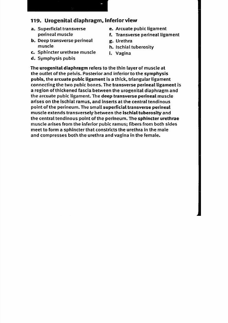
8/13/2019 113264016 Anatomia Omului
http://slidepdf.com/reader/full/113264016-anatomia-omului 255/298
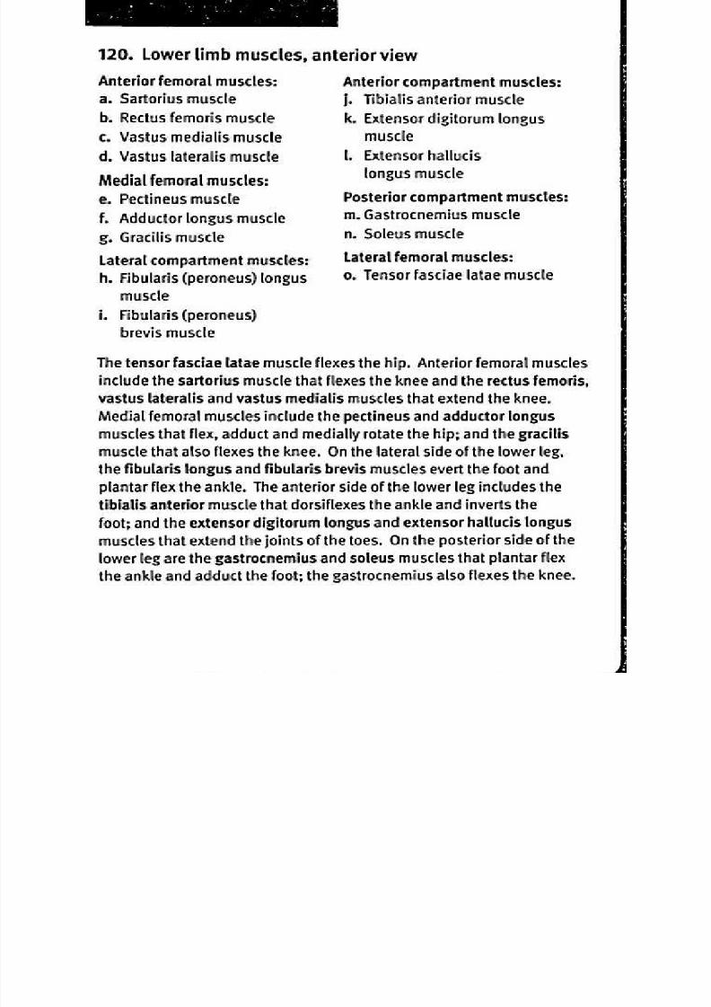
8/13/2019 113264016 Anatomia Omului
http://slidepdf.com/reader/full/113264016-anatomia-omului 256/298
121. Muscles of the lower limb, posterior view
Posterior femoral muscles:
a Biceps femoris muscle
b. Semitendinosus muscle
c. Semimembranosus muscle
Superficial posterior
compartment of the leg:
d. Plantaris muscle
e Gastrocnemius muscle
Lateral femoral (glutea0 muscles:
1. Gluteal fascia over gluteus
medius muscle
g Gluteus maximus
Medial femoral muscles:
h . Adductor magnus muscle
i. Gracilis muscle
j. Calcaneal tendon
k. Iliotibial tract
The superficial gluteus maximus muscle extends and laterally rotates
the hip, while the deeper gluteus medius muscle abducts and medially
rotates the hip. Along with other muscles, the gluteus maximus inserts
on the iliotibial tract, a layer of fascia that lies posterior to the tensor
fasciae latae muscle, interconnecting the femur, patella and tibia, and
stabilizing the knee. On the medial side, the adductor magnus muscle
abducts the hip; portions of the muscle also flex and medially rotate
or extend and laterally rotate the hip. The gracilis muscle adducts and
medially rotates the hip as well as flexing the knee. Other femoral
muscles that flex the knee include the semitendinosus muscle, the
biceps lemon s muscle that also extends and laterally rotates the
hip, and the semimembranosus muscle that also flexes and medially
rotates the hip. The gastrocnemius muscle flexes the knee, plantar
flexes the ankle, and adducts the foot. The plantaris muscle inserts
on the calcaneal tendon to flex the knee and plantar flex the ankle.
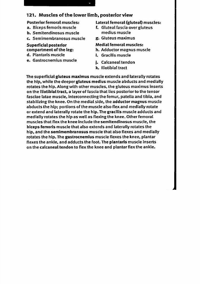
8/13/2019 113264016 Anatomia Omului
http://slidepdf.com/reader/full/113264016-anatomia-omului 257/298
122 Superficial femoral muscles, anterior view
Abdominal muscles related
to the leg:
a Psoas major muscle
b. lliacus muscle
Anterior femoral muscles:
c. Sartorius muscle
Quadriceps femoris muscle:
d. Rectus femorise. Vastus lateralis
f. Vastus medialis
Medial femoral muscles:
g.Pectineus muscle
h. Gracilis muscle
I. Adductor longus muscle
j. Inguinal ligament
k. Tendon of rectus femoris
muscle
1 Patellam. Pubic tubercle
n. Pecten pubis
The psoas major muscle originates on the lumbar vertebrae while
the iliacus muscle arises on the broad, concave superficial surface of
the ilium; both muscles insert on the lesser trochanter of the femur
and flex the hip. The quadriceps femoris muscle is composed of
four muscles that attach at the patella and continue as the patellar
ligament to insert on the tibial tuberosity and act to extend the
knee. The rectus femoris arises on the ilium; it flexes the hip as well
as the knee. The vastus medialis, vastus lateralis and the vastus
intermedius all originate on the femur. Like the rectus femoris, the
sartorius muscle arises on the iliac spine; it inserts on the tibia
and flexes the knee as well as flexing and laterally rotating the hip.
Three medial femoral muscles adduct and rotate the hip medially.
The adductor longus muscle arises on the ramus of the pubis and
the pectineus muscle originates along the pectin pubis, a ridge
on the superior side of the ramus of the pubis bone; both insert
along the femur and also flex the hip. The gracilis arises on the
ramus of the pubis, inserts on the tibia and also flexes the knee.
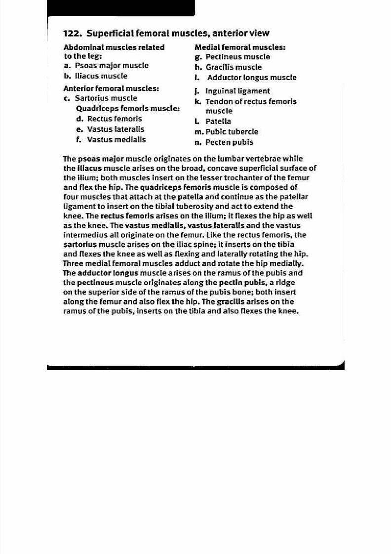
8/13/2019 113264016 Anatomia Omului
http://slidepdf.com/reader/full/113264016-anatomia-omului 258/298
123. Deep fem oral mu scles, anterior viewa Pecten pubis
Medial femoral muscles:
b. Obturator externus muscle
c. Adductor brevis muscle
d. Adductor magnus muscle
e Gracilis muscle
Anterior femoral muscle:
f. Vastus intermedius muscle
g Adductor hiatus
h. Patella
I. Iliofemoral ligament
j. Greater trochanter of femur
The vastus intermedius muscle is deep to the rectus femoris; it
arises on the upper part of the femur, inserts on the patella along
with the other quadriceps tendons, continuing on as the patellar
ligament, and acts to flex the knee. The obturator extemus muscle
arises on the obturator foramen, inserts on the fossa medial tothe greater trochanter on the posterior side of the femur and
rotates the hip laterally. The adductor brevis and adductor magnus
muscles originate on the inferior ramus of the pubis and insert on
the femur; both adduct the hip. In addition, the adductor brevis
flexes the hip while different portions of the adductor magnus
may either flex or extend the hip. The adductor hiatus is a space
near the insertion of the adductor magnus where blood vessels
pass. The gracilis muscle also arises on the inferior ramus of
the pubis; it inserts on the medial surface of the tibia below the
medial condyle, and both flexes the knee and adducts the hip.

8/13/2019 113264016 Anatomia Omului
http://slidepdf.com/reader/full/113264016-anatomia-omului 259/298
124. Medial femoral muscles, medial view
a Right h ip bone
Medial femoral muscles:
b. Pectineus muscle
c. Obturator externus muscle
d. Adductor longus muscle
e. Gracilis muscle
1. Adductor magnus muscle
g Femur
h . Tibia
On the medial aspect of the thigh, the most superficial muscle is
the gracilis; it extends from the pubis part of the hip bone to the
tibia and flexes the knee as well as adducts the hip. The adductor
magnus is a large, powerful muscle that arises on the pubis and
ischium and inserts along the femur. The adductor magnus adducts
the hip; in addition, the superior portion flexes the thigh while
the inferior portion extends it. The adductor longus and pectineus
muscles extend between the pubis bone and the femur; they act to
flex and adduct the hip, and assist in medial rotation. The obturator
extemus muscle extends between the margin of the obturator
foramen and the posterior surface of the greater trochanter of the
femur; it rotates the thigh laterally as well as assists in adduction.
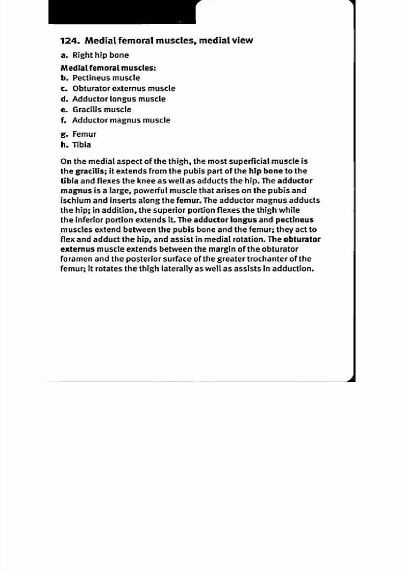
8/13/2019 113264016 Anatomia Omului
http://slidepdf.com/reader/full/113264016-anatomia-omului 260/298
125. Latera l femoral muscles, lateral view
Lateral femoral (gluteal)
muscles:a. Gluteus medius muscle
b. Gluteus maximus muscle
c. Tensor fasciae latae muscle
d. Iliotibial tract
e. Femur
1. Tibia
g. Iliac crest
On the lateral aspect of the hip, the gluteus maximus muscle is the
most superficial of the gluteal muscles; it arises along the posterior
iliac crest, the sacrum and the coccyx. The fibers of the gluteusmaximus pass inferiorly and laterally to insert on the iliotibial tract—a
band of collagen tissue that passes down along the thigh to insert on
the tibia. The gluteus maximus is the major extensor of the hip joint
and also acts in lateral rotation. The deeper gluteus medius muscle
originates on the ilium, below the iliac crest, and inserts on the greater
trochanter of the femur. The gluteus medius is the major abductor of
the hip; portions of it may assist in rotating the hip either mediallyor laterally. The tensor fasciae latae muscle arises on the anterior
iliac crest and the fasciae lata, and inserts on the iliotibial tract. The
tensor fasciae latae abducts the thigh and rotates the hip medially.

8/13/2019 113264016 Anatomia Omului
http://slidepdf.com/reader/full/113264016-anatomia-omului 261/298
126. Gluteal muscles, posterior view
a. Posterior superior iliac spine
b. Gluteus medius muscle
c. Piriformis muscle
d. Superior gemellus muscle
e. Obturator internus muscle
f. Greater trochanter of femur
g. Inferior gemellus muscle
h. Quadratus femoris muscle
i. Lesser trochanter of femur
j. Sacrotuberous ligament
k. Iliotibial tract
I Glluteus maximus muscle
m. Gluteal fascia
The superficial gluteus maximus muscle arises on the iliac crest, inserts
on the iliotibial tract, and is a major extensor of the hip. Deep to the
gluteus maximus, the gluteus minimus arises from the ilium, inserts on
the greater trochanter of the femur, and abducts the hip. The piriformis
muscle originates on the anterior part of the sacrum and inserts on
the greater trochanter of the femur; it rotates the hip laterally. The
superior gemellus muscle arises on the spine of the ischium, the
obturator internus muscle originates on the obturator foramen and
the inferior gemellus muscle arises on the ischial tuberosity. These
three muscles insert together on the greater trochanter of the femur
and rotate the hip laterally. The quadratus femoris muscle arises on
the ischium, inserts on the posterior surface of the femur between
the greater and lesser trochanters, and also rotates the hip laterally.

8/13/2019 113264016 Anatomia Omului
http://slidepdf.com/reader/full/113264016-anatomia-omului 262/298
127. Pos terior fem ora l m us cles , po ster ior v iew
a. Femur
b. Biceps femoris muscle
c. Fibula
d. Tibia
e. Semimembranosus muscle
1 Semitendinosus muscle
g. Tuberosity of ischium
On the posterior aspect of the thigh, the long head of the biceps
femoris muscle arises on the posterior surface of the ischial tuberosity,
and the deeper short head arises midway down the femur, along the
linea aspera. The two heads unite and insert together on the apex onthe head of the fibula and on the lateral tibial condyle. It flexes the
knee as well as extending the thigh. The semimembranosus muscle
arises on the ischial tuberosity and inserts on the medial condyle of
the tibia; it extends the thigh, flexes the knee, and rotates the tibia
medially. The more superficial semitendinosus muscle originates
on the ischial tuberosity and inserts on the medial side of the tibia
shaft; it flexes and medially rotates the knee, and extends the thigh.
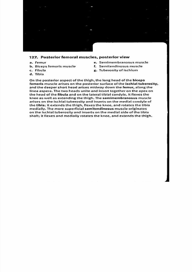
8/13/2019 113264016 Anatomia Omului
http://slidepdf.com/reader/full/113264016-anatomia-omului 263/298
128. Posterior thigh and gluteal muscles, deep dissection,
posterior view
a. Gluteus minimus muscle . Tibia
b. Inferior gemellus muscle . Semimembranosus muscle
c. Quadratus femoris muscle . Adductor magnus muscle
d. Linea aspera of femur . Obturator internus muscle
e. Biceps femoris muscle, . Superior gemellus muscle
short head . Piriformis muscle
f. Fibula
The gluteus minimus lies deep to the gluteus medius; it arises on
the outer surface of the ilium, inserts on the greater trochanter ofthe femur, and abducts the hip. The piriformis, superior gemellus,
obturator internus, inferior gemellus and quadratus femoris muscles
arise along the pelvis, insert on or near the greater trochanter of the
femur, and rotate the thigh laterally. A deep view of the posterior
thigh muscles shows the short head of the biceps femoris, as it
arises along the linea aspera, a ridge of roughened surface that runs
longitudinally along the posterior surface of the femur. The short
head is joined by the long head of the biceps femoris that arises
on the ischium and both parts insert together on the fibula and the
lateral condyle of the tibia. The adductor magnus muscle arises on
the hip bone and inserts along the length of the linea aspera, and
both adducts and medially rotates the hip. The semimembranosus
muscle arises on the ischium, inserts on the medial tibial condyle, and
flexes the knee, extends the thigh, and rotates the tibia medially.

8/13/2019 113264016 Anatomia Omului
http://slidepdf.com/reader/full/113264016-anatomia-omului 264/298
129 Thigh, transverse section
a Rectus femoris muscle . Semitendinosus muscle
b. Femur . Semimembranosus muscle
c. Vastus medialis muscle . Biceps femoris muscle, long
d. Femoral artery ead
e Fem oral vein . Biceps femoris muscle, short
f. Sartorius muscle ead
g. Gracilis muscle .Vastus lateralis muscle
h. Adductor magnus muscle . Vastus intermedius muscle
A transverse section of the thigh, shown a short distance above
the knee, illustrates the relative positions of the femoral muscles.
Anterior to the femur lies the vastus medialis, vastus intermedius,
and vastus lateralis muscles. Most superficial on the anterior aspect
is the rectus femoris. These anterior muscles all insert on the patellaand act to extend the knee. On the posterior-lateral side of the femur
are the short head and long head of the biceps femoris. Between
the biceps femoris and the large adductor magnus muscles are
the superficial semitendinosus and the deeper semimembranosus
muscles; they flex the knee. On the medial aspect are the gracilis and
sartorius muscles; they, too, flex the knee. Because of the unique
combination of origin and insertion for each, some of these muscles
also rotate the hip either medially or laterally, or adduct the hip. The
femoral artery and vein both lie deep within the thigh muscles.

8/13/2019 113264016 Anatomia Omului
http://slidepdf.com/reader/full/113264016-anatomia-omului 265/298
130 Muscles of the anterior
anterior view
a. Lateral condyle of tibia
b. Head of fibula
c. Tibialis anterior muscle
d. Extensor digitorum longus
muscle
e. Extensor hallucis longus
muscle
1 Lateral malleolus
g. Tendon of fibularis
(peroneus) brevis muscle
compartment of the leg,
h. Tendons of extensor
digitorum longus muscle
i. Inferior extensor retinaculum
j. Medial malleolus
k. Tendon of tibialis anterior
muscle
1 Tendon of extensor hallucis
longus muscle
m. Distal phalanx
In anatomical terms, the lower limb is divided into two regions—the
thigh above the knee and the leg below the knee. The anterior
compartment of the leg has several muscles involved in dorsiflexion,
or movement of the foot to decrease the angle between the foot and
leg. The tibialis anterior muscle arises on the lateral condyle of the
tibia, and its tendon inserts on the medial cuneiform tarsal and first
metatarsal bones. The extensor digitorum longus muscle originates
on the lateral condyle of the tibia and the anterior surface of the fibula;
its tendons insert on the superior surface of the phalanges of toes2-5 and it extends the toes in addition to dorsiflexion of the foot. The
extensor hallucis longus muscle originates on the fibula and its tendon
inserts on the distal phalanx of the great toe; it extends the great toe
in addition to dorsiflexion of the foot. The inferior extensor retinaculum
is a band of collagen fibers that extends from the calcaneus laterally to
the medial malleolus of the tibia; it stabilizes the synovial sheaths for
the tendons of the fibularis brevis, tibialis anterior, extensor digitorum
longus, and extensor hallucis longus as they cross the ankle joint.
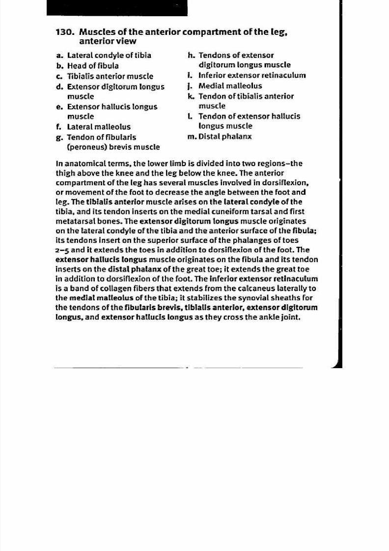
8/13/2019 113264016 Anatomia Omului
http://slidepdf.com/reader/full/113264016-anatomia-omului 266/298
131. Muscles of the lateral
lateral view
a. Fibularis (peroneus) longus
muscle
b. Fibularis (peroneus) brevis
musclec. Lateral malleolus
d. Tendon of peroneus brevis
muscle
compartment of the leg,
e. Fifth metatarsal bone
1 Tendon of peroneus longus
muscle
g. Head of fibula
he fibularis longus (also known as the peroneus longus) muscle
arises at the head of the fibula; the muscle parallels the bone and
its tendon passes posterior to the lateral malleolus of the fibula and
under the sole of the foot before inserting at the base of the first
metatarsal bone. The fibularis longus acts in plantarflexion of the
foot (increases the angle between the foot and leg) and also everts
the foot (moves the sole away from the median plane). Because
it passes under the longitudinal arch of the foot, it also serves to
support the arch. The fibularis brevis (or peroneus brevis) muscle
lies deep to the fibularis longus; it originates along the middle part
of the fibula and its tendon runs posterior to the lateral malleolus
and inserts at the base of the fifth metatarsal bone. The fibularis
brevis is also involved in plantar flexion and eversion of the foot.
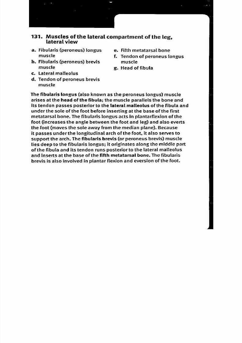
8/13/2019 113264016 Anatomia Omului
http://slidepdf.com/reader/full/113264016-anatomia-omului 267/298
132. Deep muscles of the leg, anterior view
a. Tibia
b. Interosseous membrane
c. Inferior extensor retinaculum
d. Medial malleolus
e. Tendon of extensor hallucis
longus muscle
f. Tendon of fibularis
(peroneus) brevis muscle
g. Lateral malleolus
h. Extensor hallucis longus
muscle
i. Fibularis (peroneus) brevis
muscle
j. Fibula
The interosseous membrane consists of connective tissue fibers that
attach to both the tibia and fibula, stabilizing their positions relative
to each other, dividing the anterior from the posterior compartment
of the leg, and providing attachment sites for muscles. The extensor
hallucis longus muscle arises from the middle half of both the fibula
and interosseous membrane; its tendon passes through a channel
formed by fibers of the inferior extensor retinaculum passing on
either side, then inserts at the base and dorsal surface of the distal
phalanx of the great toe. It acts both to dorsiflex the ankle and to
extend the great toe. The fibularis brevis muscle arises along the
midsection of the fibula and its tendon inserts on the fifth metatarsal
bone. The fibularis brevis plantar flexes the ankle and everts the foot.

8/13/2019 113264016 Anatomia Omului
http://slidepdf.com/reader/full/113264016-anatomia-omului 268/298
133. Muscles of the superficial posterior compartment
of the leg, posterior view
a. Plantaris muscle
b. Gastrocnemius muscle
c. Aponeurosis of
gastrocnemius muscle
d. Lateral malleolus
e. Calcaneal Achilles) tendon
1 Calcaneus
g. Medial malleolus
h. Soleus muscle
i. Femur
The gastrocnemius muscle has two heads; one arises on the
lateral condyle of the femur, the other on the femur above the
medial condyle. The deeper soleus muscle originates on the
head of the fibula and adjacent areas on the shafts of both the
tibia and fibula. The tendon of the gastrocnemius joins with the
tendon of the soleus to form the calcaneal tendon that inserts on
the large tarsus known as the calcaneus. The calcaneal tendonis commonly known as the Achilles tendon. Both muscles act
in plantar flexion of the ankle, and in adduction and inversion
of the foot. In addition, the gastrocnemius flexes the knee. The
plantaris muscle arises from the lateral supracondylar ridge
of the femur; its long tendon inserts on the calcaneal tendon,
and it weakly flexes the knee and plantar flexes the ankle.
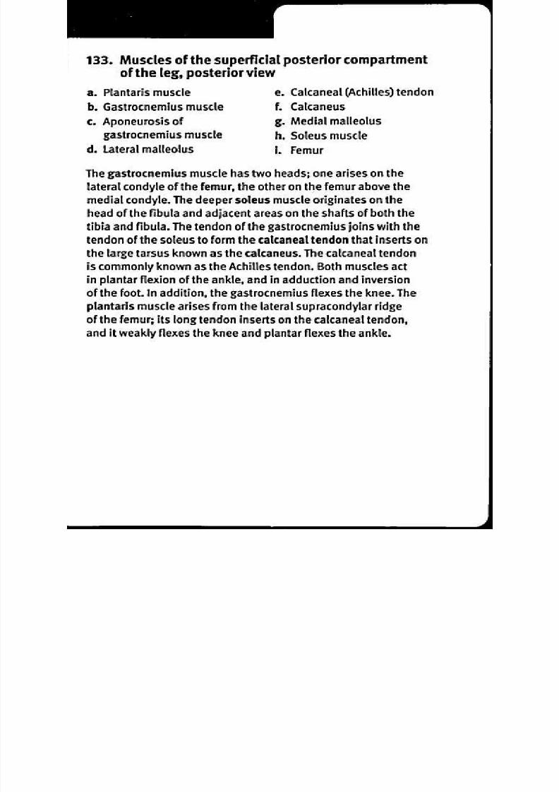
8/13/2019 113264016 Anatomia Omului
http://slidepdf.com/reader/full/113264016-anatomia-omului 269/298
134. Muscles of the deep
posterior view
a. Femur
b. Fibula
c. Flexor hallucis longus
d. Calcaneus
e. Tendon of flexor digitorum
longus muscle
f. Tendon of tibialis posterior
muscle
posterior compartment of the leg,
g. Medial malleolus
h. Flexor digitorum longus
muscle
i. Tibialis posterior muscle
j. Popliteus muscle
k. Tibia
Deep to the gracilis and soleus muscles, the flexor digitorum longus
muscle arises on the tibia, and its tendon passes behind the medial
malleolus and under the sole before it divides into four tendons
that insert on the inferior surfaces of the distal phalanges of toes
2-5. It flexes the joints of those toes. The flexor hallucis longus
arises on the fibula and the interosseous membrane; its tendon
passes behind the talus, under the sole of the foot, and inserts on
the distal phalanx of the great toe. It flexes the joints of the great
toe. The tibialis posterior arises on the interosseous membrane and
adjacent areas of the tibia and fibula; its tendon splits into slips
that insert on the navicular and second cuneiform tarsals as well
as the plantar surfaces of metatarsals 2-4. The tibialis posterioracts to adduct and invert the foot as well as to plantar flex the
ankle. The popliteus muscle originates on the lateral condyle of the
femur, inserts on the posterior surface of the shaft of the tibia, and
medially rotates the tibia at the knee (or laterally rotates the femur).

8/13/2019 113264016 Anatomia Omului
http://slidepdf.com/reader/full/113264016-anatomia-omului 270/298
135. M usc les o f the do rsal foo t
a. Middle phalanx
b. Distal phalanx
c. Extensor expansion
d. Tendons of extensor
digitorum longus muscle
e. Extensor digitorum brevis
muscle
1 Calcaneus
g. Extensor hallucis brevis
muscle
h. Tendon of extensor hallucis
longus muscle
i. Dorsal interosseous muscles
The tendons of the extensor digitorum longus muscle are joinedby other extensor tendons to form the extensor expansions that
insert on the superior surfaces of the phalanges of toes 2-5
and extend the toes at the interphalangeal joints. The extensor
digitorum brevis muscles arise on the calcaneus and insert on the
dorsal surfaces of the toes to extend the metatarsophalangeal
joints of toes 2-4. The dorsal interosseous muscles arise along
the sides of the metatarsal bones, insert on the sides of toes 2-4
and abduct toes 3 and 4 at the metatarsophalangeal joints. The
tendon of the extensor hallucis longus muscle inserts on the
dorsal surface of the phalanges of the great toe to extend the
digit. The extensor hallucis brevis muscle arises on the calcaneus
bone, inserts on the proximal phalanx of the great toe, and
extends the toe.
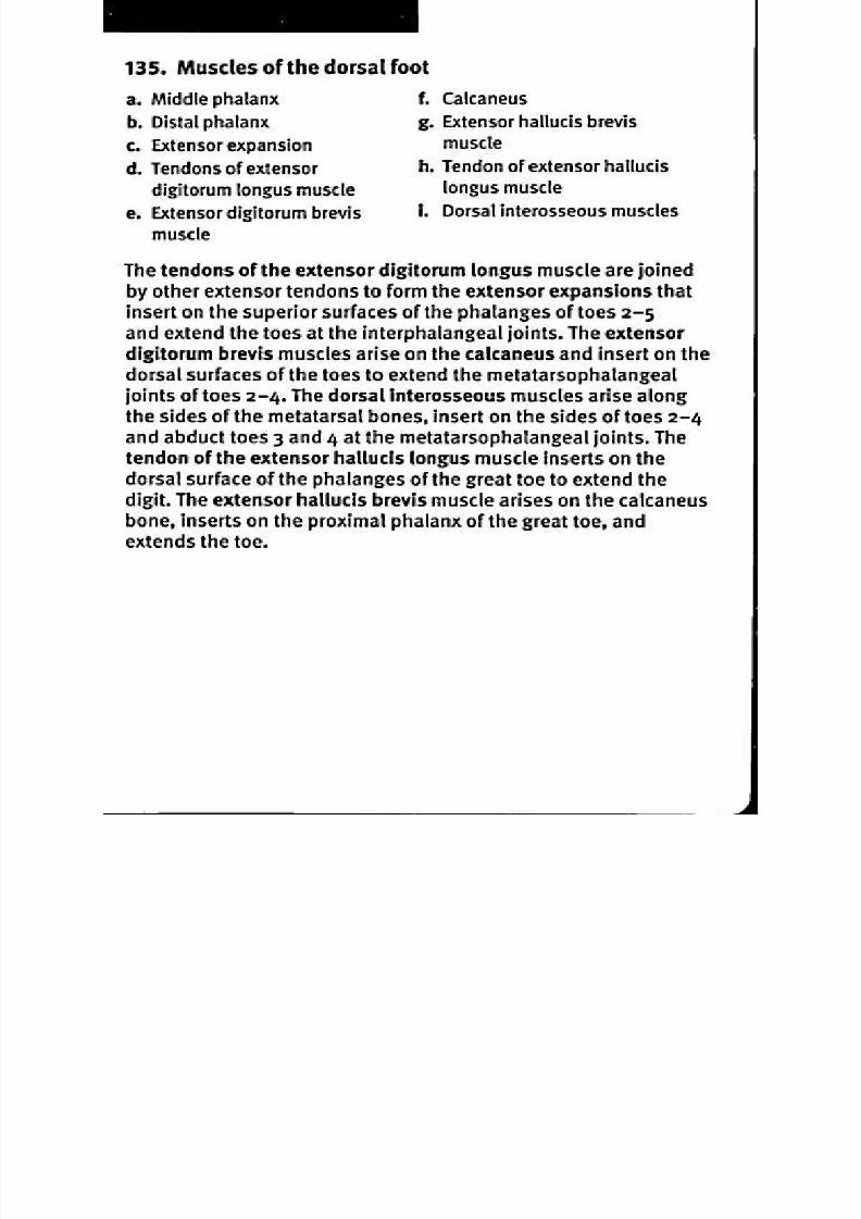
8/13/2019 113264016 Anatomia Omului
http://slidepdf.com/reader/full/113264016-anatomia-omului 271/298
136. Plantar muscles of the foot, first and second layers
a. Middle phalanx
b. Flexor digitorum brevis
muscle
c. Tendon of flexor hallucis
longus muscle
d. Lumbrical muscles
e. Tendon of flexor digitorum
longus muscle
1 Abductor hallucis muscle
g. Calcaneus
h. Quadratus plantae (flexor
accessorius) muscle
i. Abductor digiti minimi muscle
j. Proximal phalanx
k. Distal phalanx
The most superficial of the plantar muscles is the flexor digitorum
brevis muscle; it arises on the calcaneus, inserts on the sides of the
middle phalanges of toes 2-5 and flexes the toes at the proximal
interphalangeal joints. Deep to the flexor digitorum brevis, one can
see the tendons of the flexor digitorum longus muscle which insert
on the inferior surface of the distal phalanges to flex toes 2-5 at
the interphalangeal joints. The lumbrical muscles arise from the
tendons of the flexor digitorum longus and insert on the extensor
expansions of toes 2-5 to extend the interphalangeal joints but flex the
metatarsophalangeal joints. With an action similar to the lumbricals,
the quadratus plantae arises on the calcaneus and inserts on the
tendons of the extensor digitorum longus. On the lateral side of the
foot, the abductor digiti minimi muscle originates on the calcaneus,
inserts on the lateral side of the proximal phalanx of toe 5, and abducts
the toe. On the medial side, the tendon of the flexor hallucis longus
muscle inserts on the inferior surface of the distal phalanx of the
great toe, and flexes the joints of the great toe. The abductor hallucis
muscle originates on the calcaneus, inserts on the medial side of
the proximal phalanx of the great toe, and abducts the great toe.

8/13/2019 113264016 Anatomia Omului
http://slidepdf.com/reader/full/113264016-anatomia-omului 272/298
S K E L E T A L S Y S T E M ( c o n td )
29 Lumbar vertebra, superior and lateral views
30 Sacrum and coccyx, anterior view
31 Sacrum and coccyx, posterior view
32 Intervertebral discs, lateral and midsagittal views
33 Sternum, anterior view
34 Rib and vertebra, articulated, superior view; rib, posterior view
35 Rib cage, anterior view
36 Pectoral girdle and upper limb, anterior view
37 Scapula, anterior and lateral views
38 Scapula, posterior view39 Clavicle and related bones, superior view; clavicle, inferior view
40 Humerus, anterior and posterior views
41 Ulna and radius, lateral and anterior views
42 Hand, posterior (dorsal) view
43 Hand, anterior (palmar) view
44 Hip bone, lateral view
45 Pelvis, anterior view46 Differences between male and female pelvis
47 Lower limb, anterior view
48 Femur and patella, anterior and posterior views
49 Tibia and fibula, anterior and posterior views
50 Bones of the foot, dorsal view
51 Bones of the foot, lateral view
A R T I C U L A T I O N S
52 Gomphosis (peg suture)
53 Suture
54 Syndesmosis, posterior view
55 Synchondrosis
56 Symphysis
57 Synovial joint, diagrammatic sagittal section
58 Tendon sheath
59 Bursa

8/13/2019 113264016 Anatomia Omului
http://slidepdf.com/reader/full/113264016-anatomia-omului 273/298
A R T I C U L A T I O N S (c o n t d )
60 Gliding joint
61 Hinge joint
62 Rotating joint63 Ball and socket joint
64 Condyloid joint
65 Saddle joint
66 Temporomandibular joint, sagittal section
67 Shoulder joint, frontal section
68 Shoulder ligaments, anterior view
69 Elbow joint, sagittal section70 Elbow ligaments, anterior view
71 Hip joint, frontal section
72 Pelvic ligaments, posterior view
73 Knee joint, anterior view
74 Bent knee joint, anterior view with patella removed
75 Knee joint, sagittal section
76 Ankle joint, posterior view77 Ankle joint, frontal section
78 Superficial muscles of the body, anterior view
79 Superficial muscles of the body, posterior view
80 Muscle forms: fusiform and flat sheet
81 Muscle forms: pennate, circular, and multicaudal
82 Muscle forms: cylindrical, triangular, quadrilateral, biventral,
multiventral
83 Muscles of facial expression, anterior view
84 Muscles of facial expression, lateral view
85 Superficial muscles of mastication, lateral view
86 Deep muscles of mastication, lateral view
87 Muscles of the neck, anterior view
88 Suprahyoid and infrahyoid muscles of the neck, anterior view
89 Prevertebral region and root of the neck, anterior view
KAPLAN) MEDICAL
8/13/2019 113264016 Anatomia Omului
http://slidepdf.com/reader/full/113264016-anatomia-omului 274/298
90 Muscles of the neck, lateral view
91 Neck, transverse section
92 Ocular muscles, lateral view and superior view
93 Intrinsic muscles of the tongue, sagittal section
94 Extrinsic muscles of the tongue, pharynx and larynx, lateral view
95 Pharynx, posterior view
96 Superficial shoulder muscles, anterior view
97 Muscles of the shoulder, scapula and arm, anterior view
98 Deep muscles of the shoulder and arm, anterior view
99 Muscles with scapular attachments, posterior view
100 Superficial and intermediate muscles of the back, posterior view
101 Deep muscles of the back, posterior view
102 Anterior brachial muscles (flexors), lateral view
103 Posterior brachial muscles (extensors), lateral view
104 Superficial flexor muscles of the forearm, anterior view105 Superficial extensor muscles of the forearm, lateral view
(hand pronated)
106 Deep flexor muscles of the forearm, anterior view
107 Deep extensor muscles of the forearm, posterior view
108 Transverse sections of the arm
109 Muscles of the hand, posterior (dorsal) view
110 Muscles of the hand, anterior (palmar) view111 Intercostal muscles, anterior view
112 Diaphragm, anterior view
113 Diaphragm, inferior view
114 Superficial abdominal muscles, anterior view
115 Deep abdominal muscles, anterior view
116 Posterior abdominal wall muscles, anterior view
117 Pelvic diaphragm, superior view118 Perineal muscles, inferior view
119 Urogenital diaphragm, inferior view
120 Lower limb muscles, anterior view
121 Muscles of the lower limb, posterior view

8/13/2019 113264016 Anatomia Omului
http://slidepdf.com/reader/full/113264016-anatomia-omului 275/298
122 Superficial femoral muscles, anterior view
123 Deep femoral muscles, anterior view
124 Medial femoral muscles, medial view
125 Lateral femoral muscles, lateral view
126 Gluteal muscles, posterior view
127 Posterior femoral muscles, posterior view
128 Posterior thigh and gluteal muscles, deep dissection, posterior view
129 Thigh, transverse section
130 Muscles of the anterior compartment of the leg, anterior view
131 Muscles of the lateral compartment of the leg, lateral view
132 Deep muscles of the leg, anterior view
133 Muscles of the superficial posterior compartment of the leg,
posterior view
134 Muscles of the deep posterior compartment of the leg,
posterior view
135 Muscles of the dorsal foot
136 Plantar muscles of the foot, first and second layers
137 Plantar muscles of the foot, third layer
N E R V O U S S Y S T E M
138 Nervous system, anterior view
139 Neuron
140 Synapse, cutaway view
141 Central nervous system, sagittal section
142 Brain, lateral view
143 Brain, superior view
144 Brain, inferior view
145 Brain, midsagittal section
146 Brain, frontal section
147 Brain, transverse section
148 Brainstem, posterior view
149 Brainstem, lateral view
150 Limbic system
I(APLA N MEDICAL
8/13/2019 113264016 Anatomia Omului
http://slidepdf.com/reader/full/113264016-anatomia-omului 276/298
NERVO US SY STEM (cont d)
151. Functional areas of the cerebrum
152. Ventricles of the brain
153. Cerebrospinal fluid pathway
154. Meninges of the brain, frontal section
155. Facial nerve and cutaneous branches of the cervical plexus
156. Spinal cord and spinal nerves
157. Branching of a typical spinal nerve, transverse section
158. Spinal membranes and nerve roots
159. Spinal cord, transverse sections
160. Cranial nerves, inferior view
161. Emerging spinal nerves, lateral view
162. Plexuses and thoracic nerves, posterior view
163. Cervical plexus, posterior view
164. Brachial plexus, anterior view
165. Lumbar plexus, anterior view
166. Sacral plexus, posterior view
167. Abdominal sympathetic nerves, anterior view
168. Nerves of the upper limb, anterior view
169. Nerves of the lower limb, posterior view
170. Nerves of the wrist and hand, anterior (palmar) and transverse views
171. Dermatomes, anterior and posterior views
172. Autonomic nervous system, sympathetic division
173. Autonomic nervous system, parasympathetic division
S E N S O R Y S Y S T E M S
174. Skin receptors (touch), microscopic view
175.Tongue (taste), superior view
176. Papillae and taste buds of the tongue
177. Nose (smell), midsagittal view
178. Olfactory epithelium
179. Eye and lacrimal apparatus (vision), anterior view
180. Eye (vision), sagittal view
181. Optic retina, ophthalmoscopic and microscopic views

8/13/2019 113264016 Anatomia Omului
http://slidepdf.com/reader/full/113264016-anatomia-omului 277/298
S S O R Y S Y S T E M S f ro n t
182 Auditory system (hearing), anterior view
183 Middle ear, anterior view
184 Inner ear, anterior view
185 Membranous labyrinth of the inner ear, anterior view
186 Cochlea and Organ of Corti, microscopic view
E N D O C R I N E S Y S T E M
187. Male endocrine system, anterior view
188. Female endocrine system, anterior view
189. Pituitary gland (hypophysis) and pineal body, sagittal section190. Pituitary gland (hypophysis) hormones and target organs, male
191. Pituitary gland (hypophysis) hormones and target organs, female
192. Thyroid gland, anterior view
193. Parathyroid glands, posterior view
194. Pancreas, anterior view
195. Adrenal gland, anterior and sagittal section views
196. Testis, anterior view
197. Ovary, posterior view and transverse section
198. Placenta, fetal aspect, with diagrammatic transverse section
199. Cardiovascular overview, anterior view
200. Circulation (diagrammatic)
201. Blood cells, microscopic view
202. Anatomical relationships of the heart
203. Heart, anterior view
204 Heart, posterior view
205 Heart, frontal (coronal) section
206 Valves of the heart in ventricular systole, superior view
207 Valves of the heart in ventricular diastole, superior view
208 Conduction system of the heart, frontal (coronal) view
209 Artery and vein structure
KAPLAN) MEDICAL

8/13/2019 113264016 Anatomia Omului
http://slidepdf.com/reader/full/113264016-anatomia-omului 278/298
MIIIIIIIMIIIIIMIMIIIIMIIIIM210 Arterial system, anterior view
211 Carotid and vertebral arteries, lateral view
212 Branches of the aortic arch, anterior view
213 Brain arteries
214 Middle cerebral artery, lateral view
215 Cerebral arteries, sagittal view
216 Ophthalmic artery, superior view
217 Upper limb arteries, anterior view218 Lower limb arteries, anterior view
219 Abdominal and thoracic branches of the aorta, anterior view
220 Middle thorax, transverse section viewed from below
221 Digestive system arteries, anterior view
222 Pelvic arteries, male, medial view
223 Pelvic arteries, female, medial view
224Venous system, anterior view
225 Veins of the head and neck, lateral view
226 Upper limb veins, anterior view
227 Lower limb veins, anterior and posterior view
228 Azygos veins, anterior view
229 Portal system of veins, anterior view
230 Abdominal and thoracic veins of the dorsal wall, anterior view
231 Fetal circulation
L Y M P H A T I C S Y S T E M
232 Overview, anterior view
233 Lymphatic drainage areas, anterior view
234 Lymphatic capillaries, microscopic view
235 Lymphatic drainage at the root of the neck, anterior view
236 Tonsils, sagittal view237 Waldeyer s ring, anterior view
238 Spleen, anterior view
239 Lymph node, microscopic view of transverse section
240 Intestinal lacteals, anterior and microscopic views

8/13/2019 113264016 Anatomia Omului
http://slidepdf.com/reader/full/113264016-anatomia-omului 279/298
1
R E S P I R A T O R Y S Y S T E M
241. Respiratory overview, anterior view
242. Larynx, trachea and bronchi, anterior view
243 Nasal septum, medial view
244. Nasal cartilages, lateral view
245. Lateral wall of nasal cavity, medial view
246. Nasal conchae, larynx, and sinuses, coronal section
247. Larynx, anterior view
248. Larynx, sagittal section
249 Larynx, posterior view
250 Glottis, superior view
251. Trachea and bronchial tree, anterior and transverse section views
252. Lung lobes and pleura, anterior view
253. Lung hila, medial view
254. Air pathway and alveolus
255. Thorax, transverse section viewed from below
D I G E S T IV E S Y S T E M
256. Overview, anterior view
257. Overview, lateral view
258. Mouth and oral cavity, anterior view
259. Oral cavity, sagittal section
260. Oral salivary glands, anterior view
261. Salivary glands, lateral view
262 Tooth, sagittal section view
263. Upper teeth, inferior view
264. Esophagus, anterior view
265. Greater omentum, anterior view
266. Stomach, anterior view
267. Stomach, anterior cutaway view
268. Small intestine, anterior view
269. Large intestine, anterior view
270. Liver, anterior view
271. Liver, posterior view
KAPLAN) MEDICAL....
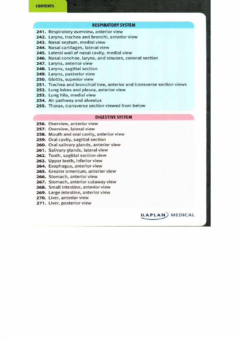
8/13/2019 113264016 Anatomia Omului
http://slidepdf.com/reader/full/113264016-anatomia-omului 280/298
Illustrator: tephanie McCann, MA, is an independent medical
and scientific illustrator. She received her MA in
Medical and Biological Illustration from The Johns
Hopkins University, and BA in Fine Arts at the
University of California, Santa Cruz. She is currently
an instructor at Santa Barbara City College, teaching
Biological Illustration (Biology Department) and
Adobe Illustrator (Multimedia Arts and Technology
Department). Stephanie s studio is located in Santa
Barbara, California.
Text oanne Kivela Tillotson, PhD, has been teachingundergraduate biology laboratories for over 14 years,
the last io of which have been at Purchase College,
State University of New York, where she has received
the Pedagogy Award for innovative use of technology
in the teaching laboratory, and the SUNY Chancellor s
Award for Excellence in Teaching. Dr. Tillotson received
her BS degree in Biology from the University ofDubuque and PhD in Biochemistry from Michigan State
University.
Chief Medical Sonia E. Reichert, MD, previously served as Director
Consultant: f Curriculum for Kaplan Medical and is currently
an Internal Medicine Resident at SUNY Downstate
at Brooklyn. Dr. Reichert is a regular participant of
National Board conferences and has many yearsof experience in the creation and development of
Kaplan Medical s online, print, DVD, and other media
related educational tools in the medical and allied
health fields. She is the leading national expert in the
content, scoring, and structure of medical licensing
and allied health exams.
Medical ailesh Harwani, MD, PhD
Proofreader:
KAPLAN MEDICAL

8/13/2019 113264016 Anatomia Omului
http://slidepdf.com/reader/full/113264016-anatomia-omului 281/298
B O D Y O R G A N I Z A T I O N
1 Anatomical position and terms of direction
2 Anatomical planes of the body
3 Anterior regions of the body4 Posterior regions of the body
5 Body cavities
6 Skeletal and visceral structures of the head and neck
7 Thoracic, abdominal and pelvic viscera, anterior view
8 Thoracic, abdominal and pelvic viscera, posterior view
9 Thoracic, abdominal and pelvic viscera, right lateral view
10 Thoracic, abdominal and pelvic viscera, left lateral view
I N T E G U M E N T A R Y S Y S T E M
11 Layers of the skin and associated structures
12 Epidermis
13 Hair
14 Fingernail
S KEL ETAL S YS TEM
15 Skeleton, anterior view
16 Skeleton, posterior view
17 Anterior view of the skull
18 Skull, lateral view
19 Skull, superior view
20 External surface of the base of the skull
21 Median sagittal section of the skull
22 Right temporal and sphenoid bones
23 Hyoid bone
24 Vertebral column, lateral view
25 Posterior view of the vertebrae
26 Atlas (Ci) and axis (C2), superior view
27 Cervical vertebra, superior and lateral views
28 Thoracic vertebra, superior and lateral views
KAPLA MEDICAL

8/13/2019 113264016 Anatomia Omului
http://slidepdf.com/reader/full/113264016-anatomia-omului 282/298
n c l u d e s
1
INT E RACT IV EC O L O R I N G
CAR D S
a
KAPLAI• MEDICAL
Anatomy Flashcards
MiC L E A R L Y L A B E L E D A N D D E T A IL E D
FU LL-CO LO R CARDS
I L L U S T R A T I O N S : S T E PH A N I E M c C A N N , M A • T E X T : J O A N N E K I VE L A T I L L O T S O N , P hD
C H I E F M E D I C A L C O N S U L T A N T : DR . S O N I A R E I C H E R T , M D

8/13/2019 113264016 Anatomia Omului
http://slidepdf.com/reader/full/113264016-anatomia-omului 283/298
8/13/2019 113264016 Anatomia Omului
http://slidepdf.com/reader/full/113264016-anatomia-omului 284/298
Kaplan's A natomy Flashcards is designed to help students of human
anatomy learn and memorize the many structures and systems within
the human body. Learning human anatomy is a challenge that is best
met through a three-part process:
• having clear visualizations of the body s anatomical structures
• gaining a thorough understanding of the relationships
between anatomical structures and their functions
• working consistently to review the anatomy in order to
internalize anatomical information for future recall
The front of each card contains a vivid and precise full-color illustration.Clear lines and labels on each image indicate the structures to be
learned, with the identification key listed on the reverse side. In
addition, each card contains a descriptive text, which enhances
understanding of the functions and relationships of each structure.
The set is organized and color-coded according to 15 anatomical
systems for ease-of-use, and can easily be carried around for learning
and review on the go.
As a bonus, this set includes io coloring cards from Kaplan s top-
selling Anatom y Coloring Boo k. Students can color each image and test
their knowledge of various anatomical structures for the ultimate in
academic retention and recall.
Kaplan s Anatomy Flashcards is the ideal human anatomy study
resource for medical and nursing students, healthcare practitioners,and anyone interested in improving their knowledge of human
anatomy.
KAPLAN)MEDICAL,....

8/13/2019 113264016 Anatomia Omului
http://slidepdf.com/reader/full/113264016-anatomia-omului 285/298
Illustrator: tephanie McCann, MA, is an independent medical
and scientific illustrator. She received her MA in
Medical and Biological Illustration from The Johns
Hopkins University, and BA in Fine Arts at the
University of California, Santa Cruz. She is currently
an instructor at Santa Barbara City College, teaching
Biological Illustration (Biology Department) and
Adobe Illustrator (Multimedia Arts and Technology
Department). Stephanie s studio is located in Santa
Barbara, California.
Text: oanne Kivela Tillotson, PhD, has been teachingundergraduate biology laboratories for over 14 years,
the last io of which have been at Purchase College,
State University of New York, where she has received
the Pedagogy Award for innovative use of technology
in the teaching laboratory, and the SUNY Chancellor s
Award for Excellence in Teaching. Dr. Tillotson received
her BS degree in Biology from the University ofDubuque and PhD in Biochemistry from Michigan State
University.
Chief Medical Sonia E. Reichert, MD, previously served as Director
Consultant: f Curriculum for Kaplan Medical and is currently
an Internal Medicine Resident at SUNY Downstate
at Brooklyn. Dr. Reichert is a regular participant of
National Board conferences and has many years
of experience in the creation and development of
Kaplan Medical s online, print, DVD, and other media
related educational tools in the medical and allied
health fields. She is the leading national expert in the
content, scoring, and structure of medical licensing
and allied health exams.
Medical ailesh Harwani, MD, PhD
Proofreader:
KAPLAN) MEDICAL

8/13/2019 113264016 Anatomia Omului
http://slidepdf.com/reader/full/113264016-anatomia-omului 286/298
O ther Books by Kaplan M edica lAnatomy Coloring Bo ok
med Essentials: High-Yield U SM LETM Step i Review
USM LETM Step i Qb ook, Fourth Edition
USM LETM S tep 2 CK Qb ook, Fourth Edition
USM LETM Step 3 Qbook, Fourth EditionUSMLETM Medical Ethics:
The loo Cases You Are Most Likely to See on the Exam
USMLETM Step CS: Complex Cases
USMLETM Flashcards:
The 2 Diagnostic Tests You Need to Know for the Exam
USM LETM Physical Findings Flashcards:The 2 Questions You Are Most Likely to See on the Exam
USM LETM Pharmacology Treatment Flashcards:
The 2 Questions You Are Most Likely to See on the Exam
8/13/2019 113264016 Anatomia Omului
http://slidepdf.com/reader/full/113264016-anatomia-omului 287/298
•
1 Anatomical position and terms of direction
2 Anatomical planes of the body
3 Anterior regions of the body
4 Posterior regions of the body
5 Body cavities
6 Skeletal and visceral structures of the head and neck
7 Thoracic, abdominal and pelvic viscera, anterior view
8 Thoracic, abdominal and pelvic viscera, posterior view
9 Thoracic, abdominal and pelvic viscera, right lateral view
10 Thoracic, abdominal and pelvic viscera, left lateral view
I N T E G U M E N T A R Y S Y S T E M
11 Layers of the skin and associated structures
12 Epidermis
13 Hair
14 Fingernail
S K E L E T A L S Y S T E M
15 Skeleton, anterior view
16 Skeleton, posterior view
17 Anterior view of the skull
18 Skull, lateral view
19 Skull, superior view
20 External surface of the base of the skull
21 Median sagittal section of the skull
22 Right temporal and sphenoid bones
23 Hyoid bone
24 Vertebral column, lateral view
25 Posterior view of the vertebrae
26 Atlas (Ci) and axis (C2), superior view
27 Cervical vertebra, superior and lateral views
28 Thoracic vertebra, superior and lateral views
KAPLA_t0 MEDICAL
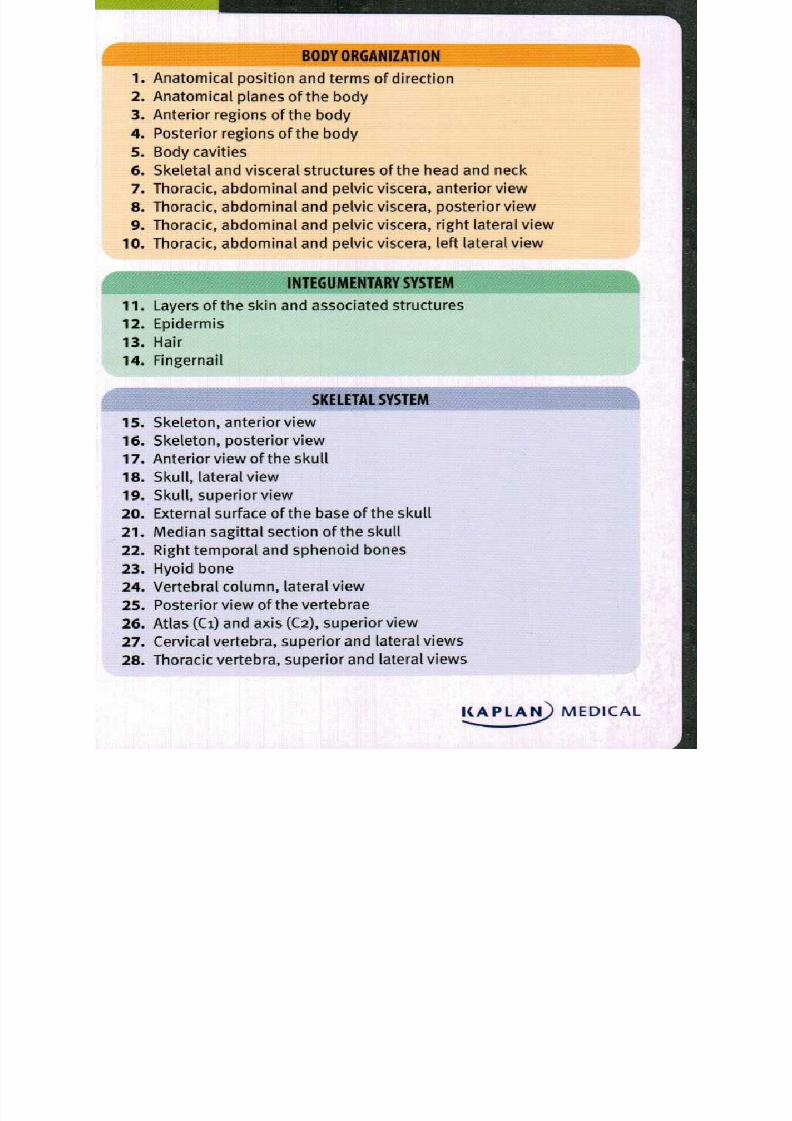
8/13/2019 113264016 Anatomia Omului
http://slidepdf.com/reader/full/113264016-anatomia-omului 288/298
S K E L E T A L S Y S T E M c o a t i )
29 Lumbar vertebra, superior and lateral views
30 Sacrum and coccyx, anterior view
31 Sacrum and coccyx, posterior view
32 Intervertebral discs, lateral and midsagittal views
33 Sternum, anterior view
34 Rib and vertebra, articulated, superior view; rib, posterior view
35 Rib cage, anterior view
36 Pectoral girdle and upper limb, anterior view
37 Scapula, anterior and lateral views
38 Scapula, posterior view
39 Clavicle and related bones, superior view; clavicle, inferior view
40 Humerus, anterior and posterior views
41 Ulna and radius, lateral and anterior views
42 Hand, posterior (dorsal) view
43 Hand, anterior (palmar) view
44 Hip bone, lateral view
45 Pelvis, anterior view
46 Differences between male and female pelvis
47 Lower limb, anterior view
48 Femur and patella, anterior and posterior views
49 Tibia and fibula, anterior and posterior views
50 Bones of the foot, dorsal view
51 Bones of the foot, lateral view
A R T I C U L A T I O N S
52 Gomphosis (peg suture)
53 Suture
54 Syndesmosis, posterior view
55 Synchondrosis
56 Symphysis
57 Synovial joint, diagrammatic sagittal section
58 Tendon sheath
59 Bursa
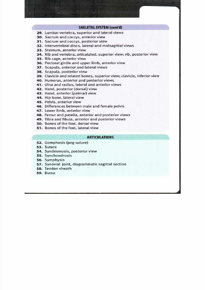
8/13/2019 113264016 Anatomia Omului
http://slidepdf.com/reader/full/113264016-anatomia-omului 289/298
D I G E S T I V E S Y S T E M ( c o n t d )
272 Gall bladder, pancreas and duodenum, anterior view
273 Gall bladder, pancreas and duodenum, anterior cutaway view
274 Rectum, coronal section view
275 Abdomen, transverse section at T12, seen from below
276 Abdomen, transverse section at Li, seen from below
U R I N A R Y S Y S T E M
277 Overview, female urinary system, anterior view278 Anatomical relationships of the urinary system, anterior view
279 Overview, male urinary system, anterior view
280 Overview, male urinary system, viewed from the left
281 Kidney, anterior view
282 Kidney, frontal section view
283 Kidney and renal artery, frontal section view
284 Female urinary bladder, frontal section view285 Nephron of the kidney, microscopic view
R E P R O D U C T IV E S Y S T E M
286 Overview male reproductive system, anterior view
287 Male pelvis, anterolateral view
288 Male reproductive system, sagittal section view
289 Testes, anterior view
290 Seminal vesicles, prostate gland and seminiferous tubules,
posterior view
291 Penis, ventral and cross section views
292 Spermatogenesis, microscopic view
293 Overview female reproductive system, anterior view
294 Female pelvis, anterior view
295 Female reproductive system, sagittal section view
296 Ovary, frontal section view
297 Female reproductive system, menstrual cycle
298 Uterus, ovaries and vagina, frontal section, posterior view
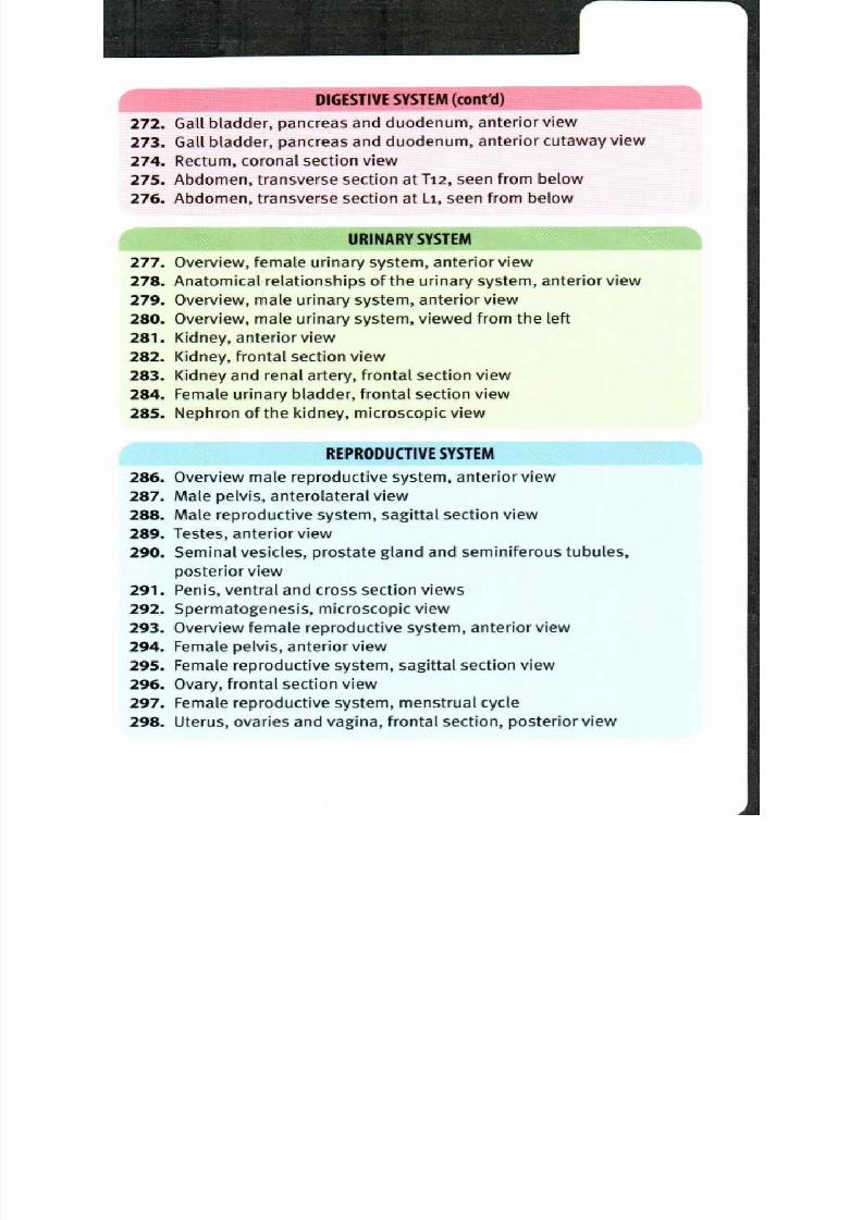
8/13/2019 113264016 Anatomia Omului
http://slidepdf.com/reader/full/113264016-anatomia-omului 290/298
R E P R O D U C T I V E S Y S T E M ( c o n t d )
299 Mammary gland and lymphatic drainage, anterior view
300Female external genitalia, inferior view
C O L O R I N G C A R D S
301. Regions of the abdomen, anterior view
302 Skeletal system, anterior view
303 Nervous system, posterier view
304 Lymphatic system
305 Muscular system
306 Digestive system
307 Cardiovascular system: arteries
308 Skeletal system: hand, posterior (dorsal) view
309 Nervous system: brain, midsagittal section
310 Respiratory system
l 1_)AtiMEDICAL
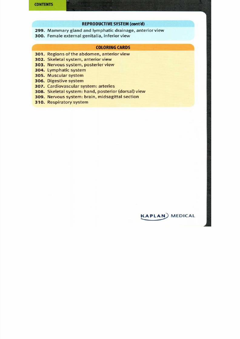
8/13/2019 113264016 Anatomia Omului
http://slidepdf.com/reader/full/113264016-anatomia-omului 291/298
This publication is designed to provide accurate and authoritative
information in regard to the subject matter covered. It is sold with the
understanding that the publisher is not engaged in rendering legal,
accounting, or other professional service. If legal advice or other expert
assistance is required, the services of a competent professional should
be sought.
© 2009 Kaplan, Inc.
Published by Kaplan Publishing, a division of Kaplan, Inc.1 L iberty Plaza, 24th F loor
New York, NY l0006
All rights reserved. The text of this publication, or any part thereof,
may not be reproduced in any manner whatsoever without written
permission from the publisher.
Printed in China
January 2009
1 9 8 7 6 5
ISBN-13: 978-1-4277-9694-3
Kaplan Publishing books are available at special quantity discounts
to use for sales promotions, employee premiums, or educational
purposes. Please email our Special Sales Department to order or for
more information at [email protected] , or write to K aplan
Publishing, i L iberty Plaza, 24th F loor, New York, NY 101306.
8/13/2019 113264016 Anatomia Omului
http://slidepdf.com/reader/full/113264016-anatomia-omului 292/298
Illustrator: tephanie M cCann, M A , is an indep enden t medical
and scientif ic i l lustrator. She received her M A in
M edical and Biological Illustration from Th e John s
H opkins University, and BA in F ine Arts at theUn iversity of C alifornia, Santa Cruz. She is currently
an instructo r at Santa Barbara City Co llege, teaching
Biological Illustration (Biology Departmen t) and
Ad obe Il lustrator (M ultimed ia Arts and Techno logy
D epartm ent). Step hanie's stud io is located in Santa
Barbara, Californ ia.
Text: oann e K ivela Tillotson, PhD , has been teaching
und ergraduate biology laboratories for over 14 years,
the last io of w hich h ave been at Pu rchase Co llege,
State University of New York, where she h as received
the Ped agogy A ward for innovative use of technology
in the teaching laboratory, and t he SU NY C hancellor's
A ward for E xcellence in Teaching. D r. Tillotson receivedher BS degree in Biology from the Un iversity of
Du buque and PhD in Biochemistry from M ichigan State
University.
Ch ief M edical Son ia E. R eichert, M D , previously served as Director
Consultant: f Curriculum for K aplan M edical and is currently
an Internal M edicine R esident at SUNY D ownstateat Broo klyn. Dr . Reichert is a regular participant o f
National Board con ference s and has many years
of experience in the creation and d evelopm ent of
K aplan M edical's online, print, DVD , and other m edia
related ed ucation al tools in th e me dical and all ied
health fields. Sh e is the leading n ational expert in th e
conten t, scoring, and structure of m edical licensingand allied health ex ams.
Medical ailesh H arwani, M D, PhD
Proofreader:

8/13/2019 113264016 Anatomia Omului
http://slidepdf.com/reader/full/113264016-anatomia-omului 293/298

8/13/2019 113264016 Anatomia Omului
http://slidepdf.com/reader/full/113264016-anatomia-omului 294/298
Kaplan's Anatom y f lashcards is designed to help students of human
anatomy learn and memorize the many structures and systems within
the human body. Learning human anatomy is a challenge that is best
met through a three-part process:
• having clear visualizations of the body's anatomical structures
• gaining a thorough understanding of the relationships
between anatomical structures and their functions
• working consistently to review the anatomy in order to
internalize anatomical information for future recall
The front of each card contains a vivid and precise full-color illustration.Clear lines and labels on each image indicate the structures to be
learned, with the identification key listed on the reverse side. In
addition, each card contains a descriptive text, which enhances
understanding of the functions and relationships of each structure.
The set is organized and color-coded according to 15 anatomical
systems for ease-of-use, and can easily be carried around for learning
and review on the go.
As a bonus, this set includes io coloring cards from Kaplan's top-
selling Anatom y Coloring Boo k. Students can color each image and test
their knowledge of various anatomical structures for the ultimate in
academic retention and recall.
Kaplan's Anatomy Flashcards is the ideal human anatomy study
resource for medical and nursing students, healthcare practitioners,
and anyone interested in improving their knowledge of human
anatomy.
CAPLAN) MEDICAL
8/13/2019 113264016 Anatomia Omului
http://slidepdf.com/reader/full/113264016-anatomia-omului 295/298
B O D Y O R G N I Z T I O N
1. An atomical posit ion and term s of direction
2. A natomical planes of the body
3. An terior regions of the bod y4. Posterior regions of the body
5. Bod y cavities
6. Skeletal and visceral structures of the head and neck
7. Th oracic, abdom inal and p elvic viscera, anterior v iew
8. Th oracic, abdom inal and p elvic viscera, posterior view
9. Thoracic, abdom inal and p elvic viscera, right lateral view
10. Tho racic, abdom inal and pelvic viscera, left lateral view
I N T E G U M E N T R Y S Y S T E M
11. L ayers of the skin and associated structures
12. Epidermis
13. Hair
14. Fingernail
S K E L E T L S Y S T E M
15. Skeleton , anterior view
16. Skeleton , posterior view
17. An terior view o f the skull
18. Sku ll, lateral view
19. Skull, superior v iew20. E xternal surface of the base of the skull
21. M edian sagittal sect ion o f the skull
22. R ight tempo ral and sphenoid bones
23. H yoid bone
24. Ve rtebral column , lateral view
25. Posterior view of the vertebrae
26. At las (Ci) and axis (C2), super ior view27. Ce rvical vertebra, super ior and lateral views
28. Thoracic vertebra, superior and lateral views
CAPLAN)MEDICAL

8/13/2019 113264016 Anatomia Omului
http://slidepdf.com/reader/full/113264016-anatomia-omului 296/298
r K E L E T A L S Y S T E M ( c o n t d )
29. Lumbar vertebra, superior and lateral views
30. Sacrum and coccyx, anterior view
31. Sacrum and coccyx, posterior view32. Intervertebral discs, lateral and midsagittal views
33. Sternum, anterior view
34. Rib and vertebra, articulated, superior view; rib, posterior view
35. Rib cage, anterior view
36. Pectoral girdle and upper limb, anterior view
37. Scapula, anterior and lateral views
38. Scapula, posterior view
39. Clavicle and related bones, superior view; clavicle, inferior view
40. Humerus, anterior and posterior views
41. Ulna and radius, lateral and anterior views
42. Hand, posterior (dorsal) view
43. Hand, anterior (palmar) view
44. Hip b one, lateral view
45. Pelvis, anterior view
46. Differences between male and female pelvis
47. Lower limb, anterior view
48. Femur and patella, anterior and posterior views
49. Tibia and Fibula, anterior and posterior views
50. Bones of the foot, dorsal view
51. Bones of the foot, lateral view
A R T I C U L A T I O N S
52. Gomphosis (peg suture)
53. Suture
54. Syndesmosis, posterior view
55. Synchondrosis
56. Symphysis
57. Synovial joint, diagrammatic sagittal section
58. Tendon sheath59. Bursa
8/13/2019 113264016 Anatomia Omului
http://slidepdf.com/reader/full/113264016-anatomia-omului 297/298
52. Go m phosis peg suture)
a Tooth
b Alveolar socket
c Enamel
d Dentin
e Pulp
f Gingiva
g Alveolar ridge
h Periodontal ligaments
A gomphosis is a fibrous synarthrotic immovable) joint holding a tooth
in its alveolar socket in the maxilla or mandible. The bulk of the tooth is
composed of dentin, a mineralized matrix secreted by cells found in the
pulp cavity. The exposed portion of the tooth is covered by a crystalline
calcium phosphate layer called enamel—the hardest substance in thehuman body. The root of the tooth is bound in place by the periodontal
l igament; it is composed of collagen fibers extending from the dentin
of the tooth to the bone surrounding the root of the tooth. A bony
alveolar ridge forms the deep socket or alveolus where the peg-like
root of the tooth is inserted. Superficial to the bone is the gingiva,
mucosal tissue tightly bound to the bone surrounding the teeth; it
provides a smooth surface to reduce friction with food.

8/13/2019 113264016 Anatomia Omului
http://slidepdf.com/reader/full/113264016-anatomia-omului 298/298