
Titlu proiect: Sprijinirea tinerilor doctoranzi cu frecven ... reserve evaluation of its...
41
Investeşte în oameni! FONDUL SOCIAL EUROPEAN Programul Operaţional Sectorial Dezvoltarea Resurselor Umane 2007-2013 Axa Prioritară: 1. Educaţia şi formarea profesională în sprijinul creşterii economice şi dezvoltării societăţii Domeniul Major de Intervenţie: 1.5. Programe doctorale şi postdoctorale în sprijinul cercetării Titlu proiect: "Sprijinirea tinerilor doctoranzi cu frecvenţă prin acordarea de burse doctorale" Cod Proiect: POSDRU/6/1.5/S/8 Beneficiar: Universitatea de Medicină şi Farmacie din Craiova
Transcript of Titlu proiect: Sprijinirea tinerilor doctoranzi cu frecven ... reserve evaluation of its...

Investeşte în oameni!
FONDUL SOCIAL EUROPEAN
Programul Operaţional Sectorial Dezvoltarea Resurselor Umane 2007-2013
Axa Prioritar ă: 1. Educaţia şi formarea profesională în sprijinul cre şterii economice şi
dezvoltării societăţii
Domeniul Major de Intervenţie: 1.5. Programe doctorale şi postdoctorale în sprijinul cercetării
Titlu proiect: "Sprijinirea tinerilor doctoranzi cu frecvenţă prin acordarea de
burse doctorale"
Cod Proiect: POSDRU/6/1.5/S/8
Beneficiar: Universitatea de Medicină şi Farmacie din Craiova
2
UNIVERSITY OF MEDICINE AND PHARMACY OF CRAIOVA
PhD THESIS Abstract
OVARIAN RESERVE:
evaluation of its prognostic markers and insights into the genetics of ovarian
insufficiency
Director:
Professor Liliana NOVAC, MD, PhD
PhD Candidate:
Adela Ştefania VOICAN
CRAIOVA 2011
3
Table of contents
1. Introduction ..................................................................... 4
2. Background ..................................................................... 6
3. A study of the ovarian reserve in primary ovarian insufficiency ............................................................................ 8
Patients and methods ........................................................... 9
Results ............................................................................... 10
Discussions and conclusions. ............................................ 13
4. Genetic aspects of ovarian insufficiency: a study of the NR5A1 gene in a large cohort of patients with idiopathic POI 16
Patients, material and methods.......................................... 16
Results ............................................................................... 19
Discussions and conclusions. ............................................ 23
5. Genetic aspects of hypogonadotropic hypogonadism - TAC3/TACR3 mutations: characterization of neuroendocrine phenotypes and novel mutations ........................................... 24
Patients, material and methods.......................................... 25
Results ............................................................................... 27
Discussions and conclusions ............................................. 34
6. Final conclusions .......................................................... 36
References. ............................................................................ 38
4
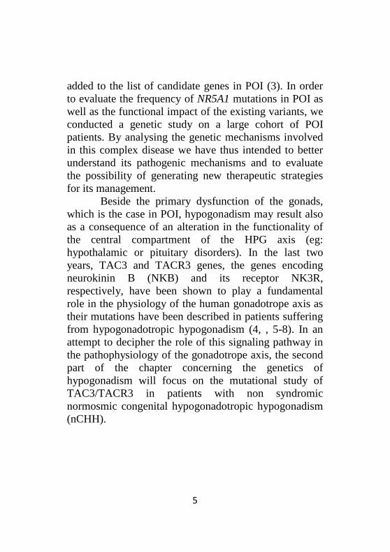
1. Introduction
Current trends in modern society are characterized by a delay of the moment when individuals plan to start a family and to procreate. However, it is well-known that aging is accompanied by a declining fecundity and postponing the moment of conception may result in a difficult procreation process
One key element influencing the reproductive outcome is represented by the ovarian reserve (OR), a notion encompassing both the number and the quality of the existing oocytes available to produce a dominant follicle late in the follicular phase of the menstrual cycle (1). Its assessment may provide essential information in women with impaired fertility but also in healthy women planning to postpone childbearing. Primary ovarian insufficiency (POI), a disorder defined by amenorrhea and sex steroid deficiency in women younger than 40 years (2) may be considered the most extreme phenotype of diminished ovarian reserve at young age. Data regarding the characteristics of OR markers in POI are scant and more evidences evaluating the role of the currently available ovarian reserve markers in POI are needed. Moreover, the aetiology of POI is currently poorly understood and despite the increasing efforts for unveiling the mechanisms responsible for its development, in the great majority of cases this disease is classified as idiopathic. Genetic component has been frequently suggested as a possible explanation of cases of nonsyndromic forms. NR5A1, a nuclear receptor also known as steroidogenic factor 1, has been recently
5
added to the list of candidate genes in POI (3). In order to evaluate the frequency of NR5A1 mutations in POI as well as the functional impact of the existing variants, we conducted a genetic study on a large cohort of POI patients. By analysing the genetic mechanisms involved in this complex disease we have thus intended to better understand its pathogenic mechanisms and to evaluate the possibility of generating new therapeutic strategies for its management.
Beside the primary dysfunction of the gonads, which is the case in POI, hypogonadism may result also as a consequence of an alteration in the functionality of the central compartment of the HPG axis (eg: hypothalamic or pituitary disorders). In the last two years, TAC3 and TACR3 genes, the genes encoding neurokinin B (NKB) and its receptor NK3R, respectively, have been shown to play a fundamental role in the physiology of the human gonadotrope axis as their mutations have been described in patients suffering from hypogonadotropic hypogonadism (4, , 5-8). In an attempt to decipher the role of this signaling pathway in the pathophysiology of the gonadotrope axis, the second part of the chapter concerning the genetics of hypogonadism will focus on the mutational study of TAC3/TACR3 in patients with non syndromic normosmic congenital hypogonadotropic hypogonadism (nCHH).
6
2. Background
Throughout the first chapter of the literature
review, entitled “The human ovary throughout life: a key player in the reproductive function”, I will briefly present the different elements influencing the ovarian development and function. Next, an analysis of the consequences of the ovarian function cessation encountered in menopause and of the controversies currently surrounding the hormone replacement therapy in menopause is detailed.
The second chapter of the literature review, entitled “Ovarian reserve – evaluation and clinical implications” , represents a review of the available methods for ovarian reserve evaluation, underlying the advantages but also the current limitations characterising each method.
The central part of the introductive section is constituted by the chapter entitled “Pathologic aspects of the ovarian reserve: primary ovarian insufficiency”. This chapter is focused on a disorder characterised by the premature alteration of the ovarian reserve, POI, and a special attention is paid to the genetic mechanisms possibly involved in its pathophysiology. Finally, a detailed presentation of one candidate gene for POI, the NR5A1 gene, is included in the last chapter of the literature review: “NR5A1 – a key player in the reproductive system”.

7
Personal contributions
8
3. A study of the ovarian reserve in primary ovarian insufficiency
Notwithstanding the important advances in the
ovarian reserve evaluation, a single test which exactly estimates the oocyte pool has not been determined yet. Moreover, data regarding the characteristics of OR markers in patients with POI are scant. In this context, given the need for more evidences regarding the value of the ovarian reserve markers in POI, our study focussed on the ovarian reserve assessment in women admitted in our clinic for infertility investigations.
We have centred our evaluation on the parameters which are currently frequently used or considered to provide the best results in the OR assessment:
- follicle stimulating hormone (FSH): the most commonly used OR biomarker in the current clinical practice (9);
- anti-műllerian hormone (AMH): the biomarker considered to provide a better estimation of ART outcome compared to other available serum markers (10);
- Inhibin B : it is regarded as a serum marker of oogenesis and may offer an improved diagnosis of ovarian function;
- follicular count (AFC): the ultrasound test considered as the test of first choice when estimating quantitative ovarian reserve before IVF (11).
9
Patients and methods In this study we have included 43 women
presenting for infertility evaluation at the Municipal Hospital of Craiova between 2000-2010. The following inclusion criteria were applied to patients included in our study: women younger than 40 years with elevated FSH levels (FSH superior to 30 UI/l), the presence of both ovaries (assessed by ultrasound), no evidence of other endocrine disorders (e.g. polycystic ovarian syndrome, hypothyroidism, hyperthyroidism or hyperprolactinemia) which might interfere with the hormonal measurements of the ovarian reserve, no endometriosis, patent fallopian tubes (assessed by hysterosalpingography) and exclusion of male causes for the couple infertility (normal semen analysis). The study was performed according to the protocol that was approved by the University of Medicine and Pharmacy of Craiova Ethics Committee and written informed consent was obtained from the recruited patients.
Hormonal assessment. Plasma FSH, LH, E2, PRL and TSH were dosed
using an electrochemiluminescence immunoassay (ECLIA), while AMH and inhibin B were dosed using an enzyme-linked immunosorbent assay (ELISA). . In order to ensure a coherent representation of the hormonal profiles found among our patients, all serum hormonal measurements were performed by the same laboratory
Echographic assessment of the ovaries. Echographic evaluation of the ovarian volume and of the presence of ovarian follicles was performed by transvaginal technique using a Voluson Expert 730TM (GE Medical Systems) equipped with a 7.5-MHz
10
transvaginal transducer. Ultrasound examinations were carried out by the same experienced specialist in each of the patients participating in the study.
Statistical analysis Qualitative variables were expressed as numbers
(percentages) while quantitative variables were expressed as means ± SD. Statistical significance was assumed when a null hypothesis could be rejected at P < 0.05. Statistical and graphical analysis was performed using the Prism version 3.02 (GraphPad Software Inc., SanDiego, CA).
Results 43 patients presenting for infertility evaluation in
the Gynaecology department were included in the study. Their mean age was of 32.3 (21-40 years) and the majority of them presented with secondary amenorrhea (41 patients, representing 95% of the entire study group). The mean FSH level was 83.4 mUI/mL (41.3-196.5 mUI/mL), while the mean E2 was 24.8 pmol/L (10-136.4 mUI/mL). As expected, the serum level of the two hormonal markers of the ovarian reserve, AMH and inhibin B, were low: 0.76µg/L and 12.4 ng/L respectively. Inhibin B levels were below the detection threshold in 25 patients (58%) while AMH levels were not detectable in 27 patients (62%). In 9 of the 16 patients presenting a detectable AMH level, this level was inferior to the normal range for the respective age group (1-8µg/L). Transvaginal ultrasound measurements included the OVVOL and AFC.. The mean ovarian volume in our study was 3.6 cm3, while the mean AFC value in our study group was 1.8.
11
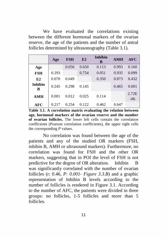
We have evaluated the correlations existing between the different hormonal markers of the ovarian reserve, the age of the patients and the number of antral follicles determined by ultrasonography (Table 3.1).
Age FSH E2 Inhibin
B AMH AFC
Age 0.056 0.650 0.113 0.993 0.160
FSH 0.293 0.754 0.051 0.935 0.099
E2 0.070 0.049 0.350 0.873 0.432
Inhibin B
0.245 0.298 0.145 0.465 0.001
AMH 0.001 0.012 0.025 0.114 2.72E-06
AFC 0.217 0.254 0.122 0.462 0.647
Table 3.1. A correlation matrix evaluating the relation between age, hormonal markers of the ovarian reserve and the number of ovarian follicles. The lower left cells contain the correlation coefficients (Pearson correlation coefficients), the upper right cells the corresponding P values.
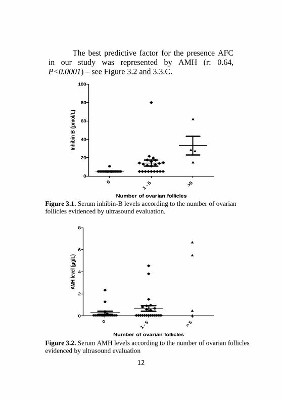
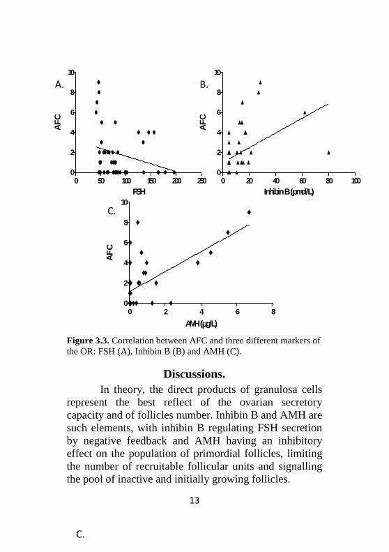
No correlation was found between the age of the patients and any of the studied OR markers (FSH, inhibin B, AMH or ultrasound markers). Furthermore, no correlation was found for FSH and the other OR markers, suggesting that in POI the level of FSH is not predictive for the degree of OR alteration. Inhibin B was significantly correlated with the number of ovarian follicles (r: 0.46, P: 0.001- Figure 3.3.B) and a graphic representation of Inhibin B levels according to the number of follicles is rendered in Figure 3.1. According to the number of AFC, the patients were divided in three groups: no follicles, 1-5 follicles and more than 5 follicles.
12
The best predictive factor for the presence AFC in our study was represented by AMH (r: 0.64, P<0.0001) – see Figure 3.2 and 3.3.C.
Number of ovarian follicles
Inhi
bin
B (p
mol
/L)
0
1 -
5 >5
0
20
40
60
80
100
Number of ovarian follicles
AMH
leve
l (µµ µµg
/L)
0
1 -
5 > 5
0
2
4
6
8
Figure 3.1. Serum inhibin-B levels according to the number of ovarian follicles evidenced by ultrasound evaluation.
Figure 3.2. Serum AMH levels according to the number of ovarian follicles evidenced by ultrasound evaluation
13
Figure 3.3. Correlation between AFC and three different markers of the OR: FSH (A), Inhibin B (B) and AMH (C).
Discussions. In theory, the direct products of granulosa cells
represent the best reflect of the ovarian secretory capacity and of follicles number. Inhibin B and AMH are such elements, with inhibin B regulating FSH secretion by negative feedback and AMH having an inhibitory effect on the population of primordial follicles, limiting the number of recruitable follicular units and signalling the pool of inactive and initially growing follicles.
AMH (µµµµg/L)
AF
C
0 2 4 6 80
2
4
6
8
10FSH
AF
C
0 50 100 150 200 2500
2
4
6
8
10
Inhibin B (pmol/L)A
FC
0 20 40 60 80 1000
2
4
6
8
10
B.
C.
A.
C.
14
Among the ultrasound markers of the OR, the ovarian volume is accepted as useful for OR prediction, even if its main role was evidenced in patients with polycystic ovary syndrome. In patients participating in our study a reduced ovarian volume was evidenced, with a mean value of 3.6 cm3, suggesting a poor ovarian reserve in our study group. AFC is considered an important screening test for the prediction of poor ovarian response in couples candidate for ART. We have considered the AFC results as follows: < 5 follicle was considered as a poor reserve, 5–7 follicles a low count and 8–12 follicles a slightly reduced reserve. The majority of the patients included in our study presented a poor reserve and a value superior to 5 follicles was found only in 4 patients.
Massin et al. have previously demonstrated that plasma levels of E2 and inhibin B are of limited value in predicting the presence of an ovarian reserve in patients with POI (12). In our study both inhibin B and AMH were correlated with the AFC determined by ultrasound. However, the inhibin B displayed a less significant correlation compared with AMH, suggesting that AMH has a superior predictive value for the presence of ovarian follicles. No correlation of ovarian reserve markers and the age or the FSH level were evidenced in women with POI, contrary to what was reported in studies of OR markers in women not presenting with POI.
Study limitations. Due to the relatively small number of patients participating in this study, these results should be considered as preliminary and further analyses on larger cohorts of patients are needed to
15
confirm them. Furthermore, the presence of ovarian follicles was assessed by ultrasound and not by ovarian biopsies, this method possibly underestimating the presence of ovarian follicles (12). However, the ovarian biopsy realisation implies an invasive technique with limited practical value in POI, being not clinically recommended in women with POI (13) .
Beside the impact manifested by the persistence of ovarian follicles on the therapeutic strategy of patients with POI, its estimation might be even more important in the evaluation of the aetiology of POI. The strategy of testing for the different genetic anomalies responsible for POI may be adapted according to the ovarian phenotype. Thus, patients in which the presence of follicles at ultrasonography was evidenced might be good candidates for genetic studies of genes involved in folliculogenesis, while patients in which no follicles were found should be screened for defects in genes involved in the ovarian development or in the early follicular development (14).
Conclusions. The present study shows that in women with
POI, inhibin B and AMH concentrations positively correlate with the number of antral follicles estimated by ultrasound, with AMH exhibiting a better predictive value. Even if none of the currently existing markers does not present an absolute predictive value, the assessment of ovarian reserve in POI patients may guide both therapeutic and etiologic diagnosis strategy.
16
4. Genetic aspects of ovarian insufficiency: a study of the NR5A1
gene in a large cohort of patients with idiopathic POI
Primary ovarian insufficiency (POI) is a
condition defined by the presence of amenorrhea, sex steroids deficiency and two recordings of serum FSH levels in the menopausal range in women younger than 40 years (2). As previously mentioned, the aetiology of POI represents a challenge for clinicians, and despite the intensive research conducted in this field, in a vast majority of cases this condition is classified as “idiopathic”. NR5A1 is a gene recently reported as possibly associated with POI (3).
The central aim of our study was to evaluate the frequency of NR5A1 variants in idiopathic POI. Furthermore, by a detailed analysis of each of the identified variants, we intended to offer the dimension of the clinical implications of NR5A1/SF-1 in POI. Additionally, given the current variety of phenotypes associating NR5A1 mutations, we assessed also the possibility of determining a genotype – phenotype correlation in these patients.
Patients, material and methods A cohort of 357 patients diagnosed with POI was
available for conducting our study. A written informed consent for genetic studies was obtained from all patients prior to further investigations. Karyotype and X-fragile genetic testing was performed in all patients
17
participating in our study and NR5A1 was therefore analysed only in patients with normal karyotype and no premutations of the FMR1 gene (Fragile X Mental Retardation gene). The control population consisted of 82 subjects from the general population.
Laboratory assays . Plasma levels of FSH and LH were measured by conventional RIA, inhibin B was measured in serum by an enzyme immunometric assay , while serum AMH concentrations were measured in duplicate using a sandwich ELISA method.
DNA extraction and sequencing. Genomic DNA was extracted from white blood cells. The entire coding region (exons 2 to 7) of NR5A1 was amplified and sequenced and sequencing products were analyzed on an automated capillary sequencer (ABI PRISM 3130xl Genetic Analyzer, Applied BiosystemsTM, Foster City, CA). Electrophoregram-derived sequences were compared to NCBI reference NM_004959.4.
In vitro mutations reproduction. Mutant NR5A1 constructs containing the S54R, P198L and previously described G35E and P129L variants were generated by site-directed mutagenesis (QuikChange II Site-Directed Mutagenesis Kit, Stratagene) using the wild-type (WT) human NR5A1 cDNA in a pCMX vector. Mutant NR5A1 coding sequence was verified by full sequencing prior to functional studies.
Cell culture. Hek293T cells were plated 48h before transfection in high-glucose Dulbecco’s minimal essential medium supplemented with 2mM glutamine, 100 IU/mL penicillin, 100 µg/mL streptomycin and 10% heat-inactivated fetal calf serum, at 37°C in a 5%CO2 atmosphere.
18
Transfections. The cells were transiently transfected in serum free OptiMEM, using Lipofectamine 2000, with WT or mutant NR5A1 pCMX, a NR5A1 responsive promoter linked to luciferase (Cyp11a-luciferase construct) and a beta-galactosidase vector as a control of transfection efficiency.
Luciferase assay. 24 h after transfection, cells were rinsed with cold PBS and harvested using the Passive Lysis Buffer (Promega). Supernatants were used for luciferase and beta-galactosidase assays. Luciferase activities were normalized to ß-galactosidase activities and expressed as percents of relative luciferase units compared with control.
Studies of WT/mutant interactions Studies of possible potential dominant negative
interactions between the WT and mutant NR5A1 were performed by transfecting increasing amounts of WT or mutant NR5A1 expression vectors with either an empty vector or WT NR5A1 and Cyp11a1 reporter in HEK293T cells. DNA quantities transfected were maintained constant by the addition of an empty vector.
Electrophoretic mobility-shift assay Nuclear extracts were prepared from HEK293T
cells transfected with WT or mutant NR5A1. Synthetic oligonucleotides corresponding to the 3’ SF1 response elements on the Cyp11a minimal promoter were generated. The probe was labeled with [32P]dCTP by Kleanow polymerase. The DNA-protein complexes were separated on polyacrylamide gels, which were dried under vacuum and exposed to X-ray film at -80°C. In competition experiments, a 125-fold molar excess of unlabeled probe was included.
19
Confirmation of the transfection: NR5A1/SF-1 RNA and protein detection
In order to confirm that the HEK293T cells used in our experiments are not natively NR5A1- expressing cells and to further test NR5A1 expression following transfections with the pCMX NR5A1, RNA extracts and protein extracts of both transfected and non-transfected cells were analysed by RT-PCR (Reverse transcription polymerase chain reaction) and Western Blot, using the High Capacity cDNA Reverse Transcription Kit (Applied Biosystems) for RT-PCR and a NR5A1/SF1 antibody from Santa Cruz Biotechnology was used in WBr experiments. Detection of the resulting complexes was performed using the Odyssey Infrared Imaging System (LiCor Biosciences) and images were further analyzed using the Odyssey Application Software, version 1.2 (LiCor Biosciences).
Results The initial POI cohort included 357 patients with
a mean age of 26.5 ± 7.8 years (11–39) at the time of the diagnosis, a median FSH level of 78.0 IU/l (30.3–284; normal range : 3–9 IU/l), a median inhibin B of 5 pg/ml (5–381; n: 60–200 ng/l) and a serum AMH below the detection threshold (0.4 ng/ml) in 113 women (77%), below the normal range in 27 women (18%) and within the normal range in 7 women (5%). Serum AMH values were negatively correlated with the age of the patients (r=-0.23, P=0.005). Serum E2 correlated with inhibin B levels (r=0.43, P<0.001). No correlation, either positive or negative, was found between AMH, inhibin B, and FSH. No significant difference was observed between
20
FSH, inhibin B, and AMH levels of patients presenting with either primary or secondary amenorrhea.
NR5A1 mutations frequency in primary ovarian insufficiency. After excluding other causes of POI, NR5A1 was sequenced in 188 patients. The overall mutation frequency resulting from our study was of 1.59% (95% Confidence Interval 0-3.4%). The missense variants identified in our study group, included two original variants (p.S54A and p.P198L) and two previously reported variants (p.P129L and p.G123A) (3, 15). These four nonsynonymous substitutions were found in a heterozygous state in 3 patients. All the three patients were of caucasian origin and presented with secondary amenorrhea and normal sexual development.
Functional studies of NR5A1 mutations. The putative impact of S54R, P129L and P198L variants on the nuclear receptor NR5A1 function was tested by electrophoretic-mobility shift assay and luciferase assays. The G123A variant, already described as having an activity similar to that of the wild-type protein (3), was not included in our functional characterisation. A loss-of-function mutation, G35E (c.104G>A and c.105C>A, p.G35E), the first identified NR5A1 human mutation, was used as a control for the experiments (16).
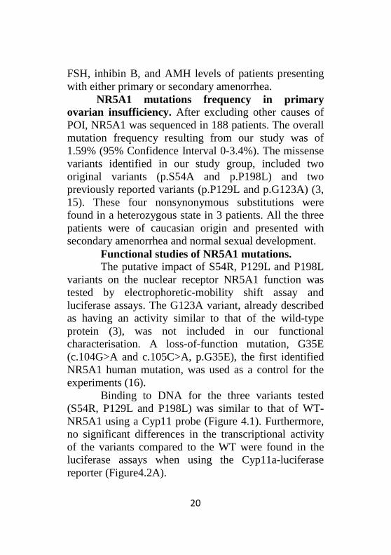
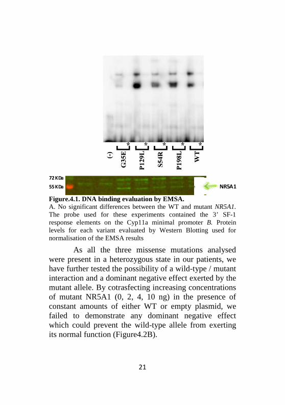
Binding to DNA for the three variants tested (S54R, P129L and P198L) was similar to that of WT-NR5A1 using a Cyp11 probe (Figure 4.1). Furthermore, no significant differences in the transcriptional activity of the variants compared to the WT were found in the luciferase assays when using the Cyp11a-luciferase reporter (Figure4.2A).
21
Figure.4.1. DNA binding evaluation by EMSA. A. No significant differences between the WT and mutant NR5A1. The probe used for these experiments contained the 3’ SF-1 response elements on the Cyp11a minimal promoter B. Protein levels for each variant evaluated by Western Blotting used for normalisation of the EMSA results
As all the three missense mutations analysed were present in a heterozygous state in our patients, we have further tested the possibility of a wild-type / mutant interaction and a dominant negative effect exerted by the mutant allele. By cotrasfecting increasing concentrations of mutant NR5A1 (0, 2, 4, 10 ng) in the presence of constant amounts of either WT or empty plasmid, we failed to demonstrate any dominant negative effect which could prevent the wild-type allele from exerting its normal function (Figure4.2B).
72 KDa
55 KDa NR5A1
22
Figure 4.2. Functional characterisation of the NR5A1variants identified in our group of POI patients Luciferase assay evaluating the transcriptional activity of NR5A1 variants (A) and the potential dominant-negative effect exerted by mutant variants (B). A. Transfection of equal amounts of empty (-), WT, or mutant (G35E, S54R, P129L, P198L) expression vector. Results are expressed as a percentage of WT-NR5A1 activity. B. Transfection of increasing amounts of empty (-), WT, or mutant expression vector (0, 2, 4, 10 ng) with 2 ng of empty vector (ng of WT-NR5A1containing vector and a Cyp11a promoter reporter (100 ng). Transfected DNA was maintained constant by transfecting additional empty vector. All experiments (A, B) have been performed by using a Cyp11 promoter linked to the luciferase gene transfected in HEK293T cells. Data represent the mean±SEM of three independent experiments each performed in quadriplaRelative light units.
0
50
100
150
(-) G35E P129L S54R P198L
- - - - - - - - - - - -- - - - - -+ + + ++ + + + + + + + + + + +
NR5A1 missense Luciferase assay
variants (A) and the negative effect exerted by mutant variants (B).
), WT, or mutant (G35E, S54R, P129L, P198L) expression vector. Results are
Transfection ), WT, or mutant NR5A1
expression vector (0, 2, 4, 10 ng) with 2 ng of empty vector (-) or 2 promoter reporter
transfecting All experiments (A, B) have been
performed by using a Cyp11 promoter linked to the luciferase gene Data represent the mean±SEM of
three independent experiments each performed in quadriplate. RLU,
WT
- - + + + +
23
Discussions and conclusions. Sequencing the NR5A1 gene in 188 patients with
POI and no other known genetic anomalies has evidenced mutations in three patients, resulting in an overall prevalence of 1.59%, significantly lower than the one initially estimated by the Lourenco et al. Furthermore, evaluating the functional impact of the three missense variants identified in our study group showed no significant difference compared to the activity of the wild-type NR5A1. However, even if the basal transactivation capacity of our variants doesn’t differ from that of the WT in our experimental system, the pathogenic link between the variants and the disease couldn’t be excluded. The absence of these variants in the control group or in the dbSNP supports the hypothesis of a causal link between NR5A1 variants and POI. NR5A1 variants may exert variable transactivation capacity depending on the specific target gene or depending on the cofactors involved in the transcriptional process. Thus, evaluating the basal transcriptional activity doesn’t cover all the vast situations encountered in the “real“ gonadal environment.
In conclusion, analyzing NR5A1 gene in a large cohort of patients diagnosed with POI showed that NR5A1 is not a major cause of idiopathic POI and we suggest that genetic investigation of NR5A1 in POI might be restricted to the limited cases presenting with familial history of POI and disorders of sexual development in male relatives
24
5. Genetic aspects of hypogonadotropic hypogonadism - TAC3/TACR3 mutations: characterization of
neuroendocrine phenotypes and novel mutations
After performing the study concerning the role played by NR5A1/SF-1 in POI, a disease in which the primary defect is localised at the gonadal level, the genetic study of the gonadotrope axis was then centered on a disease characterised also by gonadal insufficiency, but this time secondarily to a central disorder of the HPG axis: the hypogonadotropic hypogonadism. Loss-of-function mutations in TAC3 and TACR3, the genes encoding neurokinin B (NKB) and its receptor NK3R, respectively, have been described in patients with non syndromic normosmic congenital hypogonadotropic hypogonadism (nCHH), pointing to a fundamental role of this pathway in the physiology of the human gonadotrope axis (4, 5-8). By conducting a detailed neuroendocrine study of adult nCHH patients with TAC3/TACR3 mutations, we aimed to decipher the role of this signaling pathway in the pathophysiology of the gonadotrope axis. We have first screened the nCHH patients for novel TACR3 mutations, followed by the functional characterization of the newly identified mutations. We have consequently provided evidence that NKB and its receptor participate in controlling the frequency of GnRH secretory pulses.
25
Patients, material and methods From a cohort of 352 patients with congenital
hypogonadotropic hypogonadism we selected 173 patients with normosmic nCHH and TAC3/TACR3 mutations have been screened in all the patients participating in the study. The presence of gonadotropin deficiency was defined by the following elements (17): absent or incomplete puberty at age 18 years; low plasma testosterone levels in men and low to low-normal E2 levels in women plus low or normal serum gonadotropin levels; otherwise normal pituitary function; normal serum ferritin concentrations; normal MRI of the hypothalamic-pituitary region; a normal sense of smell on olfactometry, and no anosmia/hyposmia in relatives. All the participants and controls gave their written informed consent for genetic analyses, which were approved by the Paris-Sud Medical School and Bicêtre Hospital Ethics Committees.
Neuroendocrine profiling of TAC3/TACR3-mutated nCHH patients was estimated by evaluating the free alpha subunit secretion, which is considered the best surrogate for evaluating GnRH secretion in nCHH patients presenting with an apulsatile LH profile (18, 19). The FSH/LH ratio studies were performed in 11 patients with biallelic TAC3/TACR3 mutations and the results were compared with those in patients with CHH of different genetic origins (e.g. mutations in GPR54/KISS1R, GNRH1, GNRHR, KAL1, FGFR1 or PROK2/PROKR2).
Hormone assays. We measured serum levels of LH, FSH, inhibin B, testosterone and E2 levels with immunoradiometric assay, ELISA or RIA, respectively.
26
Endogenous LH and alpha-subunit secretion, analyzed with Thomas’ algorithm, was evaluated overnight at 10-minutes intervals (20-23).Serum free alpha-subunit (FAS) levels were measured, using an immunoradiometric assay (IRMA) with two monoclonal antibodies (Immunotech, Marseille, France).
DNA analysis. Genomic DNA was extracted from white blood cells by using standard procedures.
Modeling studies. The model of NK3R was generated by homology, using as template the crystal structure of rhodopsin, a hepta-transmembrane protein, with the Modeller package (version 9.8) (24). Loops differing in length between NK3R and rhodopsin were reconstructed using the structural proteins bank (rotamer library) included in the O package (25).
Directed mutagenesis. NK3R mutants were reproduced by site-directed mutagenesis using the pcDNA3.1+ plasmid encoding human TACR3 with an hemagglutinin (HA) tag localized on the extracellular NK3R N-terminal extremity (Missouri S&T cDNA Resource Center) with the QuickChange Stratagen II kit (Stratagene, La Jolla, CA)
Luciferase reporter gene assays. Tachykinin G protein-coupled receptors signal through several second messenger pathways including phosphoinositide and MAP kinase pathways (ERK 1/2) (26). We thus used the luc2P/SRE/Hygro plasmid (Promega) which can induce luciferase production in response to MAP kinase activation as a reporter gene system. HEK 293 cells were used in our experiments. After transfection, the cells were treated with seven different dilutions (from 10-10M to 10-6M) of neurokinin B and incubated for 5–6 h.
27
Then, they were harvested and assayed for β-galactosidase and luciferase activities, using a luminometer (Victor, Perkin Elmer). To standardize the transfection efficiency, the relative light units obtained in the luciferase assay were divided by the optical density obtained in the β-galactosidase assay. Maximum activity was considered at 5x10⁻⁷ M NKB. All experiments have been performed at least in triplicate.
Statistical analyse. Prism version 3.02 (GraphPad Software Inc., SanDiego, CA) was used for curve fitting from at least three independent experiments performed in triplicate.
Immunocytochemistry. After transfection with the expression vector coding for HA-TACR3, cells were treated and incubated overnight at 4°C with an anti-HA antibody (clone 3F10, Roche Applied Science), followed by an anti-rat fluorochrome-coupled secondary antibody (Dylight 549, Jackson ImmunoResearch Laboratories) for 30 min. Nuclear counterstaining was performed with 0.5 µg/mL DAPI (4′,6′ -diamidino-2-phenylindole). Fluorescent cells were observed with an Olympus Provis AX70. Images were acquired with Qcapture Pro version 5.1 (Q Imaging Inc.). For live cell staining, a Zeiss LSM-510 confocal scanning laser microscope (Carl Zeiss, Thornwood, NY) was used to acquire Z-series of focal planes using a Plan Apochromat 63 oil objective.
Results TAC3/TACR3 genes mutation studies:
molecular analysis, modeling and functional studies GNRH1, GNRHR, KISS1, KISS1R and FGFR1 mutations were not found in the 173 patients.
28
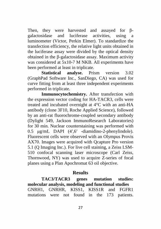
TAC3/TACR3 variants were present in 9 propositi (1 carrying a TAC3 variant and 8 with TACR3) of these 173 patients (5.2%) (Figure 5.1).
Figure 5.1 Schematic representation of the human NK3R variants found in a cohort of 173 normosmic CHH. The mutated residues are indicated by red circles
Modeling and functional studies of the missense mutations. Modeling was used to predict the potential impact of point mutations on the three-dimensional organization of NK3R. We found that the hydrophobic Tyr267 residue is located in the middle of the fifth transmembrane segment and points towards the lipid bilayer. Introduction of an asparagine residue at position 267 (Y267N mutation) places a polar residue in a highly hydrophobic environment. Thus, it is likely that the asparagine side chain, in order to adopt a buried position, will induce transmembrane reorganization (twist or rotation). It is thus likely that the Tyr267Asn mutation leads to NK3R misfolding and dysfunction. This misfolding hypothesis was tested by
29
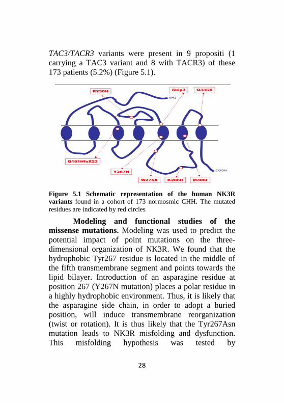
immunocytochemical studies in order to observe subcellular localization of the Trp275stop mutant. Immunocytochemical studies performed in non permeabilized cells showed the membrane location of the wild-type NK3R molecule (Figure 5.2, upper panel). In contrast, the Tyr267Asn mutant was not detected at the membrane, suggesting defective trafficking (27). In order to ascertain that the mutant was efficiently expressed, we performed immunocytochemical experiments in permeabilized cells.
Figure 5.2. Subcellular localization of ectopically expressed NK3R and Y267N mutant in non-permeabilized and permeabilized cells. Cells were transfected with the indicated expression vector and then treated for indirect immunofluorescence as described in the Methods section. The nuclei are counterstained by DAPI (blue). Upper panel: Z-stack projection of NK3R distribution in non-permeabilized cells obtained by confocal microscopy. Lower panel: fluorescence micrographs of fixed and permeabilized cells. Note the absence of Y267N NK3R mutant at the membrane (upper) despite its efficient expression in the cell (lower) whereas wild-type NK3R is localized at the plasma membrane.
30
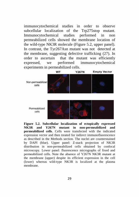
Compared to wild-type, the NK3R mutant was localized in the perinuclear region suggesting a misfolding (Figure 5.2, lower panel). Thus, the substitution of Tyr267 by asparagine impaired the proper targeting of the receptor to the cell surface. To quantify and compare the stimulation effect of neurokinin B on wild-type and mutant NK3R, we performed dose-response curves for SRE luciferase assay in HEK 293 cells. Consistent with immunocytochemical and modeling studies, functional analyses clearly showed that mutated NK3R failed to stimulate a p44/42 MAP-sensitive SRE reporter gene contrary to the wild-type receptor which showed a clear dose-response stimulation in the presence of neurokinin B (Figure 5.3).
Figure 5.3. Functional characterisation of Y267N variant. NKB dose response of the reporter luc2P/SRE. Increasing concentrations of NKB led to an increase in the luciferase activity of wild-type NK3R (black circles). The mutant NK3R (red triangles) did not significantly enhance luciferase activity.
Similar modeling and functional studies were performed to characterize the other variants identified in our cohort. In the case of Lys286Arg, Met306Ile and Arg230His, no meaningful difference between the variants and the wild-type NK3R were found, suggesting that these variants did not affect the structure and the
31
function of NK3R. Therefore they can not be considered as causative of the disease.
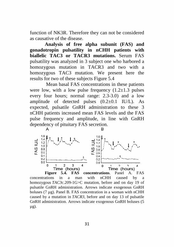
Analysis of free alpha subunit (FAS) and gonadotropin pulsatility in nCHH patients with biallelic TAC3 or TACR3 mutations. Serum FAS pulsatility was analyzed in 3 subject one who harbored a homozygous mutation in TACR3 and two with a homozygous TAC3 mutation. We present here the results for two of these subjects Figure 5.4
Mean basal FAS concentrations in these patients were low, with a low pulse frequency (1.2±1.3 pulses every four hours; normal range: 2.3-3.0) and a low amplitude of detected pulses (0.2±0.1 IU/L). As expected, pulsatile GnRH administration to these 3 nCHH patients increased mean FAS levels and the FAS pulse frequency and amplitude, in line with GnRH dependency of pituitary FAS secretion.
Figure 5.4. FAS concentrations. Panel A. FAS
concentrations in a man with nCHH caused by a homozygous TAC3c.209-1G>C mutation, before and on day 19 of pulsatile GnRH administration. Arrows indicate exogenous GnRH boluses (7 µg). Panel B. FAS concentration in a woman with nCHH caused by a mutation in TACR3, before and on day 13 of pulsatile GnRH administration. Arrows indicate exogenous GnRH boluses (5 µg).
32
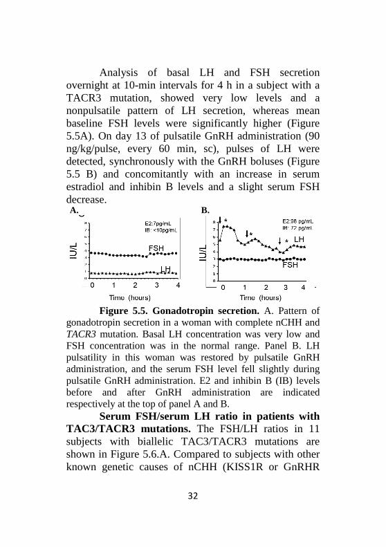
Analysis of basal LH and FSH secretion overnight at 10-min intervals for 4 h in a subject with a TACR3 mutation, showed very low levels and a nonpulsatile pattern of LH secretion, whereas mean baseline FSH levels were significantly higher (Figure 5.5A). On day 13 of pulsatile GnRH administration (90 ng/kg/pulse, every 60 min, sc), pulses of LH were detected, synchronously with the GnRH boluses (Figure 5.5 B) and concomitantly with an increase in serum estradiol and inhibin B levels and a slight serum FSH decrease.
Figure 5.5. Gonadotropin secretion. A. Pattern of
gonadotropin secretion in a woman with complete nCHH and TACR3 mutation. Basal LH concentration was very low and FSH concentration was in the normal range. Panel B. LH pulsatility in this woman was restored by pulsatile GnRH administration, and the serum FSH level fell slightly during pulsatile GnRH administration. E2 and inhibin B (IB) levels before and after GnRH administration are indicated respectively at the top of panel A and B.
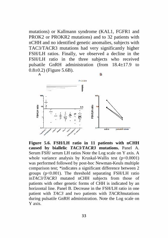
Serum FSH/serum LH ratio in patients with TAC3/TACR3 mutations. The FSH/LH ratios in 11 subjects with biallelic TAC3/TACR3 mutations are shown in Figure 5.6.A. Compared to subjects with other known genetic causes of nCHH (KISS1R or GnRHR
B. A.
33
mutations) or Kallmann syndrome (KAL1, FGFR1 and PROK2 or PROKR2 mutations) and to 32 patients with nCHH and no identified genetic anomalies, subjects with TAC3/TACR3 mutations had very significantly higher FSH/LH ratios. Finally, we observed a decline in the FSH/LH ratio in the three subjects who received pulsatile GnRH administration (from 18.4±17.9 to 0.8±0.2) (Figure 5.6B).
Figure 5.6. FSH/LH ratio in 11 patients with nCHH caused by biallelic TAC3/TACR3 mutations. Panel A. Serum FSH/ serum LH ratios Note the Log scale on Y axis. A whole variance analysis by Kruskal-Wallis test (p<0.0001) was performed followed by post-hoc Newman-Keuls multiple comparison test; *indicates a significant difference between 2 groups (p<0.001). The threshold separating FSH/LH ratio inTAC3/TACR3 mutated nCHH subjects from those of patients with other genetic forms of CHH is indicated by an horizontal line. Panel B. Decrease in the FSH/LH ratio in one patient with TAC3 and two patients with TACR3mutations during pulsatile GnRH administration. Note the Log scale on Y axis.
34
Discussion We found here TAC3/TACR3 mutations in
5.2% of our nCHH population, a prevalence similar to that described by Gianetti et al (7). In this series, all the subjects with nCHH and biallelic TAC3/TACR3 mutations were born to healthy heterozygous parents. This reinforces the autosomal recessive transmission of these two genetic forms of nCHH, as reported by Topaloglu et al. and our team(4, 6).
Analysis of gonadotropin concentrations in patients with biallelic TAC3/TACR3 mutations showed very weak and apulsatile LH secretion, contrasting with preserved FSH concentrations. This profile points to the existence of low-frequency (and probably low amplitude) endogenous GnRH secretion(6). We actually observed slow FAS secretion in all three patients studied, strongly supporting this hypothesis. Moreover, pulsatile GnRH administration at a physiological frequency re-established pulsatile LH and FAS secretion, and decreased the FSH/LH ratio. These pattern of response to pulsatile GnRH treatment are different from those reported in hypothalamic CHH patients, where GnRH leads to a rise in FSH, sometimes to supraphysiologic levels (28), further underlining the originality of the TAC3/TACR 3mutated nCHH patients' neuroendocrine phenotype. Our data strongly suggest that the gonadotrope deficiency in subjects with TAC3/TACR3 mutations is linked to a slowing of the frequency of endogenous GnRH secretion. It is therefore likely that neurokinin B, via its receptor NK3R, acts on the hypothalamus to regulate, either directly or indirectly (via kisspeptin/dynorphin/NKB neurons) (29, 30), the
35
frequency of GnRH release into the hypothalamo-pituitary portal system.
The existence of a high FSH/LH ratio prior to any treatment in a significant number of patients with biallelic TAC3/TACR3 mutations, contrary to patients with other genetic causes of CHH, suggests that this ratio could serve as a diagnostic marker to prescreen for TAC3/TACR3 mutations in untreated patients with nCHH. This would narrow down the number of subjects in whom sequencing is necessary and would thus reduce the cost of genetic studies, although larger studies are needed before recommending this diagnostic approach.
All 11 patients with biallelic TAC3/TACR3 mutations that we have analyzed to date had nCHH persisting into adulthood (from 18 yrs-old to 46 yrs-old). These results, in keeping with data reported by Topaloglu et al. (4) , Guran et al. (5) and Fukami et al. (8), indicate that NKB and its receptor NK3R are crucial for physiological GnRH secretion after puberty, and not only during fetal life as suggested by Gianetti et al.(7). Furthermore only one documented case of reversible nCHH in patients with TAC3 biallelic mutations have been reported to date, indicating that this phenomenon does not predominate in this genetic form of CHH.
In conclusion, TAC3/TACR3 mutations are an important genetic cause of nCHH that should be particularly searched in patients with a high serum FSH/LH ratio. Patients with nCHH and biallelicTAC3/TACR3 mutations represent a useful model for deciphering the physiological role of NKB and its receptor NK3R in the gonadotrope axis.
36
6. Final conclusions The three major contributions of my PhD research
work are as follows: - A study of the ovarian reserve in primary ovarian insufficiency. Knowing exactly the ovarian phenotype of the patients with POI, defined by either a persistance of ovarian follicles or conversely by a completely absence of follicular endowment, impacts on both the therapeutic and diagnostic strategy in these patients. We have thus evaluated different markers of the ovarian reserve in women with POI and we showed that AMH and inhibin B are the only markers correlating with the presence of AFC in ultrasound evaluation, with AMH presenting a superior predictive value compared to inhibin B.
- Genetic aspects of primary ovarian insufficiency: a study of the NR5A1 gene in a large cohort of patients with idiopathic primary ovarian insufficiency. Understanding the etiology of POI is an area of high interest and research and the role played by genetic factors is increasingly studied. Evaluating new candidate genes may provide key elements in the understanding of POI and may impact of the development of new theraputic strategies. We have thus studied the role of NR5A1/SF-1 in POI etiology . By sequencing NR5A1 gene in a large cohort of POI patients and further performing a molecular characterisation of the identified mutations we have evidenced that these mutations play a limited role in the etiology of POI, contrary to what was initially suggested and we consider that genetic investigation of NR5A1 in POI should be restricted to the limited cases presenting
37
with familial history of POI and disorders of sexual development in male relatives. - Genetic aspects of hypogonadotropic hypogonadism - TAC3/TACR3 mutations: characterization of neuroendocrine phenotypes and novel mutations. The molecular mechanisms governing the central control of the gonadal function are not fully understood. Patients with nCHH and biallelicTAC3/TACR3 mutations represent a useful model for deciphering the physiological role of NKB and its receptor NK3R in the gonadotrope axis. In our study we have demonstrated that TAC3/TACR3 mutations are an important genetic cause of hypogonadotropic hypogonadism that should be particularly searched in patients with a high serum FSH/LH ratio.
Overall, my PhD research studied different elements of uncertainity surrounding the ovarian reserve in POI, starting from its assessement and its best predictive markers in the case of patients with POI, continuing with a genetic study of one candidate gene – NR5A1 and ending with the genetic and molecular study of central mechanisms responsible for an alteration of the gonadal development and function.
38
References. 1. Roudebush WE, Kivens WJ, Mattke JM 2008 Biomarkers of
Ovarian Reserve. Biomark Insights 3:259-268 2. De Vos M, Devroey P, Fauser BC 2010 Primary ovarian
insufficiency. Lancet 376:911-921 3. Lourenco D, Brauner R, Lin L, De Perdigo A, Weryha G,
Muresan M, Boudjenah R, Guerra-Junior G, Maciel-Guerra AT, Achermann JC, McElreavey K, Bashamboo A 2009 Mutations in NR5A1 associated with ovarian insufficiency. N Engl J Med 360:1200-1210
4. Topaloglu AK, Reimann F, Guclu M, Yalin AS, Kotan LD, Porter KM, Serin A, Mungan NO, Cook JR, Ozbek MN, Imamoglu S, Akalin NS, Yuksel B, O'Rahilly S, Semple RK 2009 TAC3 and TACR3 mutations in familial hypogonadotropic hypogonadism reveal a key role for Neurokinin B in the central control of reproduction. Nat Genet 41:354-358
5. Guran T, Tolhurst G, Bereket A, Rocha N, Porter K, Turan S, Gribble FM, Kotan LD, Akcay T, Atay Z, Canan H, Serin A, O'Rahilly S, Reimann F, Semple RK, Topaloglu AK 2009 Hypogonadotropic hypogonadism due to a novel missense mutation in the first extracellular loop of the neurokinin B receptor. J Clin Endocrinol Metab 94:3633-3639
6. Young J, Bouligand J, Francou B, Raffin-Sanson ML, Gaillez S, Jeanpierre M, Grynberg M, Kamenicky P, Chanson P, Brailly-Tabard S, Guiochon-Mantel A 2010 TAC3 and TACR3 defects cause hypothalamic congenital hypogonadotropic hypogonadism in humans. J Clin Endocrinol Metab 95:2287-2295
7. Gianetti E, Tusset C, Noel SD, Au MG, Dwyer AA, Hughes VA, Abreu AP, Carroll J, Trarbach E, Silveira LF, C osta EM, de Mendonca BB, de Castro M, Lofrano A, Hall JE, Bolu E, Ozata M, Quinton R, Amory JK, Stewart SE, Arlt W, Cole TR, Crowley WF, Kaiser UB, Latronico AC, Seminara SB 2010 TAC3/TACR3 mutations reveal preferential activation of gonadotropin-releasing hormone release by neurokinin B in neonatal life followed by reversal in adulthood. J Clin Endocrinol Metab 95:2857-2867
39
8. Fukami M, Maruyama T, Dateki S, Sato N, Yoshimura Y, Ogata T 2010 Hypothalamic dysfunction in a female with isolated hypogonadotropic hypogonadism and compound heterozygous TACR3 mutations and clinical manifestation in her heterozygous mother. Horm Res Paediatr 73:477-481
9. Macklon NS, Fauser BC 2005 Ovarian reserve. Semin Reprod Med 23:248-256
10. Hazout A, Bouchard P, Seifer DB, Aussage P, Junca AM, Cohen-Bacrie P 2004 Serum antimullerian hormone/mullerian-inhibiting substance appears to be a more discriminatory marker of assisted reproductive technology outcome than follicle-stimulating hormone, inhibin B, or estradiol. Fertil Steril 82:1323-1329
11. Hendriks DJ, Kwee J, Mol BW, te Velde ER, Broekmans FJ 2007 Ultrasonography as a tool for the prediction of outcome in IVF patients: a comparative meta-analysis of ovarian volume and antral follicle count. Fertil Steril 87:764-775
12. Massin N, Gougeon A, Meduri G, Thibaud E, Laborde K, Matuchansky C, Constancis E, Vacher-Lavenu MC, Paniel B, Zorn JR, Misrahi M, Kuttenn F, Touraine P 2004 Significance of ovarian histology in the management of patients presenting a premature ovarian failure. Hum Reprod 19:2555-2560
13. Nelson LM 2009 Clinical practice. Primary ovarian insufficiency. N Engl J Med 360:606-614
14. Bachelot A, Rouxel A, Massin N, Dulon J, Courtillot C, Matuchansky C, Badachi Y, Fortin A, Paniel B, Lecuru F, Lefrere-Belda MA, Constancis E, Thibault E, Meduri G, Guiochon-Mantel A, Misrahi M, Kuttenn F, Touraine P 2009 Phenotyping and genetic studies of 357 consecutive patients presenting with premature ovarian failure. Eur J Endocrinol 161:179-187
15. Bashamboo A, Ferraz-de-Souza B, Lourenco D, Lin L, Sebire NJ, Montjean D, Bignon-Topalovic J, Mandelbaum J, Siffroi JP, Christin-Maitre S, Radhakrishna U, Rouba H, Ravel C, Seeler J, Achermann JC, McElreavey K 2010 Human male infertility associated with mutations in NR5A1 encoding steroidogenic factor 1. Am J Hum Genet 87:505-512
40
16. Achermann JC, Ito M, Ito M, Hindmarsh PC, Jameson JL 1999 A mutation in the gene encoding steroidogenic factor-1 causes XY sex reversal and adrenal failure in humans. Nat Genet 22:125-126
17. Brioude F, Bouligand J, Trabado S, Francou B, Salenave S, Kamenicky P, Brailly-Tabard S, Chanson P, Guiochon-Mantel A, Young J 2010 Non-syndromic congenital hypogonadotropic hypogonadism: clinical presentation and genotype-phenotype relationships. Eur J Endocrinol 162:835-851
18. Whitcomb RW, O'Dea LS, Finkelstein JS, Heavern DM, Crowley WF, Jr. 1990 Utility of free alpha-subunit as an alternative neuroendocrine marker of gonadotropin-releasing hormone (GnRH) stimulation of the gonadotroph in the human: evidence from normal and GnRH-deficient men. J Clin Endocrinol Metab 70:1654-1661
19. Winters SJ, Troen P 1988 Alpha-subunit secretion in men with idiopathic hypogonadotropic hypogonadism. J Clin Endocrinol Metab 66:338-342
20. Salenave S, Chanson P, Bry H, Pugeat M, Cabrol S, Carel JC, Murat A, Lecomte P, Brailly S, Hardelin JP, Dode C, Young J 2008 Kallmann's syndrome: a comparison of the reproductive phenotypes in men carrying KAL1 and FGFR1/KAL2 mutations. J Clin Endocrinol Metab 93:758-763
21. Bouligand J, Ghervan C, Tello JA, Brailly-Tabard S, Salenave S, Chanson P, Lombes M, Millar RP, Guiochon-Mantel A, Young J 2009 Isolated familial hypogonadotropic hypogonadism and a GNRH1 mutation. N Engl J Med 360:2742-2748
22. de Roux N, Young J, Misrahi M, Genet R, Chanson P, Schaison G, Milgrom E 1997 A family with hypogonadotropic hypogonadism and mutations in the gonadotropin-releasing hormone receptor. N Engl J Med 337:1597-1602
23. Bry-Gauillard H, Meduri G, Abirached F, Constancis E, Brailly S, Chanson P, Young J 2008 Primary amenorrhea revealing an occult progesterone-secreting ovarian tumor. Fertil Steril 90:1198 e1191-1195
24. Eswaran J, Debreczeni JE, Longman E, Barr AJ, Knapp S 2006 The crystal structure of human receptor protein tyrosine
41
phosphatase kappa phosphatase domain 1. Protein Sci 15:1500-1505
25. Jones TA, Zou JY, Cowan SW, Kjeldgaard M 1991 Improved methods for building protein models in electron density maps and the location of errors in these models. Acta Crystallogr A 47 ( Pt 2):110-119
26. Schmidlin F, Roosterman D, Bunnett NW 2003 The third intracellular loop and carboxyl tail of neurokinin 1 and 3 receptors determine interactions with beta-arrestins. Am J Physiol Cell Physiol 285:C945-958
27. Conn PM, Janovick JA 2009 Trafficking and quality control of the gonadotropin releasing hormone receptor in health and disease. Mol Cell Endocrinol 299:137-145
28. Pitteloud N, Hayes FJ, Dwyer A, Boepple PA, Lee H, Crowley WF, Jr. 2002 Predictors of outcome of long-term GnRH therapy in men with idiopathic hypogonadotropic hypogonadism. J Clin Endocrinol Metab 87:4128-4136
29. Wakabayashi Y, Nakada T, Murata K, Ohkura S, Mogi K, Navarro VM, Clifton DK, Mori Y, Tsukamura H, Maeda K, Steiner RA, Okamura H 2010 Neurokinin B and dynorphin A in kisspeptin neurons of the arcuate nucleus participate in generation of periodic oscillation of neural activity driving pulsatile gonadotropin-releasing hormone secretion in the goat. J Neurosci 30:3124-3132
30. Rance NE, Krajewski SJ, Smith MA, Cholanian M, Dacks PA 2010 Neurokinin B and the hypothalamic regulation of reproduction. Brain Res 1364:116-128
31. Navarro VM, Gottsch ML, Chavkin C, Okamura H, Clift on DK, Steiner RA 2009 Regulation of gonadotropin-releasing hormone secretion by kisspeptin/dynorphin/neurokinin B neurons in the arcuate nucleus of the mouse. J Neurosci 29:11859-11866